. 2011 Apr;32(4):568-75.
doi: 10.1093/carcin/bgr021. Epub 2011 Feb 8.

2'-hydroxyflavanone inhibits proliferation, tumor vascularization and promotes normal differentiation in VHL-mutant renal cell carcinoma

Affiliations

2'-hydroxyflavanone inhibits proliferation, tumor vascularization and promotes normal differentiation in VHL-mutant renal cell carcinoma

Lokesh Dalasanur Nagaprashantha et al. Carcinogenesis. 2011 Apr.

Abstract

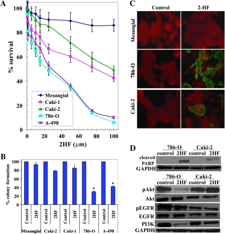
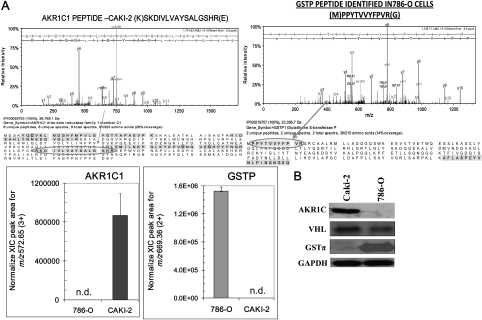
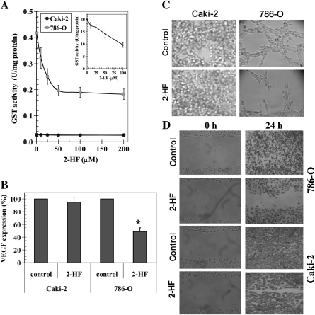
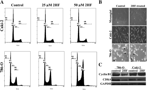
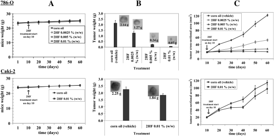
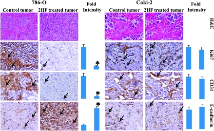
Renal cell carcinoma (RCC) is one of the top ten cancers prevalent in USA. Loss-of-function mutations in the von Hippel-Lindau (VHL) gene constitute an established risk factor contributing to 75% of total reported cases of RCC. Loss-of-VHL leads to a highly vascularized phenotype of renal tumors. Intake of citrus fruits has been proven to reduce the risk of RCC in multicenter international studies. Hence, we studied the effect of 2'-hydroxyflavanone (2HF), an active anticancer compound from oranges, in RCC. Our in vitro investigations revealed that 2HF suppresses VHL-mutant RCC to a significantly greater extent than VHL-wild-type RCC by inhibiting epidermal growth factor receptor signaling, which is increased due to VHL mutations in RCC. Our results also revealed for the first time, that 2HF inhibits glutathione S-transferase pi activity. 2HF reduced cyclin B1 and CDK4 levels and induced G2/M phase arrest in VHL-mutant RCC. Importantly, 2HF inhibited the angiogenesis in VHL-mutant RCC by decreasing vascular endothelial growth factor expression. Our in vivo studies in mice xenografts confirmed our in vitro results as evident by decreased levels of proliferation marker, Ki67 and angiogenic marker, CD31, in 2HF-treated mice xenografts of VHL-mutant RCC. 2HF also increased the expression of E-cadherin in VHL-mutant RCC, which would be of significance in restoring normal epithelial phenotype. Collectively, our in vitro and in vivo results revealed the potent antiproliferative, anti-angiogenic and prodifferentiation properties of 2HF in VHL-mutant RCC, sparing normal cells, which could have significant implications not only in the specific management of VHL-mutant RCC but also towards other VHL syndromes.


Figures

Fig. 1.
Fig. 1.
Enhanced anticancer effects of 2HF in VHL-mutant RCC sparing normal cells. Drug sensitivity assays were performed by MTT assay using 2HF at 72 h posttreatment to determine IC50. Values are presented as mean ± standard deviation from two separate determinations with eight replicates each (n = 16) (panel A). Colony-forming assay was performed and the colonies were counted using Innotech Alpha Imager HP as detailed in Materials and Methods. *P < 0.001 compared with control (panel B). For TUNEL apoptosis assay, cells were grown on coverslips and treated with 50 μM 2HF for 24 h. TUNEL assay was performed using Promega fluorescence detection kit and examined using Zeiss LSM 510 META laser scanning fluorescence microscope with filters 520 and >620 nm. Photographs taken at identical exposure at ×400 magnification are presented. Apoptotic cells showed green fluorescence (panel C). Effect of 2HF on poly ADP-ribose polymerase cleavage, EGFR, PI3K and Akt activation: VHL-wild-type (Caki-2) and VHL-mutant (786-O) control and 50 μM 2HF-treated cells were lysed and analyzed by western blot for poly ADP-ribose polymerase cleavage, pEGFR (Y1068), pAkt (S473) and PI3K (Y458/199) by using specific antibodies. Membranes were stripped and reprobed for glyceraldehyde 3-phosphate dehydrogenase as a loading control (panel D).
Fig. 2.
Fig. 2.
Differential expression of AKR1C1 and GSTπ in RCC as detected by LC-MS and MS/MS. Caki-2 and 786-O cells were subjected to proteomic analyses as described in Methods section. MS spectra were searched against a human protein database by Mascot software (Matrix Science) and label-free quantification: upper panel shows the MS/MS spectrum of one of the sequenced, isoforms-specific tryptic peptides for the respective proteins with the sequence coverage displayed below. The bar diagrams indicating the quantitative levels of AKR1C1 and GSTπ, respectively, were based on integration of extracted ion chromatograms, n = 4 in each group, for the triply and doubly charged isoform-specific tryptic peptides; n.d. denotes that the peptide was not detected in the samples (panel A). Differential protein expression was confirmed by performing western blot using 50 μg of cell lysates and antibodies against AKR1C, GSTπ and VHL. for glyceraldehyde 3-phosphate dehydrogenase was used as internal loading control. The experiment was repeated three times and similar results were obtained (panel B).
Fig. 3.
Fig. 3.
2HF inhibits GSTπ activity and angiogenesis. GST activity towards GSH and 1-chloro 2,4-dinitro benzene and its inhibition by 2HF was performed in 28  000g crude supernatant prepared from Caki-2 and 786-O cells. Recombinant purified GSTπ was used as a control (panel A and inset). The inhibitory effect of 2HF on GST was studied at a fixed concentration of GSH and GSH and 1-chloro 2,4-dinitro benzene (1 mM each) and varying concentrations of inhibitor. The enzymes were preincubated with the inhibitor for 5 min at 37°C prior to the addition of the substrates (panel A). VEGF expression in control and 2HF-treated cells by enzyme-linked immunosorbent assay kit (R&D System) (panel B). Effect of 2HF (50 μM) on tube formation of Caki-2 and 786-O cells on matrigel was assessed (panel C) and wound-healing assay shows that the 2HF inhibits 786-O cell migration as detailed in Materials and Methods (panel D).
Fig. 4.
Fig. 4.
Effect of 2HF on cell cycle progression in RCC. Inhibitory effect of 2HF on cell cycle distribution was determined by fluorescence activated cell sorting analysis. Experimental details are given in the Methods section. The stained cells were analyzed using the Beckman Coulter Cytomics FC500, Flow Cytometry Analyzer. The experiment was repeated three times and similar results were obtained (panel A). The cell morphology was observed and imaged at ×40 magnification in phase contrast microscope (Axio observer A1; Carl Zeiss microimaging, Thornwood, NY). Arrows in the panel point towards cells arrested in or completing cytokinesis (panel B). VHL-wild-type (Caki-2) and VHL-mutant (786-O) control and 50 μM 2HF-treated cells were processed for western blot analysis for cyclin B1 and CDK4 expression by using specific antibodies. Membranes were stripped and reprobed for GAPDH as a loading control (panel C).
Fig. 5.
Fig. 5.
Effect of oral administration of 2HF on tumor regression of RCC in nude mice: For 786-O RCC, mice were divided into four groups treated with corn oil (i.e. vehicle) and 2HF 0.0025, 0.005 and 0.01% (wt/wt) (equivalent to 25, 50 and 100 mg/kg body wt, respectively). For Caki-2 RCC, mice were divided into two groups treated with corn oil, and 2HF 0.01% (wt/wt) (equivalent to 100 mg/kg body wt). Experimental details are given in the Methods section. Animals were examined daily for signs of tumor growth and body weights were recorded (panels A). Photographs of animals were taken at day 1, day 10, day 20, day 40 and day 60 after subcutaneous injection are shown for all groups (data not shown). Weights and photographs of tumors were also taken at day 60 (panels B). Tumors were measured in two dimensions using calipers (panels C).
Fig. 6.
Fig. 6.
Histopathologic analyses of the markers of proliferation, angiogenesis and differentiation in tumor sections after 2HF treatment. Control and 2HF-treated RCC-bearing nude mice tumor sections were used for histopathologic analyses. Presented are hematoxylin and eosin stained sections, immuno-histochemistry analyses for Ki-67 expression (marker of cellular proliferation), CD31 (angiogenesis marker) and E-cadherin (tumor suppressor) from tumors in mice of control and 2HF-treated groups. Statistical significance of difference was determined by two-tailed Student’s t-test. P < 0.001, 786-O 2HF-treated compared with control. However, these differences were not significant in Caki-2. Immunoreactivity is evident as a dark brown stain, whereas non-reactive areas display only the background color. Sections were counterstained with hematoxylin (blue). Photomicrographs at ×40 magnification were acquired using Olympus Provis AX70 microscope. Percent staining was determined by measuring positive immunoreactivity per unit area. Arrows represent the area for positive staining for an antigen. The intensity of antigen staining was quantified by digital image analysis. Bars represent mean ± standard error (n = 5); *P < 0.001 compared with control.

References

    1. Linehan WM, et al. Genetic basis of cancer of the kidney: disease-specific approaches to therapy. Clin. Cancer Res. 2004;10:6282S–6289S. - PubMed
    1. Atkins MB, et al. Innovations and challenges in renal cell carcinoma: summary statement from the Second Cambridge Conference. Clin. Cancer Res. 2007;13:667s–670s. - PubMed
    1. Kuroda N, et al. Review of renal oncocytoma with focus on clinical and pathobiological aspects. Histol. Histopathol. 2003;18:935–942. - PubMed
    1. Hunt JD, et al. Renal cell carcinoma in relation to cigarette smoking: meta-analysis of 24 studies. Int. J. Cancer. 2005;114:101–108. - PubMed
    1. Van Dijk BA, et al. Cigarette smoking, von Hippel-Lindau gene mutations and sporadic renal cell carcinoma. Br. J. Cancer. 2006;95:374–377. - PMC - PubMed

Publication types

MeSH terms

Substances