Review
. 2011 Apr 7;117(14):3720-32.
doi: 10.1182/blood-2010-07-273417. Epub 2011 Feb 8.

Interleukin-1 in the pathogenesis and treatment of inflammatory diseases

Affiliations
Review

Interleukin-1 in the pathogenesis and treatment of inflammatory diseases

Charles A Dinarello. Blood. .

Abstract

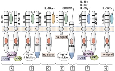
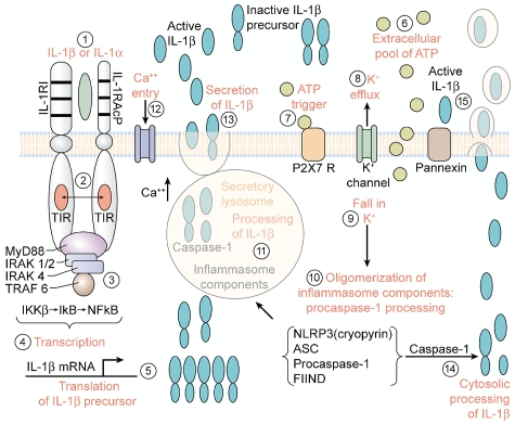
More than any other cytokine family, the IL-1 family of ligands and receptors is primarily associated with acute and chronic inflammation. The cytosolic segment of each IL-1 receptor family member contains the Toll-IL-1-receptor domain. This domain is also present in each Toll-like receptor, the receptors that respond to microbial products and viruses. Since Toll-IL-1-receptor domains are functional for both receptor families, responses to the IL-1 family are fundamental to innate immunity. Of the 11 members of the IL-1 family, IL-1β has emerged as a therapeutic target for an expanding number of systemic and local inflammatory conditions called autoinflammatory diseases. For these, neutralization of IL-1β results in a rapid and sustained reduction in disease severity. Treatment for autoimmune diseases often includes immunosuppressive drugs whereas neutralization of IL-1β is mostly anti-inflammatory. Although some autoinflammatory diseases are due to gain-of-function mutations for caspase-1 activity, common diseases such as gout, type 2 diabetes, heart failure, recurrent pericarditis, rheumatoid arthritis, and smoldering myeloma also are responsive to IL-1β neutralization. This review summarizes acute and chronic inflammatory diseases that are treated by reducing IL-1β activity and proposes that disease severity is affected by the anti-inflammatory members of the IL-1 family of ligands and receptors.


Figures

Figure 1
Figure 1
Signaling and inhibition of signaling by IL-1Rs. (A) IL-1α (either precursor or mature) or mature IL-1β bind to the IL-1RI and with the IL-1RAcP forms the receptor heterodimeric complex. The TIR domain of each receptor chain approximate recruit MyD88, followed by phosphorylation of IL-1R–associated kinases (IRAKs) and inhibitor of NFκB kinase β (IKKβ), resulting in a signal to the nucleus. (B) In the brain and spinal cord, the variant IL-1RAcPb can form the heterodimeric complex with IL-1α or IL-1β and IL-1RI, but this complex fails to recruit MyD88, and there is inhibition of the IL-1 signal. The failure to recruit MyD88 may be because of an altered TIR domain (indicated as TIRb). (C) IL-1Ra binds to IL-1RI, but there is no signal because there is failure to form a complex with IL-1RAcP. (D) IL-1β binds to the IL-1RII, but lacking a cytoplasmic segment, there is no signal. (E) Because of an altered TIR domain (indicated as TIRb), SIGIRR inhibits IL-1 and TLR signaling. SIGIRR can form a complex with IL-33 (not shown) and inhibit IL-33 signaling. (F) IL-1Rrp2 binds IL-36α, IL-36β, or IL-36γ and forms a complex with IL-1RAcP. The TIR domain of each receptor chain approximates and recruits MyD88 similar to that shown in panel A. (G) IL-36Ra binds to IL-1Rrp2 but fails to form a complex with IL-1RAcP. Thus, IL-36Ra prevents the binding of IL-36α, IL-36β, or IL-36γ to IL-1Rrp2, and IL-36Ra is the natural receptor antagonist IL-36.
Figure 2
Figure 2
Steps in the processing and release of IL-1 induced by IL-1. (1) Primary blood monocytes, tissue macrophages or dendritic cells are activated by either mature IL-1β or the IL-1α precursor with the formation of the IL-1 receptor complex heterodimer comprised of the IL-1RI with IL-1RAcP. (2) Approximation of the intracellular TIR domains. (3) Recruitment of MyD88 and phosphorylation of IL-1R–associated kinases (IRAKs) and inhibitor of NFκB kinase β (IKKβ). (4) Transcription of IL-1β. (5) Translation into the IL-1β mRNA takes place on polysomes. IL-1β mRNA is not bound to actin microfilaments but rather intermediate filaments. (6) ATP released from the activated monocyte/macrophage accumulates extracellularly. (7) Activation of the P2X7 receptor by ATP. (8) Efflux of potassium from the cell after ATP binding to P2X7 receptor. (9) Fall in intracellular levels of potassium. (10) The fall in intracellular potassium levels triggers the assembly of the components of the caspase-1 inflammasome with the conversion of procaspase-1 to active caspase-1. (11) Caspase-1 is found in the secretory lysosome together with the IL-1β precursor and lysosomal enzymes. Active caspase-1 cleaves the IL-1β precursor in the secretory lysosome, generating the active, carboxyl-terminal mature IL-1β. (12) An influx of calcium with an increase in intracellular calcium levels. The rise in intracellular calcium activates phosphatidylcholine-specific phospholipase C and calcium-dependent phospholipase A. (13) The release of mature IL-1β, the IL-1β precursor, and the contents of the secretory lysosomes by exocytosis in the absence of cell death. (14) Processing of the IL-1β precursor in the cytosol. Rab39a, a member of the GTPase family, contributes to the secretion of by helping traffic IL-1β from the cytosol into a vesicle compartment. Exocytosis is another mechanism described in mouse macrophages. (15) Mature IL-1β exists the cells via loss in membrane integrity, associated with the release of lactic dehydrogenase or microvesicles. TRAF indicates TNF receptor-associated factor.
Figure 3
Figure 3
Non–caspase-1 extracellular processing of the IL-1β precursor. The 269-aa-long IL-1β precursor is shown with the caspase-1 site at aspartic acid (D) at position 116. Extracellular protease sites are indicated by their amino acid recognition sites. The probable proteinase-3 site was derived from combinatorial methods of the specific substrate.
Figure 4
Figure 4
Role of IL-1α in necrosis versus apoptosis. (1 left and right) In healthy cells of mesenchymal origin, the IL-1α precursor is found diffusely in the cytoplasm but also in the nucleus where it binds to chromatin. (2 right) During normal cell turnover, an apoptotic signal drives cytoplasmic IL-1α into the nucleus and is no longer a dynamic in the cell. The cell shrinks. (2 left) Cells exposed to hypoxia begin to die, and nuclear IL-1α moves out of the nucleus into the cytoplasm. Taking on water, the cell swells as the necrotic process begins. (3 left) As the necrotic process continues, there is loss of membrane integrity, and cytoplasmic contents containing the IL-1α precursor leak out. (3 right) Tissue macrophages take up the apoptotic cell into endocytotic vesicles. (4) In the vesicles, the apoptotic cell is digested, and there is no inflammatory response from the macrophage. (5 left) The IL-1α precursor is released into the extracellular compartment and binds to IL-1RI expressed on adjacent cells or to resident tissue macrophages. The tissue macrophage responds with synthesis of the IL-1β precursor as well as increased in caspase-1. From step 3 left, ATP is also released on cell death and activates the P2X7 receptor for activation of caspase-1. (6) Caspase-1 is activated by the inflammasome, cleaves the IL-1β precursor, and mature, active IL-1β is released. Alternatively, IL-1β is released via pyroptosis. (7) Once released, IL-1β induces chemokine production, resulting in a chemoattractant gradient. (8) As the endothelium of the microcirculation expresses adhesion molecule, blood neutrophils adhere and cross into the ischemic area. (9) With the infiltration of myeloid cells (monocytes and neutrophils), there is expanded inflammation, which extends beyond the initial area of ischemia.

Similar articles

Cited by

References

    1. Dinarello CA. Biological basis for interleukin-1 in disease. Blood. 1996;87(6):2095–2147. - PubMed
    1. Opal SM, Fisher CJJ, Dhainaut JF, et al. Confirmatory interleukin-1 receptor antagonist trial in severe sepsis: a phase III, randomized, double-blind, placebo-controlled, multicenter trial. Crit Care Med. 1997;25(7):1115–1124. - PubMed
    1. Eichacker PQ, Parent C, Kalil A, et al. Risk and the efficacy of antiinflammatory agents: retrospective and confirmatory studies of sepsis. Am J Respir Crit Care Med. 2002;166(9):1197–1205. - PubMed
    1. Lust JA, Lacy MQ, Zeldenrust SR, et al. Induction of a chronic disease state in patients with smoldering or indolent multiple myeloma by targeting interleukin 1beta-induced interleukin 6 production and the myeloma proliferative component. Mayo Clin Proc. 2009;84(2):114–122. - PMC - PubMed
    1. Simon A, van der Meer JW. Pathogenesis of familial periodic fever syndromes or hereditary autoinflammatory syndromes. Am J Physiol Regul Integr Comp Physiol. 2007;292(1):R86–98. - PubMed

Publication types

MeSH terms