. 2011 Feb 8;4(159):ra8.
doi: 10.1126/scisignal.2001446.

The crystal structure of a self-activating G protein alpha subunit reveals its distinct mechanism of signal initiation

Affiliations

The crystal structure of a self-activating G protein alpha subunit reveals its distinct mechanism of signal initiation

Janice C Jones et al. Sci Signal. .

Abstract

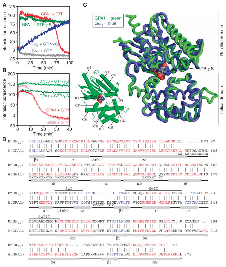
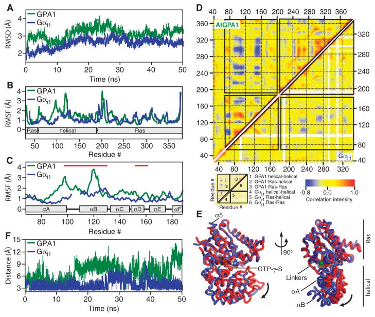
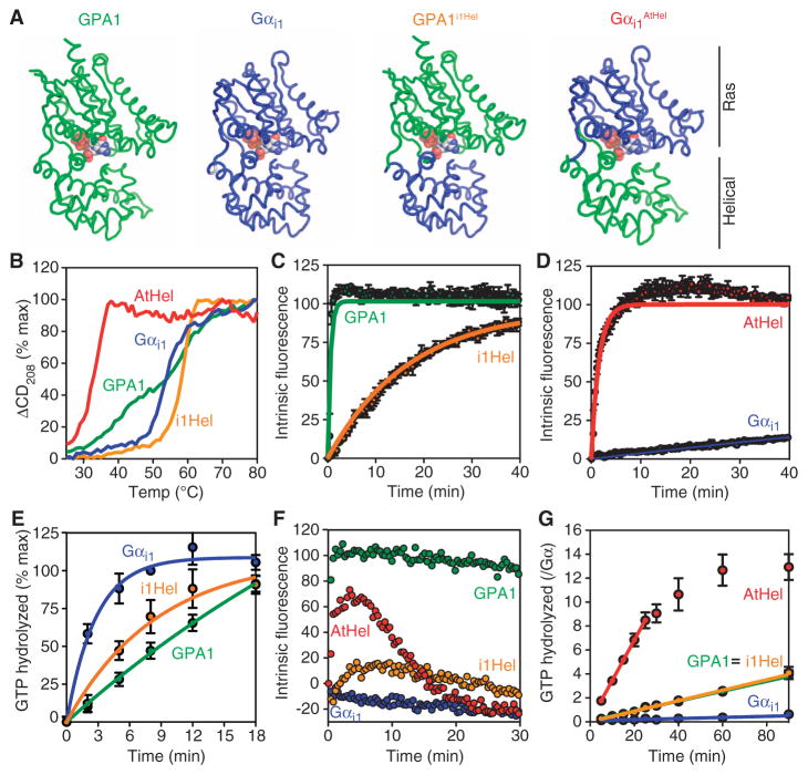
In animals, heterotrimeric guanine nucleotide-binding protein (G protein) signaling is initiated by G protein-coupled receptors (GPCRs), which activate G protein α subunits; however, the plant Arabidopsis thaliana lacks canonical GPCRs, and its G protein α subunit (AtGPA1) is self-activating. To investigate how AtGPA1 becomes activated, we determined its crystal structure. AtGPA1 is structurally similar to animal G protein α subunits, but our crystallographic and biophysical studies revealed that it had distinct properties. Notably, the helical domain of AtGPA1 displayed pronounced intrinsic disorder and a tendency to disengage from the Ras domain of the protein. Domain substitution experiments showed that the helical domain of AtGPA1 was necessary for self-activation and sufficient to confer self-activation to an animal G protein α subunit. These findings reveal the structural basis for a mechanism for G protein activation in Arabidopsis that is distinct from the well-established mechanism found in animals.


Conflict of interest statement

Competing interests: The authors declare that they have no competing interests.

Figures

Fig. 1
Fig. 1
The crystal structure of a self-activating G protein α subunit and its comparison to that of Gαi1. (A) Fluorescence-based measurement of the binding and hydrolysis of GTP. Purified AtGPA1 (GPA1) or Gαi1 (400 nM) was equilibrated at 23°C before GTP (1 μM) or nonhydrolyzable GTP-γ-S (1 μM) was added to the cuvette, and the change in intrinsic fluorescence (arbitrary units) of the G protein α subunit was monitored. Data are representative of three experiments. (B) Fluorescence-based measurement of the GTP-binding and hydrolysis properties of wild-type AtGPA1 and a truncated form of AtGPA1 that lacks the N-terminal 36–amino acid residues (ΔN36), as described for (A), but with 400 nM GTP or GTP-γ-S. (C) Superposition of the crystal structures of AtGPA1–GTP-γ-S (green) and Gαi1–GTP-γ-S (blue; PDB ID: 1GIA). (D) Structure-based sequence alignment of AtGPA1–GTP-γ-S and Rattus norvegicusi1–GTP-γ-S. Vertical lines between sequences denote residues at equivalent locations in the two structures. Residues not modeled in any of the three molecules in the asymmetric unit of AtGPA1 are marked with “o.” Residues not modeled in at least two of the three molecules are marked with an “x.” Red residues constitute α helices, whereas blue residues constitute β strands. Abbreviations for the amino acid residues are as follows: A, Ala; C, Cys; D, Asp; E, Glu; F, Phe; G, Gly; H, His; I, Ile; K, Lys; L, Leu; M, Met; N, Asn; P, Pro; Q, Gln; R, Arg; S, Ser; T, Thr; V, Val; W, Trp; and Y, Tyr.
Fig. 2
Fig. 2
All-atom MD simulations with AtGPA1 and Gαi1. All of the MD simulations were conducted with apo-Gαi1 (PDB# 1CIP, without GTP-γ-S) and apo-AtGPA1 (this report). (A) RMSDs from the starting crystal structures as a function of simulation time were calculated from the MD simulation. (B) RMSFs as a function of residue number. (C) RMSF for the G protein α-helical domains of AtGPA1 and Gαi1. Red lines mark residues that were not included in the crystallographic model for monomers B and C of AtGPA1 because of their poor electron density. (D) Covariance matrices for pairs of residues in apo-AtGPA1 and apo-Gαi1. The boxes in the main plot outline correlated movements between the Ras and helical domains as indicated in the schematic. Positively correlated movements are indicated in red, whereas negatively correlated movements are indicated in blue. (E) Structure and dynamics of AtGPA1 based on the most frequent modes of motion from movie S1. The crystal structure of AtGPA1 is depicted in red, and the ending structure for the third most frequent mode of motion is depicted in blue. (F) Distance between the side chains of Asp150 (D150) and Lys270 (K270) of Gαi1 and the comparable residues Asp162 (D162) and Lys288 (K288) of AtGPA1 during the 50-ns simulation.
Fig. 3
Fig. 3
Kinetics and unfolding properties of G protein α subunit chimeras. (A) Cartoon representations of the chimeras used in this study. (B) Temperature-induced unfolding of the indicated G protein α subunits as monitored by CD spectroscopy at 208 nm. (C and D) GTP-γ-S binding rates were measured from intrinsic fluorescence changes as described in Fig. 1 with GTP-γ-S (2 μM). (E) Single-turnover GTP hydrolysis. Purified His-tagged G protein α subunits (900 nM) were loaded with [γ-32P]GTP before the reaction was started by addition of Mg2+. Hydrolyzed 32PO4 was extracted with charcoal and quantified. (F) Fluorescence-based GTP binding and hydrolysis were measured as described in Fig. 1 with GTP (400 nM). (G) Steady-state GTP hydrolysis. Purified G protein α subunits (400 nM) were incubated with [γ-32P]GTP (10 μM) for the indicated times before hydrolyzed 32PO4 was extracted with charcoal and quantified. For all of the panels, data are representative of at least two experiments. Error bars indicate SEM.

References

    1. Sprang SR. G protein mechanisms: Insights from structural analysis. Annu Rev Biochem. 1997;66:639–678. - PubMed
    1. Kimple RJ, Kimple ME, Betts L, Sondek J, Siderovski DP. Structural determinants for GoLoco-induced inhibition of nucleotide release by Gα subunits. Nature. 2002;416:878–881. - PubMed
    1. Chen Z, Singer WD, Sternweis PC, Sprang SR. Structure of the p115RhoGEF rgRGS domain-Gα13/i1 chimera complex suggests convergent evolution of a GTPase activator. Nat Struct Mol Biol. 2005;12:191–197. - PubMed
    1. Marotti LA, Jr, Newitt R, Wang Y, Aebersold R, Dohlman HG. Direct identification of a G protein ubiquitination site by mass spectrometry. Biochemistry. 2002;41:5067–5074. - PubMed
    1. Johnston CA, Taylor JP, Gao Y, Kimple AJ, Grigston JC, Chen JG, Siderovski DP, Jones AM, Willard FS. GTPase acceleration as the rate-limiting step in Arabidopsis G protein-coupled sugar signaling. Proc Natl Acad Sci USA. 2007;104:17317–17322. - PMC - PubMed

Publication types

MeSH terms

Substances