. 2011 Mar 16;30(6):1162-72.
doi: 10.1038/emboj.2011.20. Epub 2011 Feb 8.

The conserved factor DE-ETIOLATED 1 cooperates with CUL4-DDB1DDB2 to maintain genome integrity upon UV stress

Affiliations

The conserved factor DE-ETIOLATED 1 cooperates with CUL4-DDB1DDB2 to maintain genome integrity upon UV stress

Enric Castells et al. EMBO J. .

Abstract

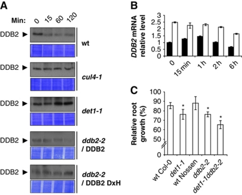
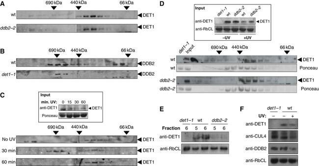
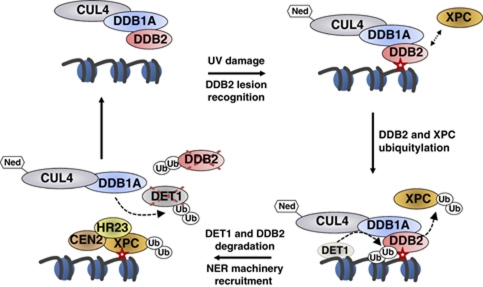
Plants and many other eukaryotes can make use of two major pathways to cope with mutagenic effects of light, photoreactivation and nucleotide excision repair (NER). While photoreactivation allows direct repair by photolyase enzymes using light energy, NER requires a stepwise mechanism with several protein complexes acting at the levels of lesion detection, DNA incision and resynthesis. Here we investigated the involvement in NER of DE-ETIOLATED 1 (DET1), an evolutionarily conserved factor that associates with components of the ubiquitylation machinery in plants and mammals and acts as a negative repressor of light-driven photomorphogenic development in Arabidopsis. Evidence is provided that plant DET1 acts with CULLIN4-based ubiquitin E3 ligase, and that appropriate dosage of DET1 protein is necessary for efficient removal of UV photoproducts through the NER pathway. Moreover, DET1 is required for CULLIN4-dependent targeted degradation of the UV-lesion recognition factor DDB2. Finally, DET1 protein is degraded concomitantly with DDB2 upon UV irradiation in a CUL4-dependent mechanism. Altogether, these data suggest that DET1 and DDB2 cooperate during the excision repair process.


Conflict of interest statement

The authors declare that they have no conflict of interest.

Figures

Figure 1
Figure 1
DET1 dosage influences UV-C sensitivity upon recovery in both light and dark conditions. (A) mycDET1 overexpression in DET1 OE transgenic lines. Equal amounts of whole protein extracts from epitope-tagged mycDET1 lines OE-1, OE-2 and OE-3 were loaded on a 10% SDS–PAGE and analysed by immunoblot using an anti-myc antibody. The same blot was analysed with an anti-tubulin antibody as loading control. (B, C) Root growth inhibition upon UV-C exposure. Four-day-old seedlings with the indicated genotypes were exposed to UV-C (600 J/m2) and immediately returned to normal light conditions (B) or to complete darkness (C). Relative root growth was determined 24 h after irradiation by comparison with the respective non-irradiated control of the same genotype (100% root growth). Error bars represent standard deviations from three replicate experiments (n>20). Asterisks indicate t-test significant differences at P⩽0.05 relative to wild-type control at same dose.
Figure 2
Figure 2
DET1 is required for synthesis-dependent repair of UV-induced DNA lesions. (A) Modulation of DET1 level affects the removal of 6,4PP and CPD photoproducts by light-independent DNA repair. Fourteen-day-old wild-type (Col-0), uvr3phrI, cul4-1, det1-1 and DET1 OE-3 seedlings were irradiated with UV-C (1000 J/m2) and harvested immediately after irradiation or allowed to repair for 24 h in darkness. Serial dilutions of genomic DNA were subjected to immunodot-blot analysis using anti-CPD (white bars) or anti-6,4PP (black bars) antibodies, respectively, and normalized relative to 5-methylcytosine content. For each genotype, the percent of CPDs and 6,4PPs remaining after 24 h was calculated relative to the initial level immediately after UV irradiation. Error bars represent standard deviations from three independent experiments. Asterisks indicate t-test significant differences at P⩽0.05 relative to wild-type controls at the same dose. (B) Arabidopsis DET1-defective plants are impaired in synthesis-dependent repair of UV-induced DNA lesions in vitro. Cell extracts from wild-type and det1-1 mutant plants were incubated for 0, 1 or 2 h with UV-C damaged (+UV) and undamaged control plasmid (−UV) in the presence of DIG-dUTP to monitor synthesis-dependent DNA repair efficiency.
Figure 3
Figure 3
Genetic interactions between det1-1 and rad10 and their effect on UV-C sensitivity. (A, B) Root growth inhibition upon UV-C exposure. Four-day-old seedlings with the indicated genotypes were exposed to UV-C (600 J/m2) and immediately returned to normal light conditions (A) or to complete darkness (B) for 24 h. Relative root growth was determined 24 h after irradiation by comparison with the respective non-irradiated control of the same genotype (100% root growth). Error bars represent standard deviations from three replicate experiments (n>20). Asterisks indicate t-test significant differences at P⩽0.05 relative to wild-type controls at the same dose, while plus signs (+) indicate no significant difference with respect to the det1-1 mutant.
Figure 4
Figure 4
CUL4 interconnects with DET1 and triggers DET1 protein degradation upon UV-C exposure. (A) CUL4 and DET1 mutations are epistatic for UV-C sensitivity. Seedlings with the indicated genotypes were exposed to UV-C (600 J/m2) and immediately placed in complete darkness. Relative root growth was determined 24 h after irradiation by comparison with respective non-irradiated controls of the same genotype (100% root growth). Error bars represent standard deviations from three replicate experiments (n>20). Asterisks indicate t-test significant differences at P⩽0.05 relative to wild-type controls at the same dose. (B) Immunoblot analysis of endogenous DET1 content before (0) or 15, 60 and 120 min after UV-C exposure (3000 J/m2) in wild-type and cul4-1 seedlings. Coomassie blue staining (lower panels) is shown as loading controls. Asterisks indicate cross-reacting bands. (C) Analysis of DET1 expression upon UV-C exposure. Quantitative RT–PCR analysis was used to monitor DET1 mRNA levels in 10-day-old wild-type seedlings (Col-0) harvested at the indicated times after UV-C exposure (900 J/m2). Error bars indicate standard deviations from two biological replicates. (D) Immunoblot analysis of mycDET1 protein content before (0) or 30 and 60 min after UV-C exposure (3000 J/m2). Equal amounts of whole protein extracts were loaded on a 10% SDS–PAGE and analysed by immunoblot using anti-DET1 antibody. The same blot was probed with anti-RbCL antibody as a loading control.
Figure 5
Figure 5
DDB2 degradation following UV exposure is dependent on DET1. (A) Immunoblot analysis of endogenous DDB2 content upon UV-C exposure (3000 J/m2) in seedlings from wild-type, cul4-1, det1-1 and ddb2-2 mutant expressing native DDB2 protein or DDB2 protein with mutated WDxR motif (WDxH). Coomassie blue staining is shown as loading control (lower panels). (B) Analysis of DDB2 gene expression upon UV-C exposure. Quantitative RT–PCR analysis was used to monitor DDB2 mRNA levels in 10-day-old wild-type Col-0 (black bars) and det1-1 (white bars) seedlings harvested at the indicated times after UV-C exposure (600 J/m2). Error bars indicate standard deviations from two biological replicates. (C) DDB2 and DET1 mutations show additive effects for UV-C sensitivity. Seedlings with the indicated genotypes were exposed to 600 J/m2 of UV-C and immediately placed in complete darkness. Relative root growth was determined 24 h after irradiation by comparison with respective non-irradiated controls of the same genotype (100% root growth). Error bars represent standard deviations from three replicate experiments (n>20). Asterisks indicate t-test significant differences at P⩽0.05 relative to wild-type controls at the same dose.
Figure 6
Figure 6
A high-molecular weight complex containing DET1 coelutes transiently with DDB2 following UV-C exposure. (A, B) DET1 and DDB2 fractionate in different protein complexes under normal light conditions. (A) Size-exclusion chromatography analysis of DET1 complex in protein extracts from wild-type (Nossen) and ddb2-2 mutant seedlings. Fractions were analysed by immunoblot using DET1 and DDB2 antibodies. Arrows indicate elution of molecular-weight standards in the same conditions. (B) Size-exclusion chromatography analysis of DDB2 complex in protein extracts from wild-type (Col-0) and det1-1 mutant. (C) A DET1 high-molecular weight complex is transiently detected upon UV-C irradiation. Experiment was performed as in (A) except that seedlings were exposed to UV-C (900 J/m2) and harvested after 30 or 60 min for protein extraction and size-exclusion chromatography. The inset shows DET1 protein content in input samples before chromatography separation. (D) Size-exclusion chromatography of protein extracts from wild-type Nossen and ddb2-2 mutant plants irradiated at 900 J/m2 UV-C and harvested 30 min after UV exposure. The inset shows DET1 input content in wild-type, det1-1 and ddb2-2 seedlings harvested before (−UV) or 30 min after UV-C exposure (+UV). Ponceau staining is used as control for equal fractionation of the two samples. Arrows indicate elution of molecular-weight standards in the same conditions. (E) Side-by-side analysis of fractions 5 and 6 containing DET1 UV-induced HMW complex. Fractions 5 and 6 (50 μl) from size-exclusion separation of wild-type Nossen, ddb2-2 and det1-1 extracts were analysed by immunoblot using a DET1 antibody. The same blot was probed with anti-RbCL antibody as a loading control. (F) Protein content of DET1, DDB2 and CUL4 in det1-1 mutant and wild type before and 30 min after UV-C exposure. Whole cell protein extracts (40 μg) were separated on 10% SDS–PAGE and analysed with the indicated antibodies.
Figure 7
Figure 7
A working model for DET1 cooperation with CUL4–DDB1DDB2 in DNA repair. UV-DNA photoproduct (red star) is detected and bound by DDB2, which focalizes neddylated (Ned) CUL4–DDB1 ubiquitin ligase on the damaged region. In mammals, XPC is recruited and poly-ubiquitylated by CUL4–DDB1DDB2, thereby enhancing its binding to DNA. DDB2 protein is subsequently ubiquitylated and degraded by a CUL4–DDB1- and DET1-dependent mechanism. This involves the formation of a transient large DET1 complex presumably containing CUL4 in addition to DDB1, which is a stable partner of DET1 through direct protein–protein interaction. DET1 would then also be targeted by a CUL4–DDB1 ubiquitin ligase for degradation. Proteolysis of DDB2 and DET1 may allow their eviction from chromatin to facilitate recruiting the NER machinery, which is initiated by binding of the heterotrimeric XPC–HR23–CEN2 (plant RAD4-RAD23-CEN2) factor onto the lesion. DET1 has the capacity to bind histone H2B, represented here on an adjacent nucleosome for simplicity, but whether it acts as a soluble or histone-bound factor for DDB2 degradation remains to be determined.

Similar articles

Cited by

References

    1. Al Khateeb WM, Schroeder DF (2009) Overexpression of Arabidopsis damaged DNA binding protein 1A (DDB1A) enhances UV tolerance. Plant Mol Biol 70: 371–383 - PubMed
    1. Angers S, Li T, Yi X, MacCoss MJ, Moon RT, Zheng N (2006) Molecular architecture and assembly of the DDB1-CUL4A ubiquitin ligase machinery. Nature 443: 590–593 - PubMed
    1. Barneche F, Winter V, Crevecoeur M, Rochaix JD (2006) ATAB2 is a novel factor in the signalling pathway of light-controlled synthesis of photosystem proteins. EMBO J 25: 5907–5918 - PMC - PubMed
    1. Benvenuto G, Formiggini F, Laflamme P, Malakhov M, Bowler C (2002) The photomorphogenesis regulator DET1 binds the amino-terminal tail of histone H2B in a nucleosome context. Curr Biol 12: 1529–1534 - PubMed
    1. Bernhardt A, Lechner E, Hano P, Schade V, Dieterle M, Anders M, Dubin MJ, Benvenuto G, Bowler C, Genschik P, Hellmann H (2006) CUL4 associates with DDB1 and DET1 and its downregulation affects diverse aspects of development in Arabidopsis thaliana. Plant J 47: 591–603 - PubMed

Publication types

MeSH terms

LinkOut - more resources