. 2011 Feb 8:2:181.
doi: 10.1038/ncomms1182.

The Ufm1-activating enzyme Uba5 is indispensable for erythroid differentiation in mice

Affiliations
Free PMC article

The Ufm1-activating enzyme Uba5 is indispensable for erythroid differentiation in mice

Kanako Tatsumi et al. Nat Commun. .
Free PMC article

Abstract

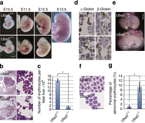
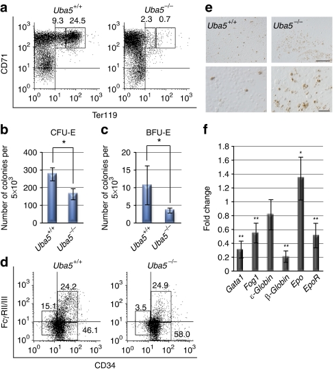
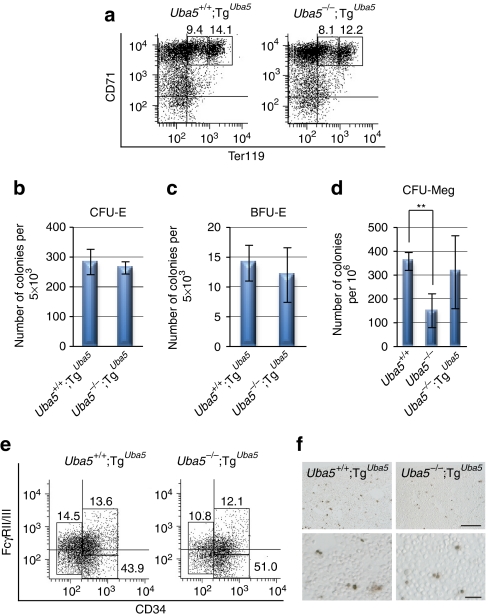
Post-translational protein modifications are systems designed to expand restricted genomic information through functional conversion of target molecules. Ubiquitin-like post-translational modifiers regulate numerous cellular events through their covalent linkages to target protein(s) by an enzymatic cascade analogous to ubiquitylation consisting of E1 (activating), E2 (conjugating) and E3 (ligating) enzymes. In this study, we report the essential role of Uba5, a specific activating enzyme for the ubiquitin-like modifier, Ufm1, in erythroid development. Mice lacking Uba5 exhibited severe anaemia, followed by death in utero. Although Uba5 was dispensable for the production of erythropoietin, its genetic loss led to impaired development of megakaryocyte and erythroid progenitors from common myeloid progenitors. Intriguingly, transgenic expression of Uba5 in the erythroid lineage rescued the Uba5-deficient embryos from anaemia and prolonged their survival, demonstrating the importance of Uba5 in cell-autonomous erythroid differentiation. Our results suggest that one of the ubiquitin-like protein modification systems, the Ufm1 system, is involved in the regulation of haematopoiesis.


Figures

Figure 1
Figure 1. Loss of Uba5 leads to death in utero because of severe anaemia.
(a) Morphological examination of Uba5−/− mice embryos. Representative photographs of Uba5+/+ and Uba5−/− embryos at various developmental stages. Uba5−/− embryo at E12.5 exhibited severe anaemia. Scale bar, 2 mm. (b) Haematoxylin and eosin staining of fetal liver sections of Uba5+/+ and Uba5−/− mice at E11.5. Scale bars: left panel, 1 mm; right panel, 20 μm. (c) Number of erythrocytes in fetal livers of Uba5+/+ and Uba5−/− mice at E11.5. (d) Immunohistochemistry of fetal liver sections of Uba5+/+ and Uba5−/− embryos at E11.5 with anti-ɛ-globin and anti-β-globin antibodies. Arrows: abnormal multinucleated erythrocytes. Scale bar, 20 μm. (e) Representative photographs of Uba5+/+ and Uba5−/− embryos at E11.5. The circulating erythrocyte volume is reduced in the yolk sac of Uba5−/− embryos. Scale bar, 2 mm. (f) Wright–Giemsa staining of peripheral blood cytospin cells prepared from the indicated genotypes at E11.5. Scale bar, 20 μm. (g) Percentage of abnormal erythrocytes shown in f. Bar graphs in c and g show the mean±s.d. values of five mice from each group. Statistical analysis was carried out using the unpaired t-test. *P<0.05 (Welch test).
Figure 2
Figure 2. Uba5 is indispensable for the development of erythroid cells.
(a) Representative FACS analysis. Mouse fetal liver cells were freshly isolated from E11.5 embryos and labelled with a FITC-conjugated anti-CD71 monoclonal antibody and PE-conjugated anti-Ter119 antibody. Left gate: proerythroblast population (Ter119med CD71high), right gate: basoerythroblast population (Ter119high CD71high). Axes mean log10 fluorescent intensity. (b) Quantification of erythroid colony-forming units (CFU-Es) and (c) erythroid burst-forming units (BFU-Es). Colony assays were carried out with cells prepared from fetal liver at E11.5. Uba5 deletion was associated with decreased erythroid progenitor activity. Data are means±s.d. of five mice from each group. Statistical analysis was performed using the unpaired t-test. *P<0.05 (Welch test). (d) Representative FACS analysis. The myeloid progenitors (LinIL-7RSca-1c-Kit+) prepared from fetal liver cells of the indicated genotype mice at E11.5 were subfractionated into presumptive common myeloid progenitors (CD34highFcγRII/IIIlow), granulocyte/macrophage progenitors (CD34highFcγRII/IIIhigh) and megakaryocyte/erythroid progenitors (CD34lowFcγRII/IIIlow) by FACS. Axes mean log10 fluorescent intensity. (e) TUNEL staining of fetal livers of Uba5+/− and Uba5−/− mice at E12.5. Scale bars: top panel, 100 μm; bottom panel, 20 μm. (f) Quantification of mRNA levels of erythroid-related genes, including Gata1, Fog1 and EPO, in Uba5-deficient liver at E11.5. Total RNAs were prepared from wild-type or Uba5-deficient livers and reverse transcribed into their respective cDNAs, which were used as templates for quantitative real-time PCR. Values were expressed relative to the amount of the respective mRNA in the wild-type livers. The experiments were conducted three times. Data are means±s.d. of three experiments. Statistical analysis was carried out by the unpaired t-test. *P<0.05 and **P<0.01 (Welch test).
Figure 3
Figure 3. Correction of anaemia in Uba5-deficient mice by expression of Uba5 in erythroid lineage.
(a) Embryonic morphology of Uba5 rescue mice. Representative photographs of Uba5+/+;TgUba5 and Uba5−/−;TgUba5 embryos at various embryonic stages. Scale bar, 2 mm. (b) Haematoxylin and eosin-stained fetal liver sections of Uba5+/+;TgUba5 and Uba5−/−;TgUba5 mice at E11.5. Scale bars: left panel, 1 mm; right panel, 20 μm. (c) Number of erythroid cells in fetal livers of Uba5+/+;TgUba5 and Uba5−/−;TgUba5 mice at E11.5. Data are means±s.d. of five mice from each group. (d) Wright–Giemsa-stained samples of peripheral blood cytospin cells prepared from control and rescued mice at E11.5. Scale bar, 20 μm. (e) Percentage of abnormal erythrocytes shown in d. Data are means±s.d. of five mice in each group.
Figure 4
Figure 4. Suppression of defective differentiation of both erythrocytes and megakaryocytes following expression of Uba5 in erythroid lineage.
(a) FACS analysis. Mouse fetal liver cells were freshly isolated from control and rescued mice at E11.5 and labelled with an FITC-conjugated anti-CD71 monoclonal antibody and PE-conjugated anti-Ter119 antibody. Left gate: proerythroblast population (Ter119med CD71high), right gate: basoerythroblast population (Ter119high CD71high). Axes mean log10 fluorescent intensity. (b) Quantification of erythroid colony-forming units (CFU-Es) and (c) erythroid burst-forming units (BFU-Es). Colony assays were carried out with cells prepared from control and rescued fetal liver at E11.5. The number of CFU-E (b) and BFU-E (c) colonies remained unchanged in both genotypes. Data are means±s.d. of five mice from each group. (d) Colony formation assay for megakaryocytes (CFU-Meg). The assays were carried out with cells prepared from the fetal liver of the indicated genotypes at E11.5. Uba5 deletion was accompanied by a weaker megakaryocytic differentiation activity, and this defect was suppressed by forced expression of Uba5 in megakaryocytic lineage. Data are means±s.d. of Uba5+/− (n=3), Uba5−/− (n=4) and Uba5−/−;TgUba5 (n=3). Statistical analysis was carried out using the unpaired t-test. **P<0.01 (Welch test). (e) FACS analysis. The myeloid progenitors (LinIL-7RSca-1c-Kit+) prepared from liver cells of control and rescued fetuses at E11.5 were subfractionated into presumptive common myeloid progenitors (CD34highFcγRII/IIIlow), granulocyte/macrophage progenitors (CD34highFcγRII/IIIhigh) and megakaryocyte/erythroid progenitors (CD34lowFcγRII/IIIlow) by FACS. Axes mean log10 fluorescent intensity. (f) TUNEL staining of the fetal livers of control and rescue mice at E12.5. Scale bars: top panel, 100 μm; bottom panel, 20 μm.

Similar articles

Cited by

References

    1. Hershko A. & Ciechanover A. The ubiquitin system. Annu. Rev. Biochem. 67, 425–479 (1998). - PubMed
    1. Goldberg A. L. Protein degradation and protection against misfolded or damaged proteins. Nature 426, 895–899 (2003). - PubMed
    1. Schulman B. A. & Harper J. W. Ubiquitin-like protein activation by E1 enzymes: the apex for downstream signalling pathways. Nat. Rev. Mol. Cell Biol. 10, 319–331 (2009). - PMC - PubMed
    1. Hochstrasser M. Origin and function of ubiquitin-like proteins. Nature 458, 422–429 (2009). - PMC - PubMed
    1. Iwasaki H. & Akashi K. Myeloid lineage commitment from the hematopoietic stem cell. Immunity 26, 726–740 (2007). - PubMed

Publication types