. 2011 Jun;31(6):1461-74.
doi: 10.1038/jcbfm.2011.9. Epub 2011 Feb 9.

Inhibition of VEGF signaling pathway attenuates hemorrhage after tPA treatment

Affiliations

Inhibition of VEGF signaling pathway attenuates hemorrhage after tPA treatment

Masato Kanazawa et al. J Cereb Blood Flow Metab. 2011 Jun.

Abstract

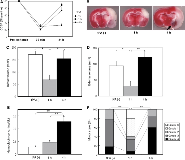
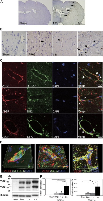
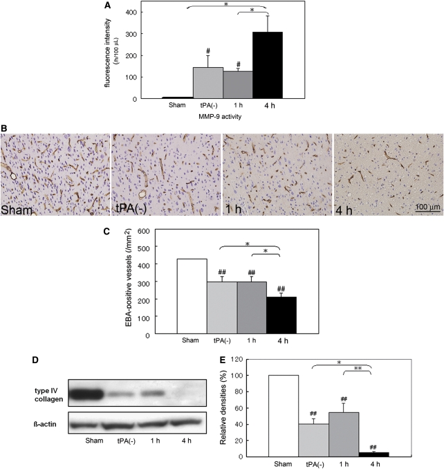
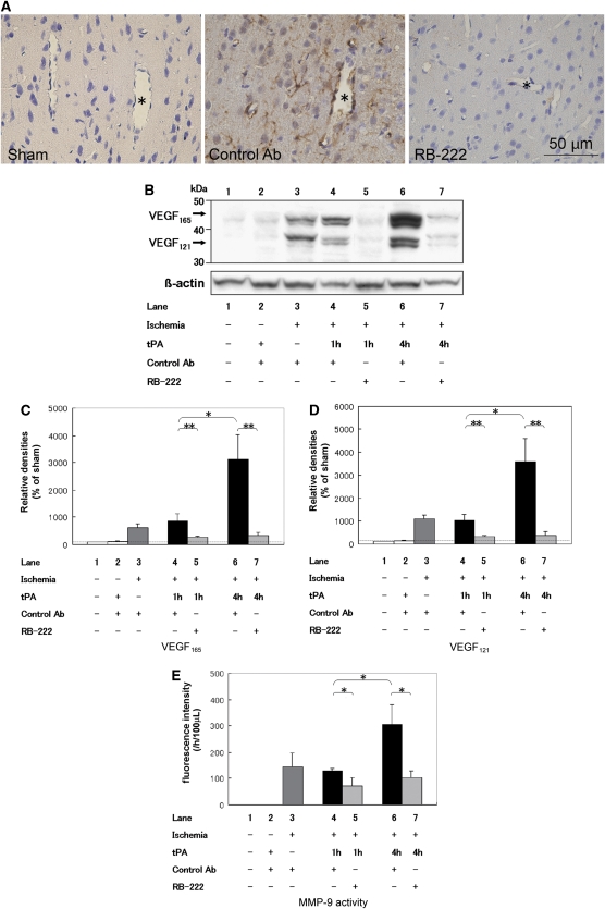
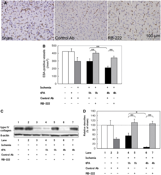
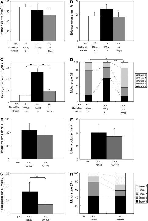
An angiogenic factor, vascular endothelial growth factor (VEGF), might be associated with the blood-brain barrier (BBB) disruption after focal cerebral ischemia; however, it remains unknown whether hemorrhagic transformation (HT) after tissue plasminogen activator (tPA) treatment is related to the activation of VEGF signaling pathway in BBB. Here, we hypothesized that inhibition of VEGF signaling pathway can attenuate HT after tPA treatment. Rats subjected to thromboembolic focal cerebral ischemia were assigned to a permanent ischemia group and groups treated with tPA at 1 or 4 hours after ischemia. Anti-VEGF neutralizing antibody or control antibody was administered simultaneously with tPA. At 24 hours after ischemia, we evaluated the effects of the antibody on the VEGF expression, matrix metalloproteinase-9 (MMP-9) activation, degradation of BBB components, and HT. Delayed tPA treatment at 4 hours after ischemia promoted expression of VEGF in BBB, MMP-9 activation, degradation of BBB components, and HT. Compared with tPA and control antibody, combination treatment with tPA and the anti-VEGF neutralizing antibody significantly attenuated VEGF expression in BBB, MMP-9 activation, degradation of BBB components, and HT. It also improved motor outcome and mortality. Inhibition of VEGF signaling pathway may be a promising therapeutic strategy for attenuating HT after tPA treatment.


Figures

Figure 1
Figure 1
Rat thromboembolic model produces hemorrhagic transformation (HT) and shows increased mortality rate after delayed 4 hours treatment of tissue plasminogen activator (tPA). (A) Cerebral cortical blood flow (CCBF) measured by laser Doppler flowmetry at preischemia and postischemia (30 minutes and 24 hours after ischemia) among the permanent ischemia group (n=23, circle), tPA 1-hour group (n=15, square), and tPA 4-hour group (n=22, triangle). CCBF was expressed as the percentage changes from preischemic baseline values. tPA treatment at 1 or 4 hours after ischemia improved CCBFs. (B) Representative figures of 2,3,5-triphenyltetrazolium chloride (TTC)-stained brain sections of rats from the permanent ischemia group (left), tPA 1-hour group (middle), and tPA 4-hour group (right). TTC staining indicates deep red staining of normal brain tissue and white nonstaining of the ischemic lesion. (CE) The volumes of infarct (C) and edema (D), and hemoglobin concentrations (E). White bars indicate permanent ischemia group; gray bars, tPA 1-hour group; black bars, tPA 4-hour group (n=6). (F) Motor scales at 24 hours after ischemia (n⩾15 per group). *P<0.05, **P<0.01.
Figure 2
Figure 2
Tissue plasminogen activator (tPA) treatment at 4 hours after ischemia promotes expression of vascular endothelial growth factor (VEGF) in the blood–brain barrier (BBB). (A) VEGF expression in the brains of the sham-operated group and permanent ischemia group. Arrows indicate VEGF expressions in the brains from the permanent ischemia group. (B) VEGF expression in the arterioles (arrows) of the brains from the sham-operated group, permanent ischemia group: tPA(−), and tPA 1 and 4 hours groups. Moreover, a secondary-only antibody control confirms that extracellular staining of VEGF after ischemia was not nonspecific. (C) Confocal microscopy studies for VEGF localization after ischemia from permanent ischemia group. Triple staining of VEGF (red), rat endothelial cell antigen-1 (RECA-1, a marker of endothelial cells), or nerve/glial antigen-2 (NG2, a marker of pericytes), or glial fibrillary acidic protein (GFAP, a marker of astrocytes; green), and 4′,6′-diamidino-2-phenylindole (DAPI, nuclear staining; blue) were performed. VEGF colocalized with endothelial cells (arrows), pericytes (arrowheads), and astrocytic foot processes (asterisks). Additionally, a secondary-only antibody control did not show any staining. (D) Three-dimensional reconstruction images also showed that VEGF colocalized with endothelial cells, pericytes, and astrocytes, which constitute the BBB. (E) Representative immunoblottings from the sham-operated group, permanent ischemia group, and tPA 1 and 4 hours groups. (F) Optical densitometric analyses for VEGF165 (left panel) and VEGF121 (right panel). Data represent relative optical densities of ischemic brain samples compared with those of sham-operated samples (n=3). #P<0.05 versus sham group, ##P<0.01 versus sham group, *P<0.05, **P<0.01.
Figure 3
Figure 3
Tissue plasminogen activator (tPA) treatment at 4 hours after ischemia promotes degradation of blood–brain barrier (BBB) components. (A) Matrix metalloproteinase-9 (MMP-9) activities of the lysates from the sham-operated or ischemic cortices at 24 hours after ischemia by fluorimetric assay using fluorescence resonance energy transfer (FRET) peptides as substrates. While MMP-9 activity was undetectable in the lysates from the sham-operated cortex, it increased after ischemic stroke, especially in the tPA 4-hour group. #P<0.05 versus sham group, *P<0.05. (B, C) Representative figures (B) and the number (C) of endothelial barrier antigen (EBA)-positive vessels from the sham-operated group, permanent ischemia group, and tissue plasminogen (tPA) 1 and 4 hours groups (n=3). The numbers of EBA-positive vessels decreased after ischemia, especially in the tPA 4-hour group. ##P<0.01 versus sham group, *P<0.05. (D, E) Representative immunoblotting (D) and optical densitometric analyses for type IV collagen (E) at 24 hours after ischemia. Data represent relative optical densities of ischemic brain samples compared with those of sham-operated samples (n=3). The expressions of type IV collagen were also decreased after ischemia, especially in the tPA 4-hour group. ##P<0.01 versus sham group, *P<0.05, **P<0.01.
Figure 4
Figure 4
RB-222 attenuates vascular endothelial growth factor (VEGF) expression and matrix metalloproteinase-9 (MMP-9) activation after tissue plasminogen activator (tPA) treatment. (A) Representative figures of VEGF expression in the sham-operated cortex, and the ischemic cortex treated with control antibody (anti-human IgG antibody) or RB-222. Asterisks indicate cavities in the arterioles. (BE) Representative immunoblotting (B) and optical densitometric analyses for VEGF165 (C) and VEGF121 (D), and MMP-9 activities (E) at 24 hours after ischemia. Lane 1: sham, lane 2: treatment with tPA and control antibody without ischemia, lane 3: treatment with control antibody at 1 hour after ischemia, lane 4: treatment with tPA and control antibody at 1 hour after ischemia, lane 5: treatment with tPA and RB-222 at 1 hour after ischemia, lane 6: treatment with tPA and control antibody at 4 hours after ischemia, lane 7: treatment with tPA and RB-222 at 4 hours after ischemia. Note that the tPA treatment without ischemia did not promote VEGF expression (B, lane 2), and that RB-222 inhibited VEGF expression (B, lanes 5 and 7) as well as MMP-9 activities (E, lanes 5 and 7) both in the tPA 1- and 4-hour groups (n=3). *P<0.05, **P<0.01.
Figure 5
Figure 5
RB-222 attenuates the degradation of blood–brain barrier (BBB) components after tissue plasminogen activator (tPA) treatment. (A, B) Representative figures (A) and the number (B) of endothelial barrier antigen (EBA)-positive vessels from the sham-operated group and tPA 4-hour group with control antibody or RB-222 at 4 hours after ischemia. **P<0.01. Representative immunoblotting (C) and optical densitometric analyses (D) for type IV collagen at 24 hours after ischemia. Lane 1: sham, lane 2: treatment with tPA and control antibody without ischemia, lane 3: treatment with control antibody at 1 hour after ischemia, lane 4: treatment with tPA and control antibody at 1 hour after ischemia, lane 5: treatment with tPA and RB-222 at 1 hour after ischemia, lane 6: treatment with tPA and control antibody at 4 hours after ischemia, lane 7: treatment with tPA and RB-222 at 4 hours after ischemia (n=3). *P<0.05, **P<0.01.
Figure 6
Figure 6
RB-222 or vascular endothelial growth factor receptor-2 (VEGFR2) inhibitor, SU1498 attenuates hemorrhagic transformation (HT) after tissue plasminogen activator (tPA) treatment. (AC) Quantification of infarct volumes (A), edema volumes (B), and hemoglobin concentrations of the lysates from the ischemic hemisphere (C) at 24 hours after ischemia. White bars indicate the permanent ischemia group treated with control antibody alone; black bars, the tPA 4-hour group treated with control antibody; gray bars, the tPA 4-hour group treated with RB-222 (n=6). **P<0.01. (D) Motor scales at 24 hours after ischemia of the permanent ischemia group treated with control antibody alone, and the tPA 4-hour group treated with control antibody or RB-222 (n⩾14). Outcomes were scored using the 5-point motor function scale: 0=no motor deficit, 1=flexion of the forelimb contralateral to the ischemic hemisphere, 2=reduced resistance against push toward the paretic side, 3=spontaneously circling toward the paretic side, and 4=death. *P<0.05. **P<0.01. (EG) Quantification of infarct volume (E), edema volume (F), and hemoglobin concentration of the lysates from the ischemic hemisphere (G) at 24 hours after ischemia. Black bars indicate the tPA 4-hour group treated with vehicle (dimethyl sulfoxide); gray bars, the tPA 4-hour group treated with SU1498 (n=6). **P<0.01. (H) Motor scales at 24 hours after ischemia of the tPA 4-hour group treated with SU1498 and vehicle (n=14).

References

    1. Al Ahmad A, Gassmann M, Ogunshola OO. Maintaining blood-brain barrier integrity: pericytes perform better than astrocytes during prolonged oxygen deprivation. J Cell Physiol. 2009;218:612–622. - PubMed
    1. Andersen M, Overgaard K, Meden P, Boysen G. Effects of citicoline combined with thrombolytic therapy in a rat embolic stroke model. Stroke. 1999;30:1464–1471. - PubMed
    1. Asahi M, Asahi K, Wang X, Lo EH. Reduction of tissue plasminogen activator-induced hemorrhage and brain injury by free radical spin trapping after embolic focal cerebral ischemia in rats. J Cereb Blood Flow Metab. 2000;20:452–457. - PubMed
    1. Bergers G, Brekken R, McMahon G, Vu TH, Itoh T, Tamaki K, Tanzawa K, Thorpe P, Itohara S, Werb Z, Hanahan D. Matrix metalloproteinase-9 triggers the angiogenic switch during carcinogenesis. Nat Cell Biol. 2000;2:737–744. - PMC - PubMed
    1. Busch E, Kruger K, Hossmann KA. Improved model of thromboembolic stroke and rt-PA induced reperfusion in the rat. Brain Res. 1997;778:16–24. - PubMed

Publication types

MeSH terms

Substances