. 2011 Jan 20;7(1):e1001054.
doi: 10.1371/journal.pcbi.1001054.

Using transcription modules to identify expression clusters perturbed in Williams-Beuren syndrome

Affiliations

Using transcription modules to identify expression clusters perturbed in Williams-Beuren syndrome

Charlotte N Henrichsen et al. PLoS Comput Biol. .

Abstract

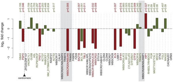
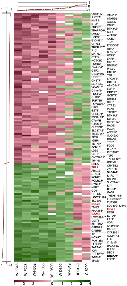
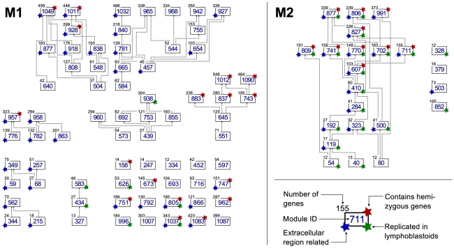
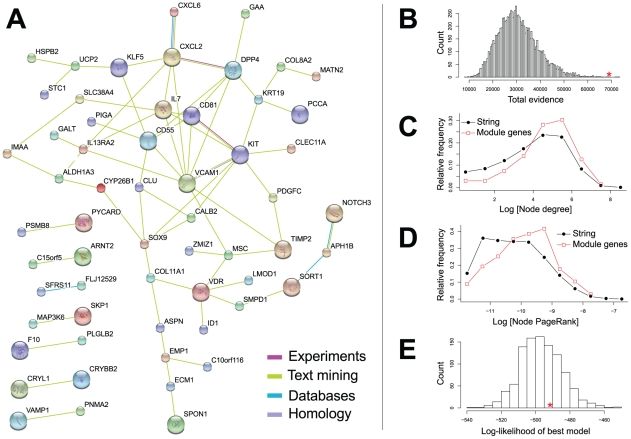
The genetic dissection of the phenotypes associated with Williams-Beuren Syndrome (WBS) is advancing thanks to the study of individuals carrying typical or atypical structural rearrangements, as well as in vitro and animal studies. However, little is known about the global dysregulations caused by the WBS deletion. We profiled the transcriptomes of skin fibroblasts from WBS patients and compared them to matched controls. We identified 868 differentially expressed genes that were significantly enriched in extracellular matrix genes, major histocompatibility complex (MHC) genes, as well as genes in which the products localize to the postsynaptic membrane. We then used public expression datasets from human fibroblasts to establish transcription modules, sets of genes coexpressed in this cell type. We identified those sets in which the average gene expression was altered in WBS samples. Dysregulated modules are often interconnected and share multiple common genes, suggesting that intricate regulatory networks connected by a few central genes are disturbed in WBS. This modular approach increases the power to identify pathways dysregulated in WBS patients, thus providing a testable set of additional candidates for genes and their interactions that modulate the WBS phenotypes.


Conflict of interest statement

The authors have declared that no competing interests exist.

Figures

Figure 1
Figure 1. Differential expression of the WBS hemizygous and flanking genes.
Genes are ordered according to their chromosomal position. Shaded areas represent the LCRs flanking the deletion. Gene names are indicated at the bottom and corresponding differential expression P-values at the top. For genes with multiple probesets the most significant P-value is considered. Red bars indicate significance (P<0.05). Genes without a P-value were not detected on the array and thus not tested.
Figure 2
Figure 2. Example of a WBS dysregulated module (#770 from the M2 module set).
This module contains 149 genes (one per line) and 9 samples (columns). Seven samples are from WBS patients (denoted with “W”), C-5290 is a control sample from our dataset, while HPGS-9 belongs to a publicly available dataset. Gene scores are plotted on the left and sample scores at the top. The 59 genes with positive gene scores (bottom lines) are downregulated (green) in the seven WBS samples and upregulated (red) in the other two. The remaining 90 genes show the opposite pattern: they are upregulated in the WBS samples and downregulated in the remaining two samples. Hemizygous gene names are emphasized in red and the names of genes mapping to HSA7 in boldface. Red asterisks indicate genes belonging to the GO category “extracellular region” while black asterisks denote genes from the “intrinsic to membrane” category.
Figure 3
Figure 3. Hierarchical diagram of the transcription modules dysregulated in WBS identified in the M1 (left) and M2 (right) modular studies.
Directed edges indicate direct subset relationships, and they always point upwards. The number of genes in a module is shown at the top left corner of the module box. Modules annotated with a red star on their top right corner contain at least one hemizygous (or flanking) gene; the ones with green stars on their bottom right corner were replicated in lymphoblastoid cell lines; blue stars on the bottom left corner indicate modules that show significant enrichment for extracellular region genes. An interactive version of this figure is available in the online supporting material at http://www.unil.ch/cbg/ISA/Fibroblasts, which allows to further query the gene content and functional enrichment of the modules.
Figure 4
Figure 4. The network of the most frequent genes in the modules, as a subset of the STRING protein interaction database.
Only genes that appear at least ten times in the dysregulated modules are considered. (A) Most frequent module genes that have at least one connection in the STRING database. Edges with evidence score higher than 0.3 are shown; their colors indicate different kinds of interaction evidence (key bottom right). (B) Most frequent module genes form a network that is denser than a random subnetwork of the same size in STRING. We generated 10,000 random subnetworks and calculated the sum of the evidence for all edges. Only five out of all random subnetworks show a higher total evidence value than the most frequent module genes indicated by a red asterisk (sum of total evidence = 69,033). (C) Distribution of the number of connections (node degree) per protein in the complete STRING network (black, filled circles), and the subnetwork of most frequent module genes (red, open squares). The subnetwork has significantly less low-degree nodes and more high-degree nodes (Wilcoxon-test P = 1.612×10⁻⁵). (D) Distribution of PageRank centrality scores in the complete STRING network and the subnetwork of most frequent module genes. The subnetwork has fewer non-central nodes and more central nodes (Wilcoxon-test P = 2.628×10⁻⁵). (E) We fitted hierarchical models to the subnetwork of the most frequent module genes, and also to 1,000 randomized networks. The network of frequent module genes (red asterisk) shows no hierarchical structure compared to the randomized networks.

Similar articles

Cited by

References

    1. Attias J, Raveh E, Ben-Naftali N, Zarchi O, Gothelf D. Hyperactive auditory efferent system and lack of acoustic reflexes in Williams syndrome. J Basic Clin Physiol Pharmacol. 2008;19:193–207. - PubMed
    1. Cherniske EM, Carpenter TO, Klaiman C, Young E, Bregman J, et al. Multisystem study of 20 older adults with Williams syndrome. Am J Hum Genet. 2004;131:255–264. - PubMed
    1. Järvinen-Pasley A, Bellugi U, Reilly J, Mills DL, Galaburda A, et al. Defining the social phenotype in Williams syndrome: a model for linking gene, the brain, and behavior. Dev Psychopathol. 2008;20:1–35. - PMC - PubMed
    1. Korenberg JR, Chen XN, Hirota H, Lai Z, Bellugi U, et al. VI. Genome structure and cognitive map of Williams syndrome. J Cognitive Neurosci. 2000;12(Suppl 1):89–107. - PubMed
    1. Meyer-Lindenberg A, Weinberger DR. Intermediate phenotypes and genetic mechanisms of psychiatric disorders. Nat Rev Neurosci. 2006;7:818–827. - PubMed

Publication types