. 2011 Jan 27;6(1):e16447.
doi: 10.1371/journal.pone.0016447.

Induction of the Wnt antagonist Dickkopf-1 is involved in stress-induced hippocampal damage

Affiliations

Induction of the Wnt antagonist Dickkopf-1 is involved in stress-induced hippocampal damage

Francesco Matrisciano et al. PLoS One. .

Abstract

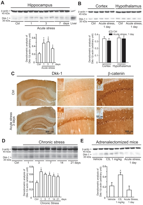
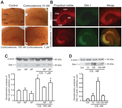
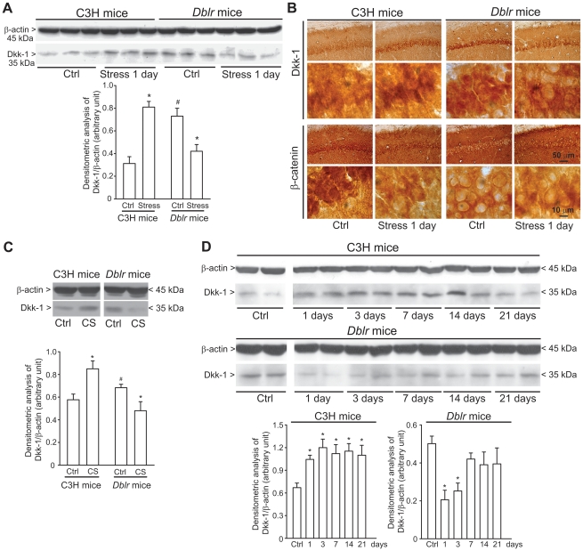
The identification of mechanisms that mediate stress-induced hippocampal damage may shed new light into the pathophysiology of depressive disorders and provide new targets for therapeutic intervention. We focused on the secreted glycoprotein Dickkopf-1 (Dkk-1), an inhibitor of the canonical Wnt pathway, involved in neurodegeneration. Mice exposed to mild restraint stress showed increased hippocampal levels of Dkk-1 and reduced expression of β-catenin, an intracellular protein positively regulated by the canonical Wnt signalling pathway. In adrenalectomized mice, Dkk-1 was induced by corticosterone injection, but not by exposure to stress. Corticosterone also induced Dkk-1 in mouse organotypic hippocampal cultures and primary cultures of hippocampal neurons and, at least in the latter model, the action of corticosterone was reversed by the type-2 glucocorticoid receptor antagonist mifepristone. To examine whether induction of Dkk-1 was causally related to stress-induced hippocampal damage, we used doubleridge mice, which are characterized by a defective induction of Dkk-1. As compared to control mice, doubleridge mice showed a paradoxical increase in basal hippocampal Dkk-1 levels, but no Dkk-1 induction in response to stress. In contrast, stress reduced Dkk-1 levels in doubleridge mice. In control mice, chronic stress induced a reduction in hippocampal volume associated with neuronal loss and dendritic atrophy in the CA1 region, and a reduced neurogenesis in the dentate gyrus. Doubleridge mice were resistant to the detrimental effect of chronic stress and, instead, responded to stress with increases in dendritic arborisation and neurogenesis. Thus, the outcome of chronic stress was tightly related to changes in Dkk-1 expression in the hippocampus. These data indicate that induction of Dkk-1 is causally related to stress-induced hippocampal damage and provide the first evidence that Dkk-1 expression is regulated by corticosteroids in the central nervous system. Drugs that rescue the canonical Wnt pathway may attenuate hippocampal damage in major depression and other stress-related disorders.


Conflict of interest statement

Competing Interests: The authors have declared that no competing interests exist.

Figures

Figure 1
Figure 1. Induction of Dkk-1 in the hippocampus of CD1 mice exposed to acute or chronic restraint stress.
A representative immunoblot of Dkk-1 in hippocampal extracts of mice at 1, 3 or 7 days following a single exposure to mild restrain stress (“acute stress”) is shown in (A). Data are means ± S.E.M. of 6–9 individual determinations. *p<0.05 (One-way ANOVA + Fisher's PLSD vs. Control (Ctrl) mice. A representative immunoblot of Dkk-1 in extracts from cortex and hypothalamus of mice at 1 day following a single exposure to mild restrain stress (“acute stress”) is shown in (B). Data are means ± S.E.M. of 5-6 individual determinations. Immunohistochemical analysis of Dkk-1 and â-catenin in control mice and mice killed 3 days after acute stress is shown in (C). Note the lack of Dkk-1 immunostaining in cell nuclei in the inset at high magnification. Immunoblot analysis of Dkk-1 in mice exposed to repeated episodes of mild restrain stress (once a day for 21 consecutive days) (“chronic stress”) is shown in (D). Mice were killed 24 h after the last exposure to stress at day 1, 3, 7, 14, and 21 (i.e. mice were killed at day 2, 4, 8, 15, and 22). Values are means ± S.E.M. of 6–9 determinations. *p<0.05 (One-way ANOVA + Fisher's PLSD) vs. controls. Immunoblot analysis of Dkk-1 levels in the hippocampus of adrenalectomized mice injected with vehicle or corticosterone (CS, 1 mg/kg, s.c.) or exposed to acute stress is shown in E. The group of adrenalictomized mice was also injected with vehicle to allow comparisons with the two previous groups. Mice were killed 24 h after treatments. Values are means ± S.E.M. of 6–7 determinations. *p<0.05 (One-way ANOVA + Fisher's PLSD) vs. unstressed mice injected with vehicle.
Figure 2
Figure 2. Exogenous corticosterone enhances Dkk-1 expression in organotypical hippocampal slice cultures and cultured hippocampal neurons.
Expression of Dkk-1 in organotypic hippocampal cultures exposed to different concentrations of corticosterone for is shown in (A and B). Cultures were incubated with corticosterone or vehicle (Ctrl) for 24 h and then processed for DAB (A) or immunofluorescent (B) analysis. In (B) propidium iodide staining is in red, Dkk-1 immunostaining in green. Note that Dkk-1 induction is visible in CA3, CA4, and CA1 hippocampal subfields. The experiment has been repeated three times with identical results. Induction of Dkk-1 by corticosterone (CS) in primary cultures of mouse or rat hippocampal neurons is shown in C and D, respectively. The time of incubation with CS was 24 h in C and 16 h in D. Note that induction of Dkk-1 by corticosterone was antagonized by mifepristone (MP, 10 µM) but not by spironolactone (SP, 10 µM). MP and SP were applied to the cultures 10 min prior to CS and mainteined in the culture medium for 16–24 h. Ctrl  =  control cultures treated with vehicle. Values are means ± S.E.M. of 5–6 individual determinations from 2 independent cultures. *p<0.05 (One-way ANOVA + Fisher's PLSD) vs. Ctrl.
Figure 3
Figure 3. Dkk-1 response to stress or corticosterone in C3H and doubleridge mice.
Immunoblot analysis of Dkk-1 in the hippocampus of C3H or doubleridge (Dblr) mice exposed to acute restraint stress and killed 24 h later is shown in (A). Note that basal Dkk-1 levels were higher in the hippocampus of doubleridge mice and that acute stress enhanced Dkk-1 levels in C3H control mice but reduced Dkk-1 levels in doubleridge mice. Values are means ± S.E.M. of 6 determinations. p<0.05 (One-way ANOVA + Fisher's PLSD) vs. the respective control values (*) or vs. unstressed (Ctrl) C3H mice (#). Immunohistochemical analysis of Dkk-1 and β-catenin in the hippocampus of control C3H and doubleridge mice and C3H and doubleridge mice killed 1 day after acute stress is shown in (B). Immunoblot analysis of Dkk-1 levels in the hippocampus of C3H or doubleridge mice subjected to chronic restraint stress (once a day for consecutive 21 days; see legend of Figure 1) is shown in (C). Values are means ± S.E.M. of 5–8 determinations. *p<0.05 (One-way ANOVA + Fisher's PLSD) vs. control mice of the respective strain. In (D), we show the differential effect of 24-h treatment with 1 µM corticosterone (CS) on Dkk-1 levels in primary cultures of hippocampal neurons prepared from C3H or doubleridge mice. Values are means ± S.E.M. of 6 determinations from 2 independent cultures. p<0.05 (One-way ANOVA + Fisher's PLSD) vs. the respective control values (*).
Figure 4
Figure 4. Chronic restraint stress causes hippocampal damage in C3H mice, but not in doubleridge mice.
Measurements of hippocampal volume in C3H and doubleridge (Dblr) mice subjected to chronic restraint stress (one a day for 21 consecutive days) is shown in (A). Mice were killed 24 h after the last stress episode. Representative serial Nissl-stained sections showing the entire extension of the hippocampus in unstressed (Ctrl) and stressed CH3 and doubleridge mice are shown on the left side. Values are means ± S.E.M. of 8 determinations. *p<0.05 (Student's t test) vs. the respective controls. Stereological counting of Nissl stained neurons are shown in (B). Arrowheads in the left pannels show the presence of piknotic cells. Values are means ± S.E.M. of 8 determinations. *p<0.05 (Student's t test) vs. the respective controls. Golgi staining of CA1 neurons with real measures of dendritic lenght is shown in (C). Camera lucida representative images of CA1 neurons of unstressed or stressed C3H or Dblr mice is shown in (D). Mice were subjected to chronic restraint stress as reported in Figure 3. Values shown in the two graphs in (E) are means ± S.E.M. of 8–9 determinations. p<0.05 (One-way ANOVA + Fisher's PLSD) vs. the respective controls (i.e. unstressed mice of the same strain) (*) or vs. unstressed (Ctrl) C3H mice (#).
Figure 5
Figure 5. Chronic stress reduces proliferation of neuroprogenitors and neurogenesis in the dentate gyrus of C3H mice, but enhances proliferation of neuroprogenitors in doubleridge mice.
Dot-blot analysis of BrdU incorporation into DNA in hippocampal extracts from unstressed or stressed C3H or doubleridge (Dblr) mice is shown in (A). Mice were subjected to chronic restraint stress (one a day for 21 consecutive days) and killed 24 h after the last stress episode. A representative dot-blotting is on the left side. Values are means ± S.E.M. of 6–8 determinations. p<0.05 (One-way ANOVA + Fisher's PLSD) vs. the respective controls (unstressed mice) (*) or vs. unstressed (Ctrl) C3H mice (#). BrdU immunostaining is shown in (B) (left side). Stereological values are means ± S.E.M. of 6 determinations. p<0.05 (One-way ANOVA + Fisher's PLSD) vs. the respective controls (unstressed mice) (*) or vs. unstressed (Ctrl) C3H mice (#). Doublecortin (DCX) immunostaining is shown in (C) (left side). Stereological values are means ± S.E.M. of 6 determinations. *p<0.05 (One-way ANOVA + Fisher's PLSD) vs. the respective controls (unstressed mice).

Similar articles

Cited by

References

    1. Krishnan V, Nestler EJ. The molecular neurobiology of depression. Nature. 2008;455:894–902. - PMC - PubMed
    1. Brouwer JP, Appelhof BC, Hoogendijk WJ, Huyser J, Endert E, et al. Thyroid and adrenal axis in major depression: a controlled study in outpatients. Eur J Endocrinol. 2005;152:185–191. - PubMed
    1. Brown ES, Varghese FP, McEwen BS. Association of depression with medical illness: does cortisol play a role? Biol Psychiatry. 2004;55:1–9. - PubMed
    1. Parker KJ, Schatzberg AF, Lyons DM. Neuroendocrine aspects of hypercortisolism in major depression. Horm Behav. 2003;43:60–66. - PubMed
    1. Sapolsky RM. Glucocorticoids and hippocampal atrophy in neuropsychiatric disorders. Arch Gen Psychiatry. 2000;57:925–935. - PubMed

Publication types

MeSH terms

Substances