. 2011 Jan 27;7(1):e1001263.
doi: 10.1371/journal.ppat.1001263.

Early target cells of measles virus after aerosol infection of non-human primates

Affiliations

Early target cells of measles virus after aerosol infection of non-human primates

Ken Lemon et al. PLoS Pathog. .

Abstract

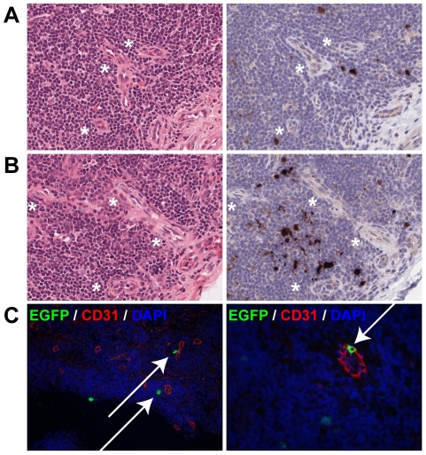
Measles virus (MV) is highly infectious, and has long been thought to enter the host by infecting epithelial cells of the respiratory tract. However, epithelial cells do not express signaling lymphocyte activation molecule (CD150), which is the high-affinity cellular receptor for wild-type MV strains. We have generated a new recombinant MV strain expressing enhanced green fluorescent protein (EGFP), based on a wild-type genotype B3 virus isolate from Khartoum, Sudan (KS). Cynomolgus macaques were infected with a high dose of rMV(KS)EGFP by aerosol inhalation to ensure that the virus could reach the full range of potential target cells throughout the entire respiratory tract. Animals were euthanized 2, 3, 4 or 5 days post-infection (d.p.i., n = 3 per time point) and infected (EGFP(+)) cells were identified at all four time points, albeit at low levels 2 and 3 d.p.i. At these earliest time points, MV-infected cells were exclusively detected in the lungs by fluorescence microscopy, histopathology and/or virus isolation from broncho-alveolar lavage cells. On 2 d.p.i., EGFP(+) cells were phenotypically typed as large mononuclear cells present in the alveolar lumen or lining the alveolar epithelium. One to two days later, larger clusters of MV-infected cells were detected in bronchus-associated lymphoid tissue (BALT) and in the tracheo-bronchial lymph nodes. From 4 d.p.i. onward, MV-infected cells were detected in peripheral blood and various lymphoid tissues. In spite of the possibility for the aerosolized virus to infect cells and lymphoid tissues of the upper respiratory tract, MV-infected cells were not detected in either the tonsils or the adenoids until after onset of viremia. These data strongly suggest that in our model MV entered the host at the alveolar level by infecting macrophages or dendritic cells, which traffic the virus to BALT or regional lymph nodes, resulting in local amplification and subsequent systemic dissemination by viremia.


Conflict of interest statement

A.D.M.E. Osterhaus wishes to declare, for the avoidance of any misunderstanding on competing interests, that he founded and is chief scientific officer of Viroclinics, a company set up in collaboration with Erasmus University. However, for clarification, no materials or support were received from the company, and no agreements were in place concerning the execution or publication of this work.

Figures

Figure 1
Figure 1. Generation and growth of rMVKSEGFP.
(A) Plasmids generated after RT-PCR, cloning and sequencing of MV RNA isolated from MVKS-infected PBMC. pMVKS is a full-length plasmid containing the complete antigenome of MVKS and pMVKSEGFP was modified by the insertion of an ATU at the promoter proximal position containing the ORF encoding EGFP. (B) rMVKS and rMVKSEGFP were rescued from Vero-SLAM cells and passaged in B-LCL. Fluorescence microscopy confirmed high levels of EGFP expression in rMVKSEGFP infected cells. (C) Growth curves of MVKS, rMVKS, rMVKSEGFP and rMVIC323EGFP in human B-LCL. Virus was harvested 24, 48, 72 and 96 hours post infection, CCID50 was determined in an endpoint titration test. Measurements shown are averages of triplicates ± SD. Key: h.p.i.: hours post infection.
Figure 2
Figure 2. Early rMVKSEGFP replication in the respiratory tract.
(A) Virus isolation performed from nose and throat swabs (left two panels), and from BAL cells (right panel). Each symbol represents an individual animal, bars indicate the geometric mean. Key: VI: virus isolation; d.p.i.: days post infection. (B) Live cell confocal microscopy performed on agarose-inflated lung slices from animals on 2 and 3 d.p.i. EGFP+ cells are shown in green, DAPI was used to counter stain nuclei (blue). Three images were collected, labeled i, ii and iii. Panels iia and iib show infected cells in one image from different orientations. Matching 3D-videos for (Bi, Bii and Biii) are available as supporting data.
Figure 3
Figure 3. Systemic rMVKSEGFP replication.
(A) MV load in PBMC and LN. The left panel shows virus isolations performed from PBMC, each symbol represents an individual animal, bars indicate the geometric means. The right panel shows the presence of MV genome in the axillary LN (crosshairs, geometric mean in blue) and in the tracheobronchial LN (triangles, geometric mean in red). Key: VI: virus isolation; RT-PCR: real-time reverse transcriptase PCR; d.p.i.: days post infection. (B) Detection of EGFP+ cells by flow cytometry from the retropharyngeal LN (left) and the tracheobronchial LN (right) on 2, 3, 4 and 5 d.p.i. Data are shown as dot plots of FL-1 (EGFP) versus FL-2 (empty channel), generated with BD FACSDiva software. In these plots autofluorescent cells usually appear on a diagonal line as they cause comparable signals in both channels. The EGFP-positive events were gated as indicated by the curvilinear line. Data of a representative animal are shown on each time point. Numbers of EGFP+ cells per million total cells are shown in each plot. (C) Representative example of macroscopic EGFP detection at 5 d.p.i. Arrow indicates the infected tonsillar tissue expressing EGFP. Key: Tg: tongue; Tn: tonsil; L: larynx.
Figure 4
Figure 4. Characterization of MV infection in BALT structures.
(A) H&E staining on lung slice from an animal euthanized on 3 d.p.i.. The number of EGFP+ foci was extremely low, the boxed area (Ai) is a BALT which was the only area on the section where EGFP+ cells were present. (Aii) shows a serial section stained with anti-GFP (black) to detect the presence of virus (see also Figure S3, annotated immunohistochemical and H&E annotated pathology scans). (B) Indirect dual immunofluorescence of the infected BALT structure, showing the presence of T-lymphocytes (CD3), DC or macrophages (CD11c, mac387) and B-lymphocytes (CD20) within the BALT. The BALT is lined by a layer of cytokeratin-positive epithelial cells, and has a blood vessel with CD31-positive endothelium running through it transversely. (C) Higher magnifications of dual immunofluorescence within the BALT indicates the presence of MV-infected T-lymphocytes (CD3), DC or macrophages (CD11c) and B-lymphocytes (CD20), Double positive cells are indicated by arrows. In panel (B) and (C), EGFP+ cells are shown in green, cell-type specific staining is shown in red. DAPI was used to counter stain nuclei in blue. (D) Dual immunofluorescence performed on uninfected BALT region. Dual labelling with cytokeratin (green) and CD3, CD11c or CD20 (red) showed that T-lymphocytes, B-lymphocytes and DC or macrophages are present in very close proximity or in direct contact with the alveolar or bronchiolar lumen (asterisks). Single colour images for (C) are available as supporting data (figure S2).
Figure 5
Figure 5. Dissemination of MV into the lymphoid organs via blood vessels.
(A, B) H&E staining (left panel) and EGFP staining (right panel) on serial sections of tonsils at 4 d.p.i. (A) and 5 d.p.i. (B). Asterisk denote the proximity of venules to MV-infected cells. (C) Dual labeling of EGFP (green) and the endothelial marker CD31 (red) performed on the tonsils from animals euthanized 5 d.p.i. The left panel shows MV-infected cells in close proximity to CD31+ endothelial cells of venules (arrows), the right panel shows an MV-infected cell migrating through the wall of the venule (arrow). DAPI was used to counter stain nuclei in blue. Single color images for (C) are available as supporting data (figure S2).

References

    1. Griffin DE. Measles virus. In: Knipe DM, Howley PM, editors. Fields Virology. Philadelphia: Lippincott Williams & Wilkins; 2007. pp. 1551–1585.
    1. WHO. Global reductions in measles mortality 2000–2008 and the risk of measles resurgence. Wkly Epidemiol Rec. 2009;84:509–516. - PubMed
    1. Smith EC, Popa A, Chang A, Masante C, Dutch RE. Viral entry mechanisms: the increasing diversity of paramyxovirus entry. FEBS J. 2009;276:7217–7227. - PMC - PubMed
    1. Dörig RE, Marcil A, Chopra A, Richardson CD. The human CD46 molecule is a receptor for measles virus (Edmonston strain). Cell. 1993;75:295–305. - PubMed
    1. Naniche D, Varior-Krishnan G, Cervoni F, Wild TF, Rossi B, et al. Human membrane cofactor protein (CD46) acts as a cellular receptor for measles virus. J Virol. 1993;67:6025–6032. - PMC - PubMed

Publication types

MeSH terms