

Dimeric SecA couples the preprotein translocation in an asymmetric manner
- PMID: 21304597
- PMCID: PMC3029384
- DOI: 10.1371/journal.pone.0016498
Dimeric SecA couples the preprotein translocation in an asymmetric manner
Abstract
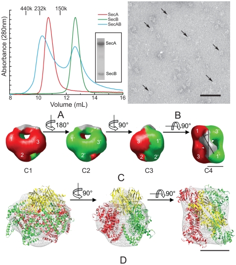
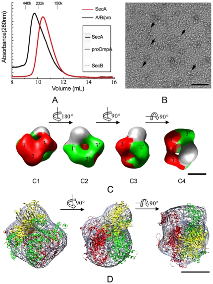
The Sec translocase mediates the post-translational translocation of a number of preproteins through the inner membrane in bacteria. In the initiatory translocation step, SecB targets the preprotein to the translocase by specific interaction with its receptor SecA. The latter is the ATPase of Sec translocase which mediates the post-translational translocation of preprotein through the protein-conducting channel SecYEG in the bacterial inner membrane. We examined the structures of Escherichia coli Sec intermediates in solution as visualized by negatively stained electron microscopy in order to probe the oligomeric states of SecA during this process. The symmetric interaction pattern between the SecA dimer and SecB becomes asymmetric in the presence of proOmpA, and one of the SecA protomers predominantly binds to SecB/proOmpA. Our results suggest that during preprotein translocation, the two SecA protomers are different in structure and may play different roles.
Conflict of interest statement
Figures
Similar articles
-
Electron microscopic visualization of asymmetric precursor translocation intermediates: SecA functions as a dimer.Sci China Life Sci. 2010 Sep;53(9):1049-56. doi: 10.1007/s11427-010-4061-x. Epub 2010 Nov 23. Sci China Life Sci. 2010. PMID: 21104364
-
Preprotein transfer to the Escherichia coli translocase requires the co-operative binding of SecB and the signal sequence to SecA.Mol Microbiol. 1998 Sep;29(5):1179-90. doi: 10.1046/j.1365-2958.1998.00997.x. Mol Microbiol. 1998. PMID: 9767586
-
Charged amino acids in a preprotein inhibit SecA-dependent protein translocation.J Mol Biol. 2009 Mar 6;386(4):1000-10. doi: 10.1016/j.jmb.2009.01.031. J Mol Biol. 2009. PMID: 19244616
-
Oligomeric states of the SecA and SecYEG core components of the bacterial Sec translocon.Biochim Biophys Acta. 2007 Jan;1768(1):5-12. doi: 10.1016/j.bbamem.2006.08.013. Epub 2006 Aug 30. Biochim Biophys Acta. 2007. PMID: 17011510 Free PMC article. Review.
-
SecA: a tale of two protomers.Mol Microbiol. 2010 Jun 1;76(5):1070-81. doi: 10.1111/j.1365-2958.2010.07176.x. Epub 2010 Apr 23. Mol Microbiol. 2010. PMID: 20444093 Review.
Cited by
-
Structural characterization of the complex of SecB and metallothionein-labeled proOmpA by cryo-electron microscopy.PLoS One. 2012;7(10):e47015. doi: 10.1371/journal.pone.0047015. Epub 2012 Oct 4. PLoS One. 2012. PMID: 23056562 Free PMC article.
-
Phospholipids induce conformational changes of SecA to form membrane-specific domains: AFM structures and implication on protein-conducting channels.PLoS One. 2013 Aug 16;8(8):e72560. doi: 10.1371/journal.pone.0072560. eCollection 2013. PLoS One. 2013. PMID: 23977317 Free PMC article.
-
SecA: a potential antimicrobial target.Future Med Chem. 2015;7(8):989-1007. doi: 10.4155/fmc.15.42. Future Med Chem. 2015. PMID: 26062397 Free PMC article. Review.
-
Defining the solution state dimer structure of Escherichia coli SecA using Förster resonance energy transfer.Biochemistry. 2013 Apr 9;52(14):2388-401. doi: 10.1021/bi301217t. Epub 2013 Mar 29. Biochemistry. 2013. PMID: 23484952 Free PMC article.
-
The basis of asymmetry in the SecA:SecB complex.J Mol Biol. 2015 Feb 27;427(4):887-900. doi: 10.1016/j.jmb.2014.12.008. Epub 2014 Dec 19. J Mol Biol. 2015. PMID: 25534082 Free PMC article.
References
-
- Rapoport TA. Protein translocation across the eukaryotic endoplasmic reticulum and bacterial plasma membranes. Nature. 2007;450:663–669. - PubMed
-
- Economou A, Wickner W. SecA promotes preprotein translocation by undergoing ATP-driven cycles of membrane insertion and deinsertion. Cell. 1994;78:835–843. - PubMed
-
- Schiebel E, Driessen AJ, Hartl FU, Wickner W. Delta mu H+ and ATP function at different steps of the catalytic cycle of preprotein translocase. Cell. 1991;64:927–939. - PubMed
-
- Triplett TL, Sgrignoli AR, Gao FB, Yang YB, Tai PC, et al. Functional signal peptides bind a soluble N-terminal fragment of SecA and inhibit its ATPase activity. J Biol Chem. 2001;276:19648–19655. - PubMed
Publication types
MeSH terms
Substances
Grants and funding
LinkOut - more resources
Full Text Sources
Molecular Biology Databases
