. 2011 Feb 3;6(2):e16816.
doi: 10.1371/journal.pone.0016816.

Nestin-GFP transgene reveals neural precursor cells in adult skeletal muscle

Affiliations

Nestin-GFP transgene reveals neural precursor cells in adult skeletal muscle

Alexander Birbrair et al. PLoS One. .

Abstract

Background: Therapy for neural lesions or degenerative diseases relies mainly on finding transplantable active precursor cells. Identifying them in peripheral tissues accessible for biopsy, outside the central nervous system, would circumvent the serious immunological and ethical concerns impeding cell therapy.

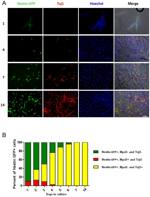
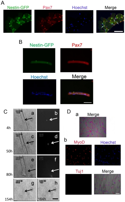
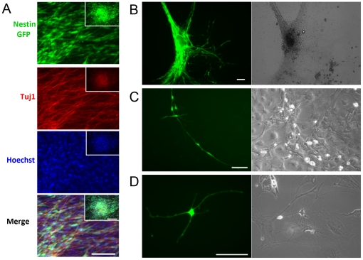
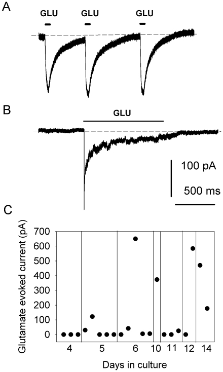
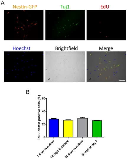
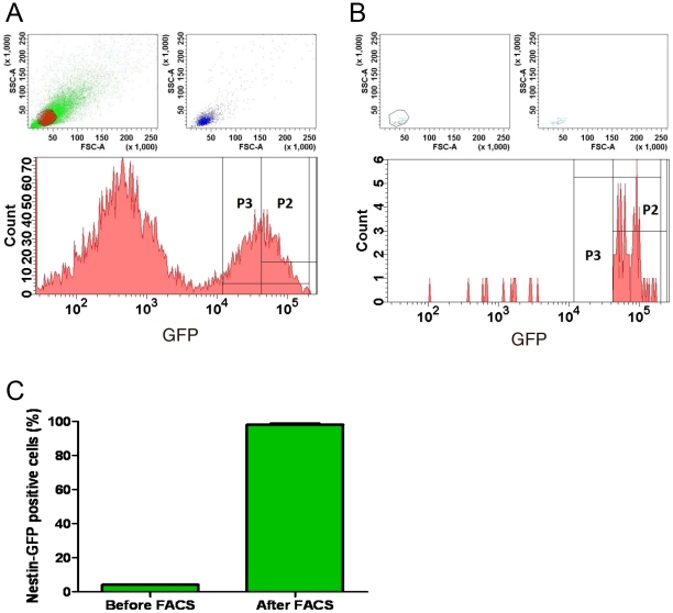
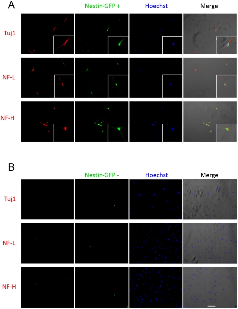
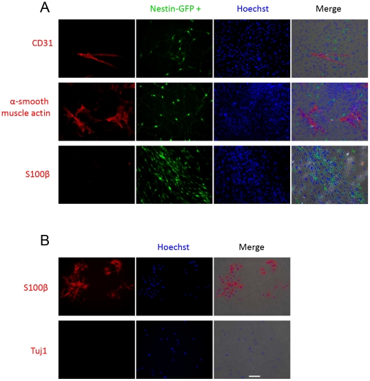
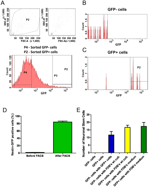
Methodology/principal findings: In this study, we isolated neural progenitor cells in cultured adult skeletal muscle from transgenic mice in which nestin regulatory elements control GFP expression. These cells also expressed the early neural marker Tuj1 and light and heavy neurofilament but not S100β, indicating that they express typical neural but not Schwann cell markers. GFP+/Tuj1+ cells were also negative for the endothelial and pericyte markers CD31 and α-smooth muscle actin, respectively. We established their a) functional response to glutamate in patch-clamp recordings; b) interstitial mesenchymal origin; c) replicative capacity; and d) the environment necessary for their survival after fluorescence-activated cell sorting.

Conclusions/significance: We propose that the decline in nestin-GFP expression in muscle progenitor cells and its persistence in neural precursor cells in muscle cultures provide an invaluable tool for isolating a population of predifferentiated neural cells with therapeutic potential.


Conflict of interest statement

Competing Interests: The authors have declared that no competing interests exist.

Figures

Figure 1
Figure 1. Nestin-GFP+ cells from FDB muscle in culture.
A. Cells were grown for 14 days and fixed at various times (1, 4, 7 and 14 days). Nestin-GFP, Tuj1, and Hoechst 33342 were merged with phase contrast. Scale bar  = 100 µm. B. Relative proportion of Tuj1+ and MyoD+ cells at different times in the nestin-GFP+ population. Notice the disappearance of MyoD expression at day 5 and the progressive increase in Tuj1+ cells to become almost the only population by days 7 and 14 in culture.
Figure 2
Figure 2. Nestin-GFP+ cells attached to myofiber (satellite cells) do not produce Tuj1+ cells with a neural phenotype.
A. A representative EDL muscle cross-section from a nestin-GFP transgenic mouse showing GFP expression and Pax7+ immunoreaction (n = 3 mice, 6 EDL muscles). Myofibers were counterstained with Hoechst. The merge image shows examples of GFP+/Pax- cells (arrows). Scale bar  = 50 µm. B. Enzymatically dissociated single FDB muscle fiber showing 2 nestin-GFP+ satellite cells that immunoreact to Pax7 and overlap with Hoechst nuclear staining. Scale bar  = 100 µm. C. FDB satellite cell time-lapse analysis. A nestin-GFP+ satellite cell attached to an FDB fiber (arrow) analyzed for more than 6 days. Snapshots of the complete record at 4, 50, 80, 154, and 162 h. Brightfield (a, c, e, g, h) and fluorescence (b, d, f). Fluorescence completely disappeared at 162 h in culture. Scale bar  = 50 µm. Da. Four-month satellite cell culture from isolated EDL fibers. Myoblasts and myotubes are stained for desmin and Hoechst, which overlap with the brightfield image. b. The culture shown in Da is MyoD+ but Tuj1-. Scale bar for all pictures in D = 100 µm.
Figure 3
Figure 3. Neurospheres derived from muscle FDB cultures.
A. Neurospheres formed on day 8 in culture are nestin-GFP+ and Tuj1+. Nuclei were stained with Hoechst 33342. B. Nestin-GFP+ cells grow as monolayers after fusing into neurospheres. C. Nestin-GFP+ cells form networks. D. Nestin-GFP+/Tuj1+ cells exhibit neural morphology, with many processes. Right panels for B, C, and D are brightfield images. Images in B-D were taken on culture day 8. Scale bar  = 100 µm for all pictures, including insets in panel A.
Figure 4
Figure 4. Glutamate-evoked membrane currents in neural multipolar cells under voltage-clamp.
A. Typical inward currents in response to three 500 mM L-glutamate pulses in neural cells from FDB muscle culture. The membrane potential was held at −60 mV. Glutamate pulses are depicted above the current traces. The dashed lines represent baseline. B. Response to glutamate application for 1 s and desensitization during agonist application. C. Glutamate-evoked current as a function of culture time (days 4–14). Data points represent individual cells.
Figure 5
Figure 5. Proliferation of neural stem cells derived from FDB skeletal muscle.
A. Unsorted nestin-GFP+ and Tuj1+ cells from FDB muscle were exposed to EdU on culture day 7. Nuclear DNA was stained with Hoechst 33342. Scale bar  = 100 µm. B. Percent of nestin-GFP+/Tuj1+ cells exhibiting EdU incorporation at days 7, 10, and 14 in culture. The fourth column corresponds to nestin-GFP+ cells sorted on culture day 7, cultured for 48 hours, and fixed (n = 3 preparations). EdU (10 µM) was administered 2 hours before cells were fixed. Data are mean ± SEM.
Figure 6
Figure 6. Nestin-GFP+ cell sorting.
Representative scatter plots for 7-day FDB cultures, before (A) and after (B) FACS. GFP fluorescence was plotted against the number of cells. We selected cells with very high GFP fluorescence (P2). A. Sorting yielded approximately 5% of total GFP events and a large population of GFP- cells. B. Re-analysis of GFP+ cells after sorting. C. Percent of nestin-GFP+ cells before and after FACS. Data are mean ± SEM.
Figure 7
Figure 7. Sorted nestin-GFP+ cells from FDB cultures show neural markers.
Sorted GFP+ (A) and GFP- (B) cells were seeded in parallel on dishes precoated with laminin and grown for 48 hours until attached. The first column shows immunostaining for Tuj1, NF-L, or NF-H, while the second shows their corresponding nestin-GFP fluorescence. Nuclei were stained with Hoechst. Merged images are shown on the far right. Scale bar  = 100 µm.
Figure 8
Figure 8. Unsorted nestin-GFP+ cells from FDB cultures do not exhibit endothelial cell, pericyte, or Schwann cell markers.
Unsorted FDB-derived cells were seeded on precoated dishes with laminin and grown for 10 days. A. A CD31 antibody identified a population of endothelial cells that did not overlap with nestin-GFP+ cells. Similarly, an α smooth-muscle actin antibody recognized a population of pericytes. Nestin-GFP+ cells do not react with the S100β antibody for Schwann cells. B. A primary Schwann cell culture shows positive immunostaining when exposed to S100β but no immunostaining when incubated with Tuj1. Scale bar  = 100 µm.
Figure 9
Figure 9. Nestin-GFP+ cell sorting and culture.
A. Representative scatter plots showing fluorescence-activated sorting of cells derived from nestin-GFP+ hindlimb muscles immediately after isolation. GFP fluorescence was plotted against the number of cells. We selected cells with very high (P2) and no GFP fluorescence (P4). Sorts of total events yielded 1.9% GFP+. Histograms of re-analysis of sorted GFP- (B) and GFP+ cells (C). Note that B shows some GFP- cells, which probably result from a decline in fluorescence with time. D. Percent of nestin-GFP+ cells before and after FACS. E. Number of nestin-GFP+ cells differentiated into neurons after culture in different conditions: nestin-GFP- or nestin-GFP+ cells cultured alone; co-cultured nestin-GFP+ and nestin-GFP- cells; nestin-GFP- or nestin-GFP+ cells co-cultured with wild-type FDB fibers; and nestin-GFP+ or nestin-GFP- cells co-cultured with medium from wild-type mouse FDB analyzed after 8 days in culture. The number of nestin-GFP+ and nestin-GFP- plated cells were approximately 5000 and 40,000 per dish, respectively, in all conditions. Only GFP+ cells with 2 or more processes were counted. Data are mean ± SEM.

Similar articles

Cited by

References

    1. Addis RC, Bulte JW, Gearhart JD. Special cells, special considerations: the challenges of bringing embryonic stem cells from the laboratory to the clinic. Clin Pharmacol Ther. 2008;83:386–389. - PMC - PubMed
    1. Li JY, Christophersen NS, Hall V, Soulet D, Brundin P. Critical issues of clinical human embryonic stem cell therapy for brain repair. Trends Neurosci. 2008;31:146–153. - PubMed
    1. Salgado AJ, Oliveira JT, Pedro AJ, Reis RL. Adult stem cells in bone and cartilage tissue engineering. Curr Stem Cell Res Ther. 2006;1:345–364. - PubMed
    1. Zipori D. The stem state: mesenchymal plasticity as a paradigm. Curr Stem Cell Res Ther. 2006;1:95–102. - PubMed
    1. Aguiari P, Leo S, Zavan B, Vindigni V, Rimessi A, et al. High glucose induces adipogenic differentiation of muscle-derived stem cells. Proc Natl Acad Sci U S A. 2008;105:1226–1231. - PMC - PubMed

Publication types

MeSH terms

LinkOut - more resources