. 2011 Feb 3;6(2):e16740.
doi: 10.1371/journal.pone.0016740.

Stage-specific expression of TNFα regulates bad/bid-mediated apoptosis and RIP1/ROS-mediated secondary necrosis in Birnavirus-infected fish cells

Affiliations

Stage-specific expression of TNFα regulates bad/bid-mediated apoptosis and RIP1/ROS-mediated secondary necrosis in Birnavirus-infected fish cells

Wei-Lun Wang et al. PLoS One. .

Abstract

Infectious pancreatic necrosis virus (IPNV) can induce Bad-mediated apoptosis followed by secondary necrosis in fish cells, but it is not known how these two types of cell death are regulated by IPNV. We found that IPNV infection can regulate Bad/Bid-mediated apoptotic and Rip1/ROS-mediated necrotic death pathways via the up-regulation of TNFα in zebrafish ZF4 cells. Using a DNA microarray and quantitative RT-PCR analyses, two major subsets of differentially expressed genes were characterized, including the innate immune response gene TNFα and the pro-apoptotic genes Bad and Bid. In the early replication stage (0-6 h post-infection, or p.i.), we observed that the pro-inflammatory cytokine TNFα underwent a rapid six-fold induction. Then, during the early-middle replication stages (6-12 h p.i.), TNFα level was eight-fold induction and the pro-apoptotic Bcl-2 family members Bad and Bid were up-regulated. Furthermore, specific inhibitors of TNFα expression (AG-126 or TNFα-specific siRNA) were used to block apoptotic and necrotic death signaling during the early or early-middle stages of IPNV infection. Inhibition of TNFα expression dramatically reduced the Bad/Bid-mediated apoptotic and Rip1/ROS-mediated necrotic cell death pathways and rescued host cell viability. Moreover, we used Rip1-specific inhibitors (Nec-1 and Rip1-specific siRNA) to block Rip1 expression. The Rip1/ROS-mediated secondary necrotic pathway appeared to be reduced in IPNV-infected fish cells during the middle-late stage of infection (12-18 h p.i.). Taken together, our results indicate that IPNV triggers two death pathways via up-stream induction of the pro-inflammatory cytokine TNFα, and these results may provide new insights into the pathogenesis of RNA viruses.


Conflict of interest statement

Competing Interests: The authors have declared that no competing interests exist.

Figures

Figure 1
Figure 1. Transcriptional profile of IPNV-infected ZF4 cells 6, 12, and 24 h p.i.
(A) Hierarchical clustering of the mRNA expression pattern, analyzed using DNA microarrays, compared with the 0 h timepoint. Red indicates up-regulation of genes, green indicates down-regulation of genes, and black is used to represent no change in expression. (B) Venn diagram detailing the number of genes up-regulated at 6, 12, and 24 h p.i. in each of three independent experiments examining IPNV-infected ZF4 cells. (C) Venn diagram detailing the number of genes down-regulated at 6, 12, and 24 h p.i. in each of three independent experiments examining IPNV-infected ZF4 cells.
Figure 2
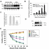
Figure 2. Treatment with TNFα-specific siRNA or AG-126 blocks TNFα expression by IPNV-infected ZF4 cells.
(A) TNFα protein expression level in IPNV-infected ZF4 cells (MOI = 1) 0, 6, 12, and 24 h p.i. The protein was detected using western blot with a polyclonal antibody specific for TNFα. Lanes 1–4: ZF4 cells were pretreated with 50 µM AG-126 and infected with IPNV for 0 (lane 1), 6 (lane 2), 12 (lane 3), or 24 (lane 4). Lanes 5–7: Untreated ZF4 cells were infected with IPNV for 6 (lane 5), 12 (lane 6), or 24 h (lane 7). The expression of actin was used as an internal control. (B) TNFα mRNA expression in IPNV-infected ZF4 cells was quantified using RT-PCR. The ZF4 cells were pre-treated with 50 µM or 100 µM AG-126 for 2 hours, infected with IPNV (MOI = 1), and incubated for 0, 3, 6, 12, or 24 h. The expression of ef1a (elongation factor 1-alpha) was used as an internal control. (C) The tnfa expression was inhibited by TNFα-specific siRNA in IPNV-infected cells. TNFα expression was efficiently inhibited by TNFα-specific siRNA after IPNV infection. Sample 1: ZF4 cells infected by IPNV. Sample 2: ZF4 cells pretreated with scrambled siRNA and then infected by IPNV. Sample 3: ZF4 cells pretreated with TNFα-specific siRNA and then infected by IPNV. The quantification of gene expression in normal versus siRNA-treated cells was calculated relative to ef1a. (D) Detection of TNFα in untreated or TNFα-specific siRNA-treated ZF4 cells after IPNV infection by western blotting. Lane 1: untreated ZF4 cells; lane 2: ZF4 cells treated with control siRNA; lane 3: ZF4 cells treated with TNFα-specific siRNA. The expression of actin was used as an internal control. (E) Cell viability of IPNV-infected ZF4 cells pre-treated with TNFα-specific siRNA or AG-126 at 0, 6, 12, 24, 36 and 48 h p.i. The viability of each sample was determined in three individual experiments. Data shown are the mean ± SD. Student's t tests indicate significant differences compared to IPNV infection only or untreated control: *, p<0.05; **, p<0.01.
Figure 3
Figure 3. A TNFα-mediated death signal regulates the expression of pro-apoptotic Bad and Bid in IPNV-infected ZF4 cells during the early-middle stage of replication.
(A) The expression level of the pro-apoptotic proteins Bad and Bid in IPNV-infected ZF4 cells (MOI = 1) at 0, 6, 12, and 24 h p.i. was determined. The proteins were detected using western blot with a polyclonal antibody specific for mouse Bad. Lanes 1–4: ZF4 cells were pretreated with TNFα-specific siRNA and infected with IPNV for 0 (lane 1), 6 (lane 2), 12 (lane 3), or 24 h (lane 4). Lanes 5–7: untreated ZF4 cells were infected with IPNV for 6 (lane 5), 12 (lane 6), or 24 h (lane 7). The expression of actin was used as an internal control. Results are expressed as the ratio of Bad/actin. The mRNA expression of bad (B) and bid (D) in IPNV-infected ZF4 cells was quantified using quantitative RT-PCR. ZF4 cells were pre-treated with TNFα-specific siRNA or AG-126 and infected with IPNV (MOI = 1) for 0, 6, 12, or 24 h. The expression of ef1a was used as an internal control. Data shown are mean ± SD. Student's t tests indicate significant differences compared to untreated control: **, p<0.01. (C) The expression level of the pro-apoptotic proteins Bid and t-Bid in IPNV-infected ZF4 cells (MOI = 1) at 0, 6 and 12 h p.i. was determined. The proteins were detected using western blot with a polyclonal antibody specific for Bid. Lanes 2–3: ZF4 cells were pretreated with TNFα-specific siRNA and infected with IPNV for 6 (lane 2) or 12 h (lane 3). Untreated ZF4 cells were infected with IPNV for 6 (lane 4) or 12 h (lane 5). Untreated ZF4 cells were infected with IPNV for 0 h (Lane 1). The expression of actin was used as an internal control. Results are expressed as the ratio of Bid/actin or t-Bid/actin.
Figure 4
Figure 4. Inhibition of TNFα production can reduce caspase activation and the number of annexin V-positive IPNV-infected cells.
(A) Detection of annexin V-positive cells following infection with IPNV. ZF4 cells were pre-treated with TNFα-specific siRNA or AG-126, infected with IPNV (MOI = 1), and incubated for 0, 6, 12, 18 and 24 h. Three individual experiments were performed for each sample. (BD) Caspase-9, -8, and -3 activities were analyzed. ZF4 cells were pre-treated with TNFα-specific siRNA or AG-126 for 2 hours, infected with IPNV (MOI = 1), and incubated for 0, 6, 12, or 24 h. Luminogenic substrate assays were performed in triplicate. Data shown are the mean ± SD. Student's t tests indicate significant differences compared to IPNV infection only: *, p<0.05.
Figure 5
Figure 5. Inhibition of TNFR1 necrotic signaling complex formation inhibits apoptosis and ROS formation in IPNV-infected cells.
(A) Detection of ROS production in TNFα-specific siRNA or AG-126 pre-treated cells after IPNV infection at 0, 6, 12, 18 or 24 h p.i. Fluorescence assays were performed in triplicate. Determination of the percentage of PI-positive cells after IPNV infection. (B) Detection of RIP1 in untreated or TNFα-specific siRNA-treated ZF4 cells by western blotting. Lane 1: untreated IPNV-infected ZF4 cells; lane 2: IPNV-infected ZF4 cells treated with scrambled siRNA; lane 3: IPNV-infected ZF4 cells treated with RIP1-specific siRNA. The expression of actin was used as an internal control. Detection of annexin V-positive cells following infection with IPNV. ZF4 cells were pre-treated with RIP1-specific siRNA (C), Nec-1 (D), DPI (E) or BHA (F), infected with IPNV (MOI = 1), and incubated for 0, 6, 12, 18 and 24 h. Three individual experiments were performed for each sample. Data shown are the mean ± SD. Student's t tests indicate significant differences compared to IPNV infection only: *, p<0.05.
Figure 6
Figure 6. Inhibition of TNF-α or TNFR1 necrotic signaling complex formation inhibits secondary necrosis in IPNV-infected cells.
Detection of PI-positive cells following infection with IPNV. ZF4 cells were pre-treated with TNFα-specific siRNA (A), AG-126 (B), RIP1-specific siRNA (C), Nec-1 (D), DPI (E), BHA (F) and z-VAD (G) then infected with IPNV (MOI = 5), and incubated for 12, 18 and 24 h. (H) Detection of ROS production in RIP1-specific siRNA-, Nec-1- or DPI-pre-treated cells after IPNV infection at 0, 6, 12, 18 or 24 h p.i. Fluorescence assays were performed in triplicate. Data shown are the mean ± SD. Student's t tests indicate significant differences compared to IPNV infection only or untreated control: *, p<0.05.
Figure 7
Figure 7. IPNV induces apoptotic and necrotic death cascades via TNFα induction.
A hypothesis of how pro-inflammatory TNFα is up-regulated by IPNV infection and how it regulates the apoptotic and necrotic death pathways. When a cell is infected with IPNV (E1-S), the virus binds to the cellular receptor, penetrates the cell, uncoats (the entry stage), and up-regulates the expression of TNFα (a) during the early replication stage (0–6 h p.i.). This up-regulation of TNFα regulates the next wave of gene expression, including the pro-apoptotic genes bad and bid, during the early-middle stage of replication (6–12 h p.i.; b), at which point the TNFR necrotic signaling complex and reactive oxygen species (ROS) are produced. During this stage, phosphatidylserine (PS) is externalized and endonuclease is released from the mitochondria, resulting in DNA restructuring and cleavage. Furthermore, the cell finally enters the post-apoptotic, necrotic stage during the middle-late stage of replication (12–18 h p.i.; c). During the late replication stage (18–24 h p.i.; d), the cells are broken down. The TNFα-mediated death signal is halted by treatment with a specific inhibitor of TNFα production. (e) TNFα-specific siRNA or AG-126, which block both the pro-apoptotic Bad/Bid-mediated death pathway and the TNFR/ROS-mediated secondary necrotic death pathway. (f) DPI, Nec-1 or RIP-1-specific siRNA, which block the activity of RIP-1 or Nox1 and block the formation of the TNFR necrotic signaling complex. (g) BHA blocks the formation of ROS.

Similar articles

Cited by

References

    1. Dobos P. Virus-specific protein synthesis in cells infected by infectious pancreatic necrosis virus. J Virol. 1977;21:242–258. - PMC - PubMed
    1. Dobos P, Hill BJ, Hallett R, Kells DT, Becht H, et al. Biophysical and biochemical characterization of five animal viruses with bisegmented double-stranded RNA genomes. J Virol. 1979;32:593–605. - PMC - PubMed
    1. Duncan R, Mason CL, Nagy E, Leong JA, Dobos P. Sequence analysis of infectious pancreatic necrosis virus genome segment B and its encoded VP1 protein: a putative RNA-dependent RNA polymerase lacking the Gly-Asp-Asp motif. Virology. 1991;181:541–552. - PMC - PubMed
    1. Duncan R, Nagy E, Krell PJ, Dobos P. Synthesis of the infectious pancreatic necrosis virus polyprotein, detection of a virus-encoded protease, and fine structure mapping of genome segment A coding regions. J Virol. 1987;61:3655–3664. - PMC - PubMed
    1. Magyar G, Dobos P. Evidence for the detection of the infectious pancreatic necrosis virus polyprotein and the 17-kDa polypeptide in infected cells and of the NS protease in purified virus. Virology. 1994;204:580–589. - PubMed

Publication types

MeSH terms