. 2011 Feb 3;7(2):e1001271.
doi: 10.1371/journal.ppat.1001271.

Inhibition of Nox2 oxidase activity ameliorates influenza A virus-induced lung inflammation

Affiliations

Inhibition of Nox2 oxidase activity ameliorates influenza A virus-induced lung inflammation

Ross Vlahos et al. PLoS Pathog. .

Abstract

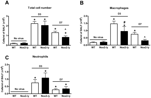
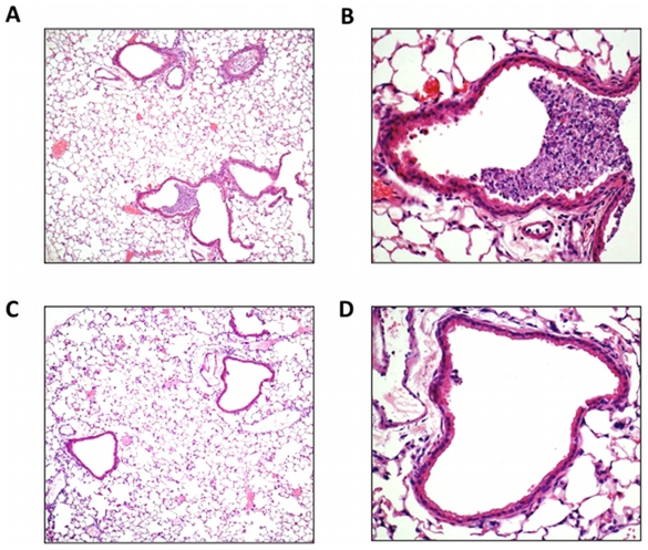
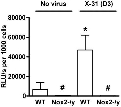
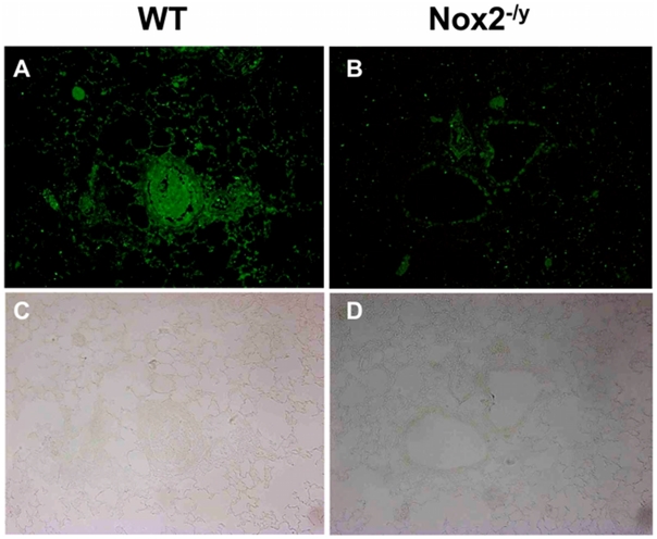
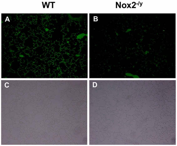
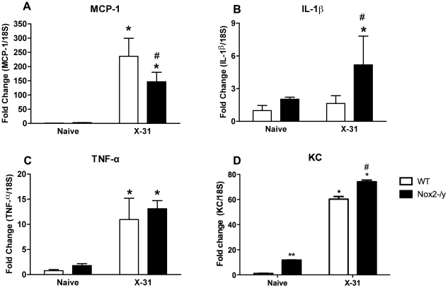
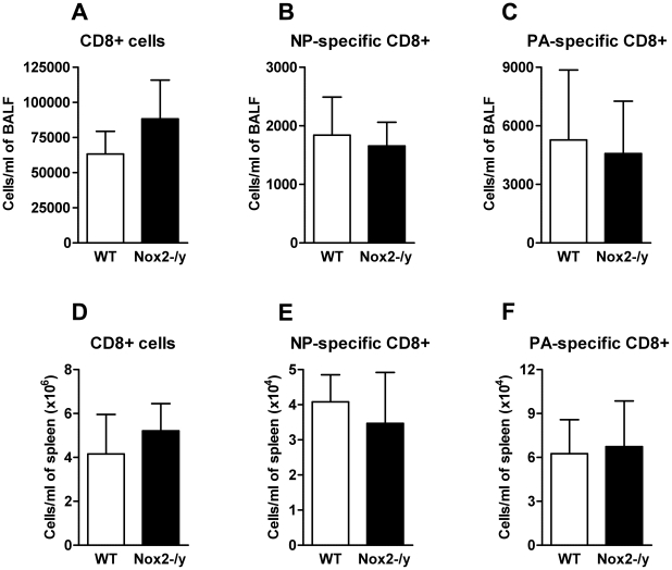
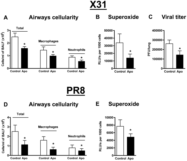
Influenza A virus pandemics and emerging anti-viral resistance highlight the urgent need for novel generic pharmacological strategies that reduce both viral replication and lung inflammation. We investigated whether the primary enzymatic source of inflammatory cell ROS (reactive oxygen species), Nox2-containing NADPH oxidase, is a novel pharmacological target against the lung inflammation caused by influenza A viruses. Male WT (C57BL/6) and Nox2(-/y) mice were infected intranasally with low pathogenicity (X-31, H3N2) or higher pathogenicity (PR8, H1N1) influenza A virus. Viral titer, airways inflammation, superoxide and peroxynitrite production, lung histopathology, pro-inflammatory (MCP-1) and antiviral (IL-1β) cytokines/chemokines, CD8(+) T cell effector function and alveolar epithelial cell apoptosis were assessed. Infection of Nox2(-/y) mice with X-31 virus resulted in a significant reduction in viral titers, BALF macrophages, peri-bronchial inflammation, BALF inflammatory cell superoxide and lung tissue peroxynitrite production, MCP-1 levels and alveolar epithelial cell apoptosis when compared to WT control mice. Lung levels of IL-1β were ∼3-fold higher in Nox2(-/y) mice. The numbers of influenza-specific CD8+D(b)NP(366)+ and D(b)PA(224)+ T cells in the BALF and spleen were comparable in WT and Nox2(-/y) mice. In vivo administration of the Nox2 inhibitor apocynin significantly suppressed viral titer, airways inflammation and inflammatory cell superoxide production following infection with X-31 or PR8. In conclusion, these findings indicate that Nox2 inhibitors have therapeutic potential for control of lung inflammation and damage in an influenza strain-independent manner.


Conflict of interest statement

The authors have declared that no competing interests exist.

Figures

Figure 1
Figure 1. Effect of H3N2 (X-31) influenza A virus infection on BALF cellularity in wild type (WT) and Nox2−/y mice.
Mice were treated with 1×10⁴ PFU of the low virulence H3N2 strain of influenza A virus and the number of (A) total cells, (B) macrophages and (C) neutrophils counted in the BALF 3 and 7 days post infection. Data are shown as mean ± SD for 6–8 mice per group. *P<0.05 vs respective no virus group, #P<0.05 vs D3 X-31-treated WT mice mice, ∧P<0.05 vs D7 X-31-treated WT mice (ANOVA and Dunnett's post hoc test).
Figure 2
Figure 2. Lung histology in response to H3N2 (X-31) influenza A virus infection.
(A–D) Hematoxylin and eosin stained paraffin sections of lungs from WT (A and B) and Nox2−/y (C and D) mice obtained at Day 3. WT mice lungs had obvious peri-bronchial inflammation and prominent bronchial infiltrates consisting of mononuclear cells and neutrophils. In contrast, Nox2−/y mice displayed considerably less bronchial cell infiltrates and peri-bronchial inflammation. Note, magnification of (A) and (C) is ×50 and (B) and (D) is ×200.
Figure 3
Figure 3. Superoxide production from BALF cells using L-O12-enhanced chemiluminescence.
BALF cells obtained from H3N2 (X-31) influenza A virus-infected WT and Nox2−/y mice 3 days post infection. Data are shown as mean ± SD relative light units per second (RLU/s) for 6–8 mice per treatment group. *P<0.05 vs WT no virus mice, #P<0.05 vs respective WT mice (ANOVA and Dunnett's post hoc test).
Figure 4
Figure 4. Peroxynitrite production in mouse lung infected with H3N2 (X-31) influenza A virus using 3-nitrotyrosine (3-NT) immunofluorescence.
Representative sections of lung tissue obtained from WT (A) and Nox2−/y mice (B) infected with X-31 were incubated with mouse monoclonal anti-3-nitrotyrosine antibody (1∶50) followed by biotinylated anti-mouse IgG reagent. (C and D) The same sections showing corresponding light microscope images. WT mice lung sections displayed strong immunofluorescence for 3-NT in the inflammatory cells that infiltrated the airways and in the alveolar tissue. In contrast, Nox2−/y mice displayed markedly less immunofluorescence for 3-NT.
Figure 5
Figure 5. Effect of H3N2 (X-31) influenza A virus infection on viral titer and body weight in wild type (WT) and Nox2−/y mice.
Mice were treated with 1×10⁴ PFU of the low virulence H3N2 strain of influenza A virus and (A) viral titer determined 3 days post infection and (B) body weight recorded for up to 7 days post infection. Data are shown as mean ± SD for 6–8 mice per group. *P<0.05 vs WT mice (Students' unpaired t test).
Figure 6
Figure 6. Cleaved caspase 3 immunofluorescence to assess lung alveolar epithelial apoptosis.
Representative sections of lung tissue obtained from WT (A) and Nox2−/y mice (B) infected with influenza A virus (H3N2; X-31) were incubated with rabbit polyclonal anti-cleaved caspase 3 antibody (1∶250) followed by goat anti-rabbit Alexa fluor 488 (Invitrogen; 1∶500) secondary antibody. (C and D) The same sections showing corresponding light microscope images. WT mice lung sections displayed strong immunofluorescence for cleaved caspase 3 in the alveolar tissue. In contrast, Nox2−/y mice displayed markedly less immunofluorescence for cleaved caspase 3.
Figure 7
Figure 7. Assessment of lung cytokines levels.
Effect of H3N2 (X-31) influenza A virus infection on (A) MCP-1, (B) IL-1β, (C) TNF-α and (D) KC mRNA expression in whole lung obtained from WT and Nox2−/y mice 3 days post infection. Responses are shown as fold change relative to 18S. Data are shown as mean ± SD of 4 individual mice. WT and Nox2−/y mice are shown as open and filled histograms, respectively. WT mice that did not receive H3N2 (i.e. naïve mice) were used as a control to demonstrate that virus infection caused an increase in MCP-1, IL-1β, TNF-α and KC. *P<0.05 vs respective naïve mice (ANOVA and Dunnett's post hoc test), #P<0.05 vs WT X-31-treated mice (ANOVA and Dunnett's post hoc test). ** P<0.05 vs WT naïve mice.
Figure 8
Figure 8. Number of the CD8+ T cell response.
Enriched CD8+ T cells from BALF (A–C) or spleen (D–F) were obtained from influenza A virus (X-31)-infected WT and Nox2−/y mice 7 days post infection. These were then stained with fluorescently labelled tetramer complexes specific for the two immunodominant CD8+ T cell epitopes (DbNP366–372 and DbPA224–232) and data analysed by flow cytometry. Data are shown as mean ± SD of 4 individual mice.
Figure 9
Figure 9. Pharmacological inhibition of Nox2 oxidase activity.
Effect of apocynin (Apo, 2.5 mg/kg) treatment on (A) BALF cellularity, (B) BALF inflammatory cell superoxide production and (C) viral titer in H3N2 (X-31) influenza A virus-infected WT mice 3 days post infection. Also, effect of apocynin treatment on (D) BALF cellularity and (E) BALF inflammatory cell superoxide production in H1N1 (PR8) influenza A virus-infected WT mice 3 days post infection. Data are shown as mean ± SD for 6–8 mice per treatment group. *P<0.05 vs WT vehicle (Control)-treated mice (ANOVA followed by Dunnett's post hoc test for (A and D) but Students' unpaired t test used for B, C and E).

Similar articles

Cited by

References

    1. La Gruta NL, Kedzierska K, Stambas J, Doherty PC. A question of self-preservation: immunopathology in influenza virus infection. Immunol Cell Biol. 2007;85:85–92. - PubMed
    1. Tumpey TM, Basler CF, Aguilar PV, Zeng H, Solorzano A, et al. Characterization of the reconstructed 1918 Spanish influenza pandemic virus. Science. 2005;310:77–80. - PubMed
    1. Oda T, Akaike T, Hamamoto T, Suzuki F, Hirano T, et al. Oxygen radicals in influenza-induced pathogenesis and treatment with pyran polymer-conjugated SOD. Science. 1989;244:974–976. - PubMed
    1. Imai Y, Kuba K, Neely GG, Yaghubian-Malhami R, Perkmann T, et al. Identification of oxidative stress and Toll-like receptor 4 signaling as a key pathway of acute lung injury. Cell. 2008;133:235–249. - PMC - PubMed
    1. Selemidis S, Sobey CG, Wingler K, Schmidt HH, Drummond GR. NADPH oxidases in the vasculature: molecular features, roles in disease and pharmacological inhibition. Pharmacol Ther. 2008;120:254–291. - PubMed

Publication types

MeSH terms