. 2011 Feb 3;7(2):e1001286.
doi: 10.1371/journal.pgen.1001286.

Nucleosomes containing methylated DNA stabilize DNA methyltransferases 3A/3B and ensure faithful epigenetic inheritance

Affiliations

Nucleosomes containing methylated DNA stabilize DNA methyltransferases 3A/3B and ensure faithful epigenetic inheritance

Shikhar Sharma et al. PLoS Genet. .

Abstract

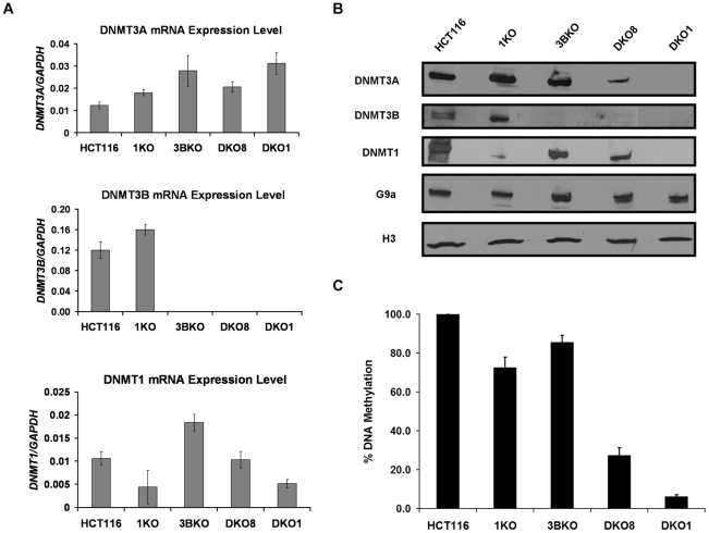
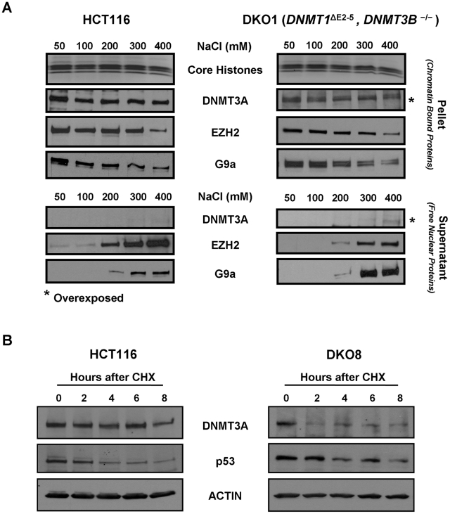
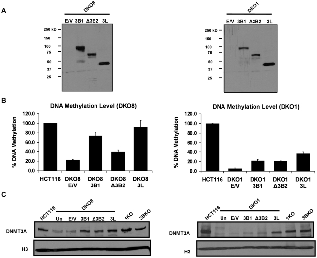
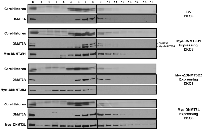
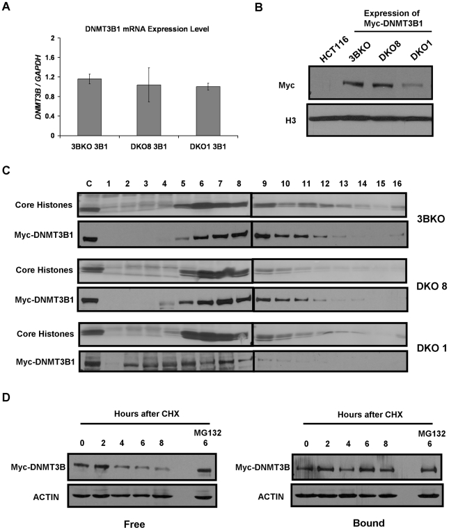
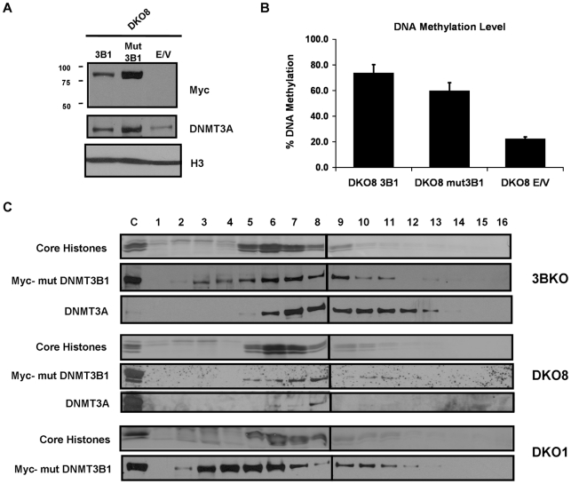
How epigenetic information is propagated during somatic cell divisions is still unclear but is absolutely critical for preserving gene expression patterns and cellular identity. Here we show an unanticipated mechanism for inheritance of DNA methylation patterns where the epigenetic mark not only recruits the catalyzing enzyme but also regulates the protein level, i.e. the enzymatic product (5-methylcytosine) determines the level of the methylase, thus forming a novel homeostatic inheritance system. Nucleosomes containing methylated DNA stabilize de novo DNA methyltransferases, DNMT3A/3B, allowing little free DNMT3A/3B enzymes to exist in the nucleus. Stabilization of DNMT3A/3B on nucleosomes in methylated regions further promotes propagation of DNA methylation. However, reduction of cellular DNA methylation levels creating more potential CpG substrates counter-intuitively results in a dramatic decrease of DNMT3A/3B proteins due to diminished nucleosome binding and subsequent degradation of the unstable free proteins. These data show an unexpected self-regulatory inheritance mechanism that not only ensures somatic propagation of methylated states by DNMT1 and DNMT3A/3B enzymes but also prevents aberrant de novo methylation by causing degradation of free DNMT3A/3B enzymes.


Conflict of interest statement

The authors have declared that no competing interests exist.

Figures

Figure 1
Figure 1. Transcription-independent decrease in DNMT3A protein level in hypomethylated DKO cells that contain severely impaired DNMT1 activity.
Figure 2
Figure 2. DNMT3A chromatin binding affinity and protein stability in WT HCT116 and DKO cells.
(A) Nuclei purified from WT HCT116 and DKO1 cells were incubated in nondenaturing extraction buffers containing 50 to 400 mM NaCl for 5 min. Equivalent volumes of both supernatant and pellet fractions were subjected to western blot analysis using specific antibodies. Ponceau S staining shows core histones transferred onto the membrane from the SDS/PAGE gel. For detecting low levels of DNMT3A in DKO1 cells, blots for both the supernatant and pellet fractions from DKO1 cells were overexposed for 5 fold more time duration compared to HCT116 cells, as indicated by *. (B) WT HCT116 and DKO8 cells were treated with cycloheximide (CHX) and the levels of DNMT3A protein remaining at different time points after treatment were determined by western blotting of nuclear extracts. p53 and actin were used as positive and loading controls, respectively. Data presented is from a single experiment, representative of two independent biological replicate experiments.
Figure 3
Figure 3. Increase in DNA methylation restores the DNMT3A protein level in DKO cells.
(A) Expression of Myc-tagged DNMT3B1, ΔDNMT3B2 and DNMT3L proteins, infected using a lentiviral system, in DKO cells was confirmed by immunoblotting of nuclear extracts using a Myc antibody. (B) DNA methylation analysis of infected DKO cells using methylation-sensitive restriction enzymes. Genomic DNA was isolated from infected cells eight weeks after infection and methylation level was estimated as described in Figure 1. Data is presented as the percentage of methylation retained compared to WT HCT116 methylation levels. Data represents mean and SEM of three independent replicate experiments. (C) Western blot analysis of nuclear extracts, prepared from infected DKO cells and different HCT116 derivative cell lines, using a DNMT3A antibody. Histone H3 was used as the loading control. Data presented in this figure is representative of two biological replicate experiments. E/V: Empty Vector.
Figure 4
Figure 4. The increased level of DNMT3A protein in infected DKO cells, which have increased levels of DNA methylation, remains tightly bound to nucleosomes.
Mononucleosomal digests prepared by extensive MNase digestion of infected DKO8 nuclei, were resolved by ultracentrifugation on a sucrose density gradient (5% to 25%) containing 300 mM NaCl. Gradients were fractioned into 16 aliquots numbered 1–16 starting from the top of the centrifuge tube. To probe the distribution of proteins in each fraction, western blotting was performed with various antibodies after TCA precipitation of proteins from each fraction. Ponceau S staining shows core histones transferred onto the membrane from the SDS/PAGE gel. Mononucleosomes peaked in fraction 6 and the small proportion of higher order oligonucleosomes remaining in the digests sedimented in later fractions. The control lanes on the gels were loaded with unfractionated nuclear extract to monitor the quality of the immunostaining of the membranes. The upper band in the DNMT3A blot for Myc-DNMT3B1 expressing DKO8 cells denotes endogenous DNMT3A. The lower band represents the residual signal of the exogenous Myc-DNMT3B1, which was probed earlier on the same membrane.
Figure 5
Figure 5. Weak nucleosome binding and selective degradation of unbound DNMT3B in the absence of elevated DNA methylation levels.
(A) RT-PCR analysis was performed using primers for Myc-DNMT3B1 to assess its mRNA levels in infected 3BKO, DKO8 and DKO1 cells. The results are normalized to GAPDH mRNA levels. Data represents mean and standard deviation of triplicate PCR reactions from a single experiment, representative of two biological replicate experiments. (B) Western blot analysis of nuclear extracts from infected 3BKO, DKO8 and DKO1 cells. Exogenous DNMT3B1 was detected with Myc antibody. (C) Nuclei extracted from infected cells were extensively digested with MNase and mononucleosomes released from them were resolved by ultracentrifugation on a sucrose density gradient (5% to 25%) containing 300 mM NaCl. The gradients were fractionated and analyzed as described previously. (D) DKO1 cells expressing Myc-DNMT3B1 were treated with cycloheximide (CHX) for different time points. The proteosome inhibitor, MG132 was added 2 hr prior to CHX treatment. Nuclei extracted from each sample were then incubated in 500 µl of ice-cold RSB containing 300 mM NaCl, 0.25 M sucrose and protease inhibitors at 4°C for 5 min. Supernatant and nuclear fractions were separated by centrifugation at low speed and equivalent protein amounts from each were subjected to western blot analysis. Data is representative of two biological replicate experiments.
Figure 6
Figure 6. DNMT3B catalytically-inactive mutant stimulates DNA methylation by increasing DNMT3A binding to nucleosomes.
Figure 7
Figure 7. Model for selective stabilization of DNMT3A/3B through anchoring to nucleosomes containing methylated DNA.
(A) In somatic cells, DNMT3A/3B remain bound to nucleosomes containing methylated DNA, enabling proper maintenance of methylated states in co-operation with DNMT1, the maintenance enzyme, which copies the methylation pattern during replication by associating with the proliferating cell nuclear antigen (PCNA). (B) When DNA methylation is lowered by genetic disruption of DNMT1 and DNMT3B in DKO cells, DNMT3A loses its ability to bind to nucleosomes which results in destabilization and subsequent degradation of the protein. (C) Restoration of DNA methylation in such hypomethylated cells, through expression of exogenous DNMT3B (WT or mut) or DNMT3L, increases DNMT3A protein levels by enabling it to bind to nucleosomes again which results in stabilization of DNMT3A protein. Exogenous DNMT3B (WT or mut) also binds strongly to nucleosomes in the presence of DNA methylation and synergistically increases methylation along with DNMT3A while the excess free DNMT3B protein, which could not anchor to the nucleosomes, gets degraded by the proteosomal machinery. mut: mutant.

Similar articles

Cited by

References

    1. Bird A. DNA methylation patterns and epigenetic memory. Genes Dev. 2002;16:6–21. - PubMed
    1. Suzuki MM, Bird A. DNA methylation landscapes: provocative insights from epigenomics. Nat Rev Genet. 2008;9:465–476. - PubMed
    1. Jones PA, Baylin SB. The epigenomics of cancer. Cell. 2007;128:683–692. - PMC - PubMed
    1. Sharma S, Kelly TK, Jones PA. Epigenetics in cancer. Carcinogenesis. 2010;31:27–36. - PMC - PubMed
    1. Okano M, Bell DW, Haber DA, Li E. DNA methyltransferases Dnmt3a and Dnmt3b are essential for de novo methylation and mammalian development. Cell. 1999;99:247–257. - PubMed

Publication types