. 2011 Feb 3;7(2):e1001288.
doi: 10.1371/journal.pgen.1001288.

Pathogenic VCP/TER94 alleles are dominant actives and contribute to neurodegeneration by altering cellular ATP level in a Drosophila IBMPFD model

Affiliations

Pathogenic VCP/TER94 alleles are dominant actives and contribute to neurodegeneration by altering cellular ATP level in a Drosophila IBMPFD model

Ya-Chu Chang et al. PLoS Genet. .

Abstract

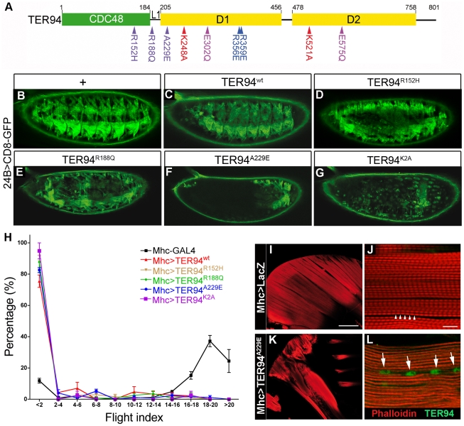
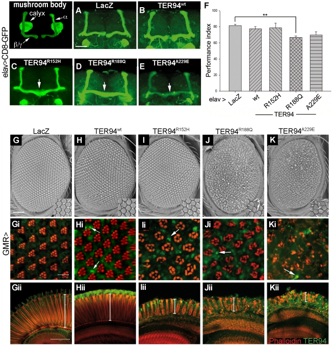
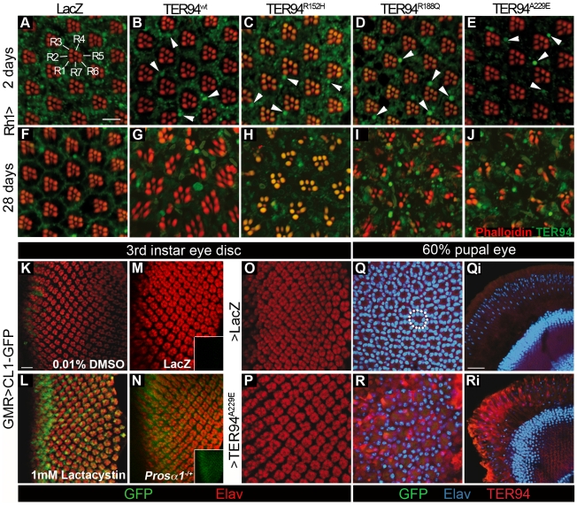
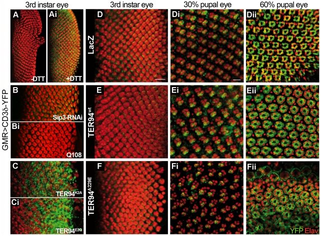
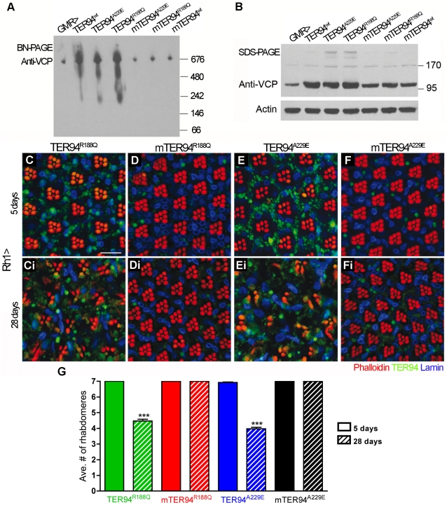
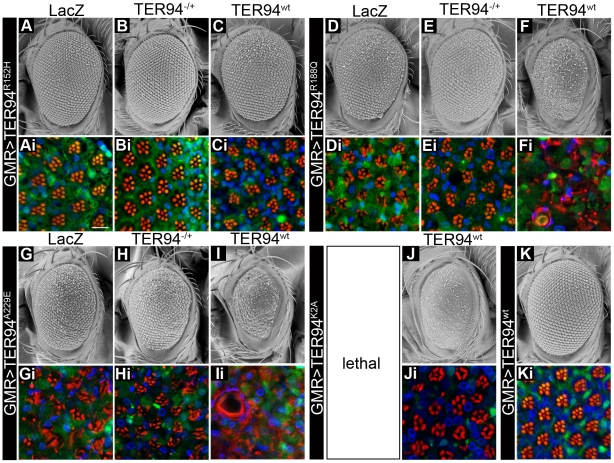
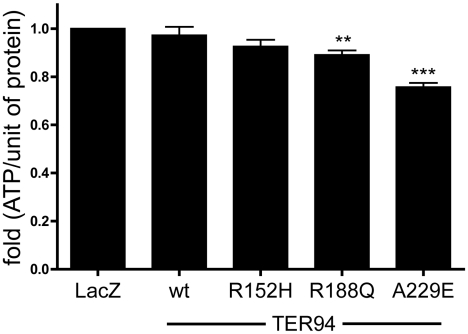
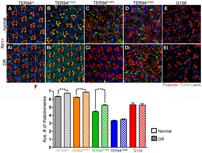
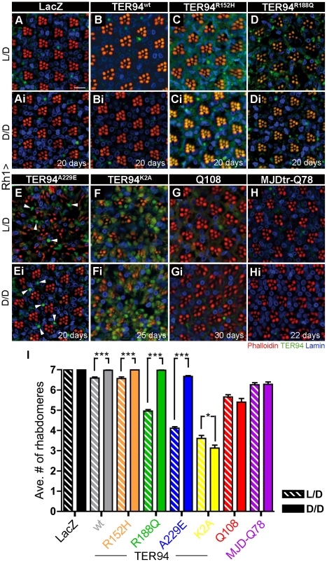
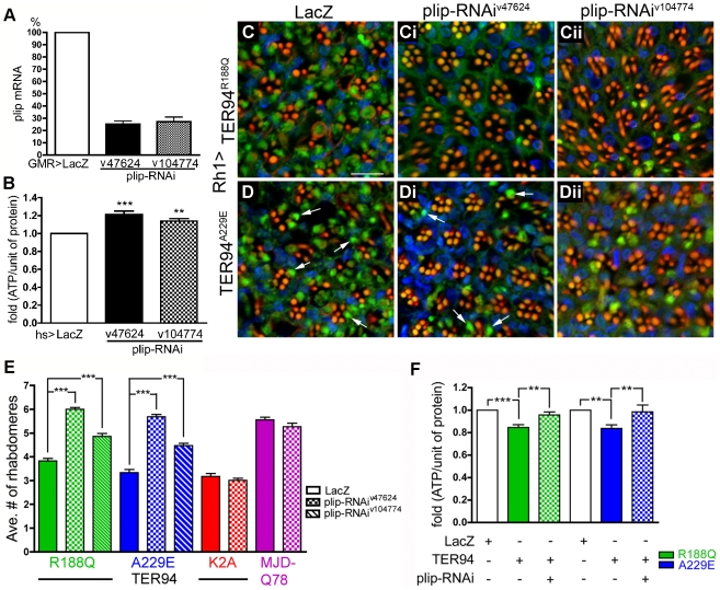
Inclusion body myopathy with Paget's disease of bone and frontotemporal dementia (IBMPFD) is caused by mutations in Valosin-containing protein (VCP), a hexameric AAA ATPase that participates in a variety of cellular processes such as protein degradation, organelle biogenesis, and cell-cycle regulation. To understand how VCP mutations cause IBMPFD, we have established a Drosophila model by overexpressing TER94 (the sole Drosophila VCP ortholog) carrying mutations analogous to those implicated in IBMPFD. Expression of these TER94 mutants in muscle and nervous systems causes tissue degeneration, recapitulating the pathogenic phenotypes in IBMPFD patients. TER94-induced neurodegenerative defects are enhanced by elevated expression of wild-type TER94, suggesting that the pathogenic alleles are dominant active mutations. This conclusion is further supported by the observation that TER94-induced neurodegenerative defects require the formation of hexamer complex, a prerequisite for a functional AAA ATPase. Surprisingly, while disruptions of the ubiquitin-proteasome system (UPS) and the ER-associated degradation (ERAD) have been implicated as causes for VCP-induced tissue degeneration, these processes are not significantly affected in our fly model. Instead, the neurodegenerative defect of TER94 mutants seems sensitive to the level of cellular ATP. We show that increasing cellular ATP by independent mechanisms could suppress the phenotypes of TER94 mutants. Conversely, decreasing cellular ATP would enhance the TER94 mutant phenotypes. Taken together, our analyses have defined the nature of IBMPFD-causing VCP mutations and made an unexpected link between cellular ATP level and IBMPFD pathogenesis.


Conflict of interest statement

The authors have declared that no competing interests exist.

Figures

Figure 1
Figure 1. Tissue-specific expression of TER94 IBMPFD mutants disrupts muscle integrity.
(A) A schematic drawing of TER94 functional domains and the alleles used in this study. Green and yellow boxes represent the N-terminal CDC48 domain and the two ATPase domains, respectively. The IBMPFD, ATP-binding defective, ATP-hydrolysis defective, and monomeric mutations are indicated in purple, red, lilac, and blue, respectively. (B–G) Fluorescent micrographs of lateral views of stage 16 embryos expressing indicated transgenes and mCD8-GFP under the control of 24B-GAL4. In all panels, anterior is to the left and dorsal is up. (H) Flight behavioral tests of Mhc>TER94 flies. Flies of indicated genotypes were used for flight behavioral tests, and results from four independent experiments were plotted. Higher percentage in smaller flight index numbers denotes flightless phenotype, whereas higher percentage in larger flight index numbers denotes normal flight behavior. Values shown represent mean ± SE (standard error of the mean). (I and K) Phalloidin-stained sections show normal dorsal longitudinal muscles (DLM) within IFM in control Mhc>LacZ (I) and irregular DLM in Mhc>TER94A229E (K). (J and L) Confocal images of Mhc>LacZ (J) and Mhc>TER94A229E (L) DLM sarcomeres stained with phalloidin and anti-VCP antibody (green). Scale bars: 100 µm (I and K), 10 µm (J and L). Genotypes: (B) 24B-GAL4,UAS-CD8-GFP/+, (C) 24B-GAL4,UAS-CD8-GFP/UAS-TER94wt, (D) 24B-GAL4,UAS-CD8-GFP/UAS-TER94R152H, (E) 24B-GAL4,UAS-CD8-GFP/UAS-TER94R188Q, (F) UAS-TER94A229E/+; 24B-GAL4,UAS-CD8-GFP/+, (G) UAS-TER94K2A/+; 24B-GAL4,UAS-CD8-GFP/+, (I and J) Mhc-GAL4/w;UAS-LacZ/+, (K and L) Mhc-GAL4/w;UAS-TER94A229E/+.
Figure 2
Figure 2. Tissue-specific expression of TER94 IBMPFD mutants impairs neuronal structure and function.
(A–E) Confocal images of mushroom bodies (labeled by mCD8-GFP) from adult brains expressing indicated transgenes under the control of elav-GAL4. A segmented adult brain illustrates the major structures of mushroom body, and arrows indicate the midline crossing of mushroom bodies in IBMPFD mutants. (F) The Pavlovian learning test of flies expressing TER94 transgenes or LacZ control. Values shown represent mean ± SE from four independent tests. **p<0.01 relative to LacZ control (Student's t test). (G–K) Scanning electron micrographs (SEM) of the 2-day old adult eyes expressing indicated transgenes under the control of GMR-GAL4. Insets show enlarged views of the bristles on the eye surface. In all SEM panels, anterior is to the left and dorsal is up. (Gi–Kii) Confocal images of tangential (Gi–Ki) and longitudinal sections (Gii–Kii) of retinas from flies of corresponding genotypes at the same age stained with phalloidin (red) and anti-VCP (green). TER94-positive structures in eyes expressing TER94 transgenes are indicated with white arrows. White lines indicate the thicknesses of the retina in the longitudinal views. Scale bars: 50 µm (A–E), 100 µm (G–K), 10 µm (Gi–Ki), 50 µm, (Gii–Kii). Genotypes: (A) UAS-GAL4,elav-GAL4C155/w; UAS-LacZ/+; UAS-mCD8-GFP/+, (B) UAS-GAL4,elav-GAL4C155/w; UAS-mCD8-GFP/UAS-TER94 wt, (C) UAS-GAL4,elav-GAL4C155/w; UAS-mCD8-GFP/UAS-TER94R152H, (D) UAS-GAL4,elav-GAL4C155/w; UAS-mCD8-GFP/UAS-TER94R188Q, (E) UAS-GAL4,elav-GAL4C155/w; UAS-TER94A229E/+; UAS-mCD8-GFP/+, (G) GMR-GAL4/UAS-LacZ, (H) UAS-TER94wt/w; GMR-GAL4/+, (I) GMR-GAL4/+; UAS-TER94R152H/+, (J) GMR-GAL4/+; UAS-TER94R188Q/+, (K) GMR-GAL4/UAS-TER94A229E.
Figure 3
Figure 3. Formation of TER94-containing structures and UPS impairment can be dissociated from IBMPFD mutant-induced toxicity.
(A–J) Confocal images of 2 day- (A–E) or 28 day-old (F–J) adult retinas expressing indicated transgenes under the control of Rh1-GAL4 stained with phalloidin (red) and anti-VCP (green). TER94-containing structures in eyes expressing TER94 transgenes are indicated with white arrowheads. The green autofluorescence in rhabdomeres is caused by eye pigment. (K–P) Larval eye discs expressing CL1-GFP and indicated transgenes (M, O, and P) or in heterozygous Prosα1 background (N) under the control of GMR-GAL4 stained with anti-Elav (red). (K and L) The GMR>CL1-GFP eye discs are treated with either 0.01% DMSO (K) or 1 mM lactacystin (L). Insets show green channel of the corresponded images (M and N). (Q-Ri) Tangential (Q and R) and longitudinal (Qi and Ri) sections of 60% pupal eyes stained for Elav (blue) and TER94 (red). A normal cluster of photoreceptors nuclei is indicated with a white dashed circle. Please note that while nuclear clusters are drastically disrupted in TER94A229E pupal eye, the CL1-GFP signal remains undetectable (R). Scale bars: 10 µm (A-J), 5 µm (K–P), 20 µm (Oi–Pi). Genotypes: (A and F) UAS-LacZ/+;Rh1-GAL4,UAS-LacZ/+, (B and G) UAS-TER94 wt/w;Rh1-GAL4,UAS-LacZ/+, (C and H) Rh1-GAL4,UAS-LacZ/UAS-TER94R152H, (D and I) Rh1-GAL4,UAS-LacZ/UAS-TER94R188Q, (E and J) UAS-TER94A229E/+;Rh1-GAL4,UAS-LacZ/+, (K and L) GMR-GAL4/+;UAS-CL1-GFP/+, (M, O, Q and Oi) GMR-GAL4/UAS-LacZ; UAS-CL1-GFP/+, (N) GMR-GAL4/Prosα1l(2)SH2342;UAS-CL1-GFP/+, (P, R and Ri) GMR-GAL4/UAS-TER94A229E; UAS-CL1-GFP/+.
Figure 4
Figure 4. Expressing TER94 IBMPFD mutant in neurons does not impair ERAD.
(A and Ai) Fluorescent images of GMR>CD3δ-YFP larval eye discs without (A) or with 5 mM DTT treatment (Ai). (B–Ci) Fluorescent images of GMR>CD3δ-YFP larval eye discs co-expressing indicated transgenes. (D–Fii) Fluorescent images of GMR>CD3δ-YFP larval (D-F) and pupal (30%, Di–Fi; 60% Dii–Fii) eye discs co-expressing LacZ (D–Dii), TER94 wt (E–Eii), and TER94A229E (F–Fii). In all panels, anti-Elav antibody (red) labels the nuclei of retinal neurons. Scale bars, 10 µm (D–F), 5 µm (Di–Fii). Genotypes: (A and Ai) UAS-CD3δ-YFP/w; GMR-GAL4/+, (B) UAS-CD3δ-YFP/w; GMR-GAL4/+; UAS-Sip3-RNAiv6870/+, (Bi) UAS-CD3δ-YFP/w; GMR-GAL4/UAS-Q108, (C)UAS-CD3δ-YFP/w; GMR-GAL4/UAS-TER94K2A, (Ci) w,UAS-CD3δ-YFP/w; GMR-GAL4/UAS-TER94E2Q, (D-Dii) UAS-CD3δ-YFP/w; GMR-GAL4/UAS-LacZ, (E-Eii) UAS-CD3δ-YFP/w; GMR-GAL4/+;UAS-TER94wt/+, (F-Fii) UAS-CD3δ-YFP/w;GMR-GAL4/UAS-TER94A229E.
Figure 5
Figure 5. Formation of hexamers is crucial for the cytotoxicity of disease proteins.
BN-PAGE (A) and SDS-PAGE (B) of lysates from GMR-GAL4-driven TER94 IBMPFD mutants (genotypes are indicated above the blots). The blots are probed with anti-VCP antibody, and anti-Actin antibody blot serves as a loading control. (C–Fi) Confocal images of 5 day- (C–F) and 28 day-old (Ci–Fi) adult retinas expressing indicated transgenes under the control of Rh1-GAL4 stained with phalloidin (red), anti-VCP (green), and anti-Lamin (blue). Scale bar: 10 µm. (G) Quantification of rhabdomere numbers per unit eye from 5 days and 28 days flies of indicated genotypes. 91 to 174 unit eyes from ≥ six eyes are scored in each group. Values shown represent mean ± SE. ***p<0.001 compares 5 days to 28 days conditions in each genotype (one-way ANOVA with Bonferroni's multiple comparison test). Genotypes: (GMR> in A and B) GMR-GAL4/+, (TER94wt in A and B) UAS-TER94wt/w; GMR-GAL4/+, (mTER94wt in A and B) GMR-GAL4/+; UAS-mTER94wt/+, (TER94R188Q in A, B and C-Ci) GMR-GAL4/+; UAS-TER94R188Q/+, (mTER94R188Q in A, B and D-Di) GMR-GAL4/+; UAS-mTER94R188Q/+, (TER94A229E in A, B and E-Ei) GMR-GAL4/UAS-TER94A229E, (mTER94A229E in A, B and F-Fi) GMR-GAL4/+; UAS-mTER94A229E/+.
Figure 6
Figure 6. TER94 IBMPFD mutants are dominant actives.
SEM (A-K) and confocal (Ai-Ki) images of 2 day-old adult eyes expressing indicated transgenes under the control of GMR-GAL4. (Ai–Ki) The adult retinas are stained with phalloidin (red), anti-VCP (green) and anti-Lamin (blue) antibodies. Scale bar: 10 µm (Ai–Ki). Genotypes: (A and Ai) GMR-GAL4/UAS-LacZ; UAS-TER94R152H/+, (B and Bi) GMR-GAL4/TER94l(2)k15502; UAS-TER94R152H/+, (C and Ci) UAS-TER94wt/w; GMR-GAL4/+; UAS-TER94R155H/+, (D and Di) GMR-GAL4/UAS-LacZ; UAS-TER94R188Q/+, (E and Ei) GMR-GAL4/TER94l(2)k15502; UAS-TER94R188Q/+, (F and Fi) UAS-TER94wt/w; GMR-GAL4/+; UAS-TER94R188Q/+, (G and Gi) GMR-GAL4,UAS-TER94A229E/UAS-LacZ, (H and Hi) GMR-GAL4,UAS-TER94A229E/TER94l(2)k15502, (I and Ii) UAS-TER94wt/w; GMR-GAL4,UAS-TER94A229E/+, (J and Ji) UAS-TER94wt/w; GMR-GAL4/UAS-TER94K2A, (K and Ki) UAS-TER94wt/w; GMR-GAL4/+.
Figure 7
Figure 7. Expression of TER94 IBMPFD mutants reduces cellular ATP level.
ATP measurement of flies expressing TER94 transgenes driven by hs-GAL4. hs>LacZ serves as control. Values are obtained from 6 independent experiments. Fold changes of the normalized ATP content are compared. Values shown represent mean ± SE. **p<0.01; ***p<0.001 (repeated measures ANOVA with Bonferroni's multiple comparison test).
Figure 8
Figure 8. Dietary restriction (DR) alleviates neurodegeneration induced by pathogenic TER94 mutants.
(A–Ei) Confocal images of adult retinas expressing indicated transgenes under the control of Rh1-GAL4 stained with phalloidin (red), anti-VCP (green), and anti-Lamin (blue) antibodies. After eclosion, these flies were raised on normal (A–E) or DR food (Ai–Ei) for 20 days (A–Di) or 35 days (E–Ei). Scale bar: 10 µm. (F) Quantification of rhabdomere numbers per unit eye from normal and DR treated flies of indicated genotypes. 155 to 360 unit eyes from ≥ six eyes are scored in each group. Values shown represent mean ± SE. ***p<0.001 compares normal to DR conditions in each genotypes (one-way ANOVA with Bonferroni's multiple comparison test). Genotypes: (A and Ai) UAS-TER94wt/w; Rh1-GAL4,UAS-LacZ/+, (B and Bi) Rh1-GAL4,UAS-LacZ/UAS-TER94R152H, (C and Ci) Rh1-GAL4,UAS-LacZ/UAS-TER94R188Q, (D and Di) UAS-TER94A229E/+; Rh1-GAL4,UAS-LacZ/+, (E and Ei) UAS-Q108/+; Rh1-GAL4,UAS-LacZ/+.
Figure 9
Figure 9. Neurodegeneration induced by pathogenic TER94 mutants can be suppressed under dark conditions.
(A–Hi) Confocal images of adult retinas expressing indicated transgenes under the control of Rh1-GAL4 stained with phalloidin (red), anti-VCP (green), and anti-Lamin (blue) antibodies. Freshly eclosed flies were raised under L/D (A–H), or D/D condition (Ai–Hi). Arrowheads in E and Ei indicate large TER94-containing structures in degenerated (E) and recovered (Ei) retinas. Scale bar: 10 µm. (I) Quantification of rhabdomere numbers per unit eye under L/D and D/D conditions. 210 to 388 unit eyes from ≥ six eyes are scored in each group. Values shown represent mean ± SE. * p<0.05; ***p<0.001 compares L/D to D/D conditions in each genotypes (one-way ANOVA with Bonferroni's multiple comparison test). Genotypes: (A and Ai) UAS-LacZ/+; Rh1-GAL4,UAS-LacZ/+, (B and Bi) UAS-TER94wt/w; Rh1-GAL4,UAS-LacZ/+, (C and Ci) Rh1-GAL4,UAS-LacZ/UAS-TER94R152H, (D and Di) Rh1-GAL4,UAS-LacZ/UAS-TER94R188Q, (E and Ei) UAS-TER94A229E/+; Rh1-GAL4,UAS-LacZ/+, (F and Fi) UAS-TER94K2A/+; Rh1-GAL4,UAS-LacZ/+, (G and Gi) UAS-Q108/+; Rh1-GAL4,UAS-LacZ/+, (H and Hi) UAS-MJDtr-Q78/+; Rh1-GAL4,UAS-LacZ/+.
Figure 10
Figure 10. Neurodegeneration induced by pathogenic TER94 mutants can be suppressed by the knockdown of plip.
(A) The knockdown of endogenous plip mRNA by plip-RNAi lines v47624 (plip-RNAiv47624) and v104774 (plip-RNAiv104774). Data from three independent RT-PCR experiments are averaged and present in respect to control. (B) Measurement of ATP content from hs>plip-RNAiv47624 or hs>plip-RNAiv104774 flies after three cycles of heat shocks at 37°C. (C–Dii) Confocal images of 20 day-old adult retinas expressing indicated transgenes under the control of Rh1-GAL4 stained with phalloidin (red), anti-VCP (green), and anti-Lamin (blue) antibodies. Scale bar: 10 µm. (E) Quantification of the effect of plip knockdown on rhabdomere numbers in TER94 IBMPFD mutants, TER94K2A, and MJD-trQ78. 121 to 358 unit eyes from more than nine eyes are scored in each group. (F) Measurement of ATP content from flies expressing TER94A229E and TER94R188Q alone, or with plip-RNAiv47624 under the control of hs-GAL4 driver. hs>LacZ serves as control. Values in A are normalized to the internal control and obtained from three independent experiments. Values in B and F are fold changes of the normalized ATP content from ≥ three independent experiments. **p<0.01; ***p<0.001 (repeated measures ANOVA with Bonferroni's multiple comparison test). Values shown in E represent mean ± SE. ***p<0.001 compares LacZ to plip-RNAi lines for each genotype (one-way ANOVA with Bonferroni's multiple comparison test). Genotypes: (C) UAS-TER94R188Q/UAS-LacZ; Rh1-GAL4,UAS-LacZ/+, (Ci) UAS-TER94R188Q/+; Rh1-GAL4,UAS-LacZ/UAS-plip-RNAiv47624, (Cii) UAS-TER94R188Q/UAS-plip-RNAiv104774; Rh1-GAL4,UAS-LacZ/+, (D) UAS-TER94A229E/UAS-LacZ; Rh1-GAL4/+, (Di) UAS-TER94A229E/+; Rh1-GAL4,UAS-LacZ/UAS-plip-RNAiv47624, (Dii) UAS-TER94A229E/UAS-plip-RNAiv104774; Rh1-GAL4,UAS-LacZ/+.

Similar articles

Cited by

References

    1. Watts GD, Wymer J, Kovach MJ, Mehta SG, Mumm S, et al. Inclusion body myopathy associated with Paget disease of bone and frontotemporal dementia is caused by mutant valosin-containing protein. Nat Genet. 2004;36:377–381. - PubMed
    1. Dai RM, Li CC. Valosin-containing protein is a multi-ubiquitin chain-targeting factor required in ubiquitin-proteasome degradation. Nat Cell Biol. 2001;3:740–744. - PubMed
    1. Jarosch E, Taxis C, Volkwein C, Bordallo J, Finley D, et al. Protein dislocation from the ER requires polyubiquitination and the AAA-ATPase Cdc48. Nat Cell Biol. 2002;4:134–139. - PubMed
    1. Latterich M, Fröhlich KU, Schekman R. Membrane fusion and the cell cycle: Cdc48p participates in the fusion of ER membranes. Cell. 1995;82:885–893. - PubMed
    1. Rabouille C, Levine TP, Peters JM, Warren G. An NSF-like ATPase, p97, and NSF mediate cisternal regrowth from mitotic Golgi fragments. Cell. 1995;82:905–914. - PubMed

Publication types

MeSH terms