. 2011 Feb 3;6(2):e16556.
doi: 10.1371/journal.pone.0016556.

Berberine improves glucose metabolism in diabetic rats by inhibition of hepatic gluconeogenesis

Affiliations

Berberine improves glucose metabolism in diabetic rats by inhibition of hepatic gluconeogenesis

Xuan Xia et al. PLoS One. .

Abstract

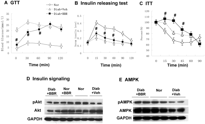
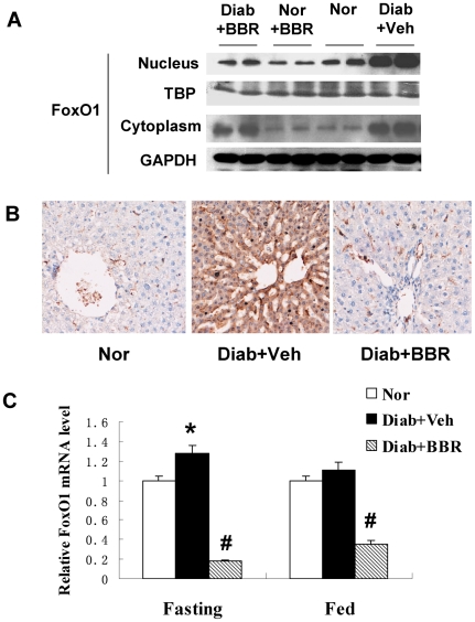
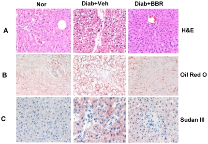
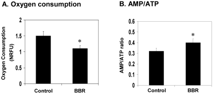
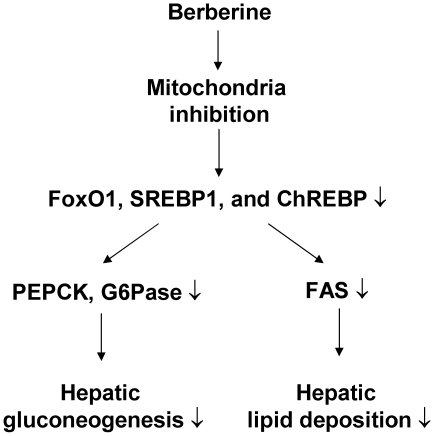
Berberine (BBR) is a compound originally identified in a Chinese herbal medicine Huanglian (Coptis chinensis French). It improves glucose metabolism in type 2 diabetic patients. The mechanisms involve in activation of adenosine monophosphate activated protein kinase (AMPK) and improvement of insulin sensitivity. However, it is not clear if BBR reduces blood glucose through other mechanism. In this study, we addressed this issue by examining liver response to BBR in diabetic rats, in which hyperglycemia was induced in Sprague-Dawley rats by high fat diet. We observed that BBR decreased fasting glucose significantly. Gluconeogenic genes, Phosphoenolpyruvate carboxykinase (PEPCK) and Glucose-6-phosphatase (G6Pase), were decreased in liver by BBR. Hepatic steatosis was also reduced by BBR and expression of fatty acid synthase (FAS) was inhibited in liver. Activities of transcription factors including Forkhead transcription factor O1 (FoxO1), sterol regulatory element-binding protein 1c (SREBP1) and carbohydrate responsive element-binding protein (ChREBP) were decreased. Insulin signaling pathway was not altered in the liver. In cultured hepatocytes, BBR inhibited oxygen consumption and reduced intracellular adenosine triphosphate (ATP) level. The data suggest that BBR improves fasting blood glucose by direct inhibition of gluconeogenesis in liver. This activity is not dependent on insulin action. The gluconeogenic inhibition is likely a result of mitochondria inhibition by BBR. The observation supports that BBR improves glucose metabolism through an insulin-independent pathway.


Conflict of interest statement

Competing Interests: The authors have declared that no competing interests exist.

Figures

Figure 1
Figure 1. BBR decreased fasting blood glucose without improvement in insulin sensitivity.
(A) Effects of BBR treatment on OGTT in diabetic rats. Oral glucose tolerance test (OGTT) was conducted with glucose 2 g.kg−1 body wt after 5 week BBR treatment (380 mg.kg−1 day−1) (n = 6). (B) Insulin release during the OGTT (n = 6). (C) ITT test (n = 6). The test was conducted after 8 hour fasting with insulin (0.75 U.kg−1 body wt). Nor, normal rats without obesity; Diab+Veh: Diabetic rats treated with vehicle; Diab+BBR, diabetic rats treated with BBR. # P<0.05, compared with Diab+Veh. (D) Insulin signaling. Phosphorylation of Akt (Ser473). Rats were challenged with insulin (0.75 U.kg−1, intraperitoneal injection) and liver was collected in 30 minutes. The Akt assay was performed in a Western blot. Loading control is GAPDH. (E) Expression and phosphorylation of AMPK (Thr172). # P<0.05, compared with Diab+Veh group. * P<0.05, compared with normal group (Nor).
Figure 2
Figure 2. Expression of gluconeogenic genes in liver.
(A) Protein levels of PEPCK and G6Pase. Total protein was made from liver tissue and used in a Western blot. (B) mRNA in fasting condition. Total RNA was extracted in from liver tissue and used in qRT-PCR. The signal was normalized with actin mRNA. (C) mRNA in fed condition. The mRNA data are presented as mean ± SEM (n = 6). # P<0.05, compared with Diab+Veh group. * P<0.05, compared with normal group (Nor).
Figure 3
Figure 3. FoxO1 expression in liver.
(A) FoxO1 protein in liver. Nuclear and cytoplasmic proteins were extracted from liver and analyzed in a Western blot. TBP (TATA-binding protein) and GAPDH proteins are loading controls in the nuclear and cytoplasmic proteins. (B) Immunohistostaining of FoxO1 protein in liver. DAB dye (brown) was used to indicate the FoxO1 protein signal; (C) mRNA expression of FoxO1. mRNA was quantified in real time RT-PCR (n = 6). # P<0.05, compared with Diab+Veh group; * P<0.05, compared with normal group (Nor).
Figure 4
Figure 4. Liver steatosis in histology.
(A) Hematoxylin and eosin (H & E) staining. (B) Oil Red O staining. (C) Sudan III staining. Pictures were taken under a microscopy with ×20 object lenses.
Figure 5
Figure 5. Lipogenesis in liver.
(A) Lipogenic transcription factor proteins. Liver total protein was used in the Western blot. SREBP1, SREBP2 and ChREBP were detected in the fasted liver with specific antibodies. Beta-actin is a loading control. (B) FAS protein. The protein was detected in a Western blot in the fasted and fed liver, respectively. (C) FAS mRNA. mRNA was detected by qRT-PCR and normalized with beta-actin mRNA (n = 6). # P<0.05, compared with Diab+Veh group; * P<0.05, compared with normal group (Nor).
Figure 6
Figure 6. Inhibition of mitochondrial function by BBR in hepatocytes.
(A) BBR decreased oxygen consumption in H4IIE hepatocytes. The cells were plated in DMEM culture medium supplemented with 10% FBS. Oxygen consumption was determined for 12 hrs after BBR (20 µM) treatment. (B) AMP/ATP ratio in H4IIE cells. The cells were treated with BBR in serum-free medium for 16 hours. AMP and ATP levels were determined using HPLC. The data are presented as mean ± SEM (n = 5). * P<0.05.
Figure 7
Figure 7. Schematic model of BBR signaling pathway.
BBR inhibits mitochondria function and decrease intracellular ATP. This leads to a reduction in gluconeogenic and lipogenic transcription factors (FoxO1, SREBP1, and ChREBP). As a result, expression of gluconeogenic genes (PEPCK and G6Pase) and lipogenic gene (FAS) are decreased. These molecular changes represent a signaling pathway for improvement of fasting glucose and liver steatosis in the BBR-treated diabetic rats.

Similar articles

Cited by

References

    1. Yin J, Zhang H, Ye J. Traditional chinese medicine in treatment of metabolic syndrome. Endocr Metab Immune Disord Drug Targets. 2008;8:99–111. - PMC - PubMed
    1. Lee YS, Kim WS, Kim KH, Yoon MJ, Cho HJ, et al. Berberine, a Natural Plant Product, Activates AMP-Activated Protein Kinase With Beneficial Metabolic Effects in Diabetic and Insulin-Resistant States. Diabetes. 2006;55:2256–2264. - PubMed
    1. Kong WJ, Zhang H, Song DQ, Xue R, Zhao W, et al. Berberine reduces insulin resistance through protein kinase C-dependent up-regulation of insulin receptor expression. Metabolism. 2009;58:109–119. - PubMed
    1. Zhang H, Wei J, Xue R, Wu JD, Zhao W, et al. Berberine lowers blood glucose in type 2 diabetes mellitus patients through increasing insulin receptor expression. Metabolism. 2010;59:285–292. - PubMed
    1. Yin J, Gao Z, Liu D, Liu Z, Ye J. Berberine Improves Glucose Metabolism through Induction of Glycolysis. Am J Physiol Endocrinol Metab. 2008;294:E148–E156. - PMC - PubMed

Publication types

MeSH terms