. 2011 Feb 3;7(2):e1001069.
doi: 10.1371/journal.pcbi.1001069.

Gene expression noise in spatial patterning: hunchback promoter structure affects noise amplitude and distribution in Drosophila segmentation

Affiliations

Gene expression noise in spatial patterning: hunchback promoter structure affects noise amplitude and distribution in Drosophila segmentation

David M Holloway et al. PLoS Comput Biol. .

Abstract

Positional information in developing embryos is specified by spatial gradients of transcriptional regulators. One of the classic systems for studying this is the activation of the hunchback (hb) gene in early fruit fly (Drosophila) segmentation by the maternally-derived gradient of the Bicoid (Bcd) protein. Gene regulation is subject to intrinsic noise which can produce variable expression. This variability must be constrained in the highly reproducible and coordinated events of development. We identify means by which noise is controlled during gene expression by characterizing the dependence of hb mRNA and protein output noise on hb promoter structure and transcriptional dynamics. We use a stochastic model of the hb promoter in which the number and strength of Bcd and Hb (self-regulatory) binding sites can be varied. Model parameters are fit to data from WT embryos, the self-regulation mutant hb(14F), and lacZ reporter constructs using different portions of the hb promoter. We have corroborated model noise predictions experimentally. The results indicate that WT (self-regulatory) Hb output noise is predominantly dependent on the transcription and translation dynamics of its own expression, rather than on Bcd fluctuations. The constructs and mutant, which lack self-regulation, indicate that the multiple Bcd binding sites in the hb promoter (and their strengths) also play a role in buffering noise. The model is robust to the variation in Bcd binding site number across a number of fly species. This study identifies particular ways in which promoter structure and regulatory dynamics reduce hb output noise. Insofar as many of these are common features of genes (e.g. multiple regulatory sites, cooperativity, self-feedback), the current results contribute to the general understanding of the reproducibility and determinacy of spatial patterning in early development.


Conflict of interest statement

The authors have declared that no competing interests exist.

Figures

Figure 1
Figure 1. Concentration profiles of the morphogenetic proteins Bicoid (Bcd) and Hunchback (Hb).
(A) A Drosophila embryo fluorescently immunostained for Bcd (green) and Hb (blue), about 30 minutes into nuclear cleavage cycle 14. Anterior left, dorsal top. Nuclei at the surface of the precellular, syncytial blastoderm are visible. (B) Fluorescence intensity against anterior-posterior (AP) position (in percent egg length (%EL), colours as in A, showing the exponential Bcd gradient and the step-like Hb pattern. From .
Figure 2
Figure 2. Model of the hb promoter.
(A) Schematic diagram of a 4776 bp fragment from the hb regulatory region determined by in vitro footprinting , , adapted from [[30], Figure S4]. The Bcd sites (red; A are strongly binding, X are weaker) and first two Hb sites can drive relatively sharp anterior expression in lacZ constructs in a WT background (e.g. Figure 7D). This region (between the green arrows) has been extensively studied as the core of the proximal promoter and is the basis of our model. (B) The reaction network based on these core binding sites, involving binding, unbinding, transcription, translation and decay. B = Bcd protein; H = Hb protein; MB and MH are their mRNAs, respectively. bcd mRNA is translated at the anterior pole. Bcd and Hb proteins diffuse. b 0–6 are the number of Bcds bound to the hb promoter; h 0–2 are the number of Hbs bound to the hb promoter. The subscript refers to the binding order based on strength. Values of the rate constants (k's) are constrained by experimental data and are given in Tables S1, S2, S3.
Figure 3
Figure 3. Wild-type (WT) expression noise.
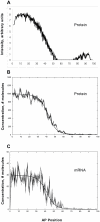
(A, B) hb protein and mRNA profiles, respectively, from the model (Figure 2B; 6 Bcd sites (3A, 3X) and 2 Hb sites, or 6B2H), with parameters fit by experimental constraints (Tables S1, S2, S3). Deterministic (no noise) results, dashed line. Stochastic results are shown over one minute (29–30 minutes into cycle 14), at 5 second intervals (comparable to the experimental heat-fixation time), to show the slow temporal variability of the noise (same format used in Figures 4, 6, 7). Noise is calculated from the differences between the deterministic and stochastic results at 30 minutes (Table 1). (C, D) Representative data from a single WT embryo, hb protein and mRNA, respectively, 30–36 minutes into cycle 14 (same time in Figures 4, 6– 8). This shows the characteristic determinate mid-embryo boundary, especially for the protein, which is also produced by the model. mRNA (B, D) has significantly higher noise than protein (see Table 1 for statistics). (E) Too-fast reaction rates are one factor that can cause noise to overwhelm determinate expression. Here, reaction rates (transcription, translation, mRNA and protein decay) have been increased ten-fold from (A, B), producing much higher fluctuation levels (protein noise 25%, mRNA noise 32% - higher than any WT results (Table 1)) and reducing the determinacy of the mid-embryo boundary. (F) Within-embryo noise contribution to between-embryo variability: 19 independent stochastic simulations of WT protein expression, with a standard deviation in boundary position of 1.0%EL (comparable to experimentally observed between-embryo variability).
Figure 4
Figure 4. The absence of hb self-regulation.
(A) Protein expression in a homozygous hb 14F mutant embryo (whose Hb protein cannot bind DNA); noise is significantly higher than WT (c.f. Figure 3C). (B) Simulation of hb14F protein expression: 6 Bcd binding sites and no Hb binding in the promoter (6B0H). Noise is higher than WT. (C) mRNA for the same simulation: noise is higher than protein (B), and higher than for WT mRNA (Figure 3B). See Table 2 for statistics.
Figure 5
Figure 5. Noise signatures for Bcd, hb mRNA and Hb protein.
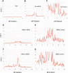
(A, B, C) Variance to mean ratio (VMR) by position, for Bcd (A), hb mRNA (B), and Hb protein (C), all from the same computation. Bcd dynamics (synthesis-diffusion-decay) produce a VMR of 1, characteristic of Poisson-distributed noise. hb mRNA (B) has a variance 2 to 3 times that for a Poisson process. Translation increases the deviation from Poisson for the protein (C). The non-Poisson character of the mRNA distribution is largely due to Hb self-feedback: compare (D), mRNA in the absence of Hb self-regulation (6B0H simulation of hb 14F, c.f. Figure 4), to (F), mRNA with self-regulation (WT 6B2H). Translation increases protein VMR about six-fold over mRNA, both without self-regulation (E) and with self-regulation (G). VMR is higher for both mRNA and protein with self-regulation, since the high protein VMR feeds back on mRNA transcription. Bcd noise has little effect on the hb mRNA or protein noise: the different VMRs point to different probability distributions (A vs. B and C), and there is negligible difference between simulations with Bcd noise (B, C) and without (F, G). I.e., the noise generated in transcription and translation dominates the noise transmitted from upstream regulator fluctuations.
Figure 6
Figure 6. The effects of Bcd binding site number and strength on noise; simulations of lacZ constructs with Bcd binding sites only.
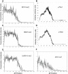
(A) A single strong Bcd site (1A, pThb3 construct); very low and noisy expression, with much posterior activation (statistics for this Figure given in Table 3). (B) Three strong sites (3A, pThb10); expression is stronger and less noisy than (A). (C) Three weak Bcd sites (3X, pThb12); expression is noisier than (B). (D) Four strong sites (4A, pThb11); expression is stronger and less noisy than (B). (E) Four weak sites (4X, pThb13); expression is noisier than (D). (F) Doubling of the 3X promoter; noise is less than (C). (G) Tripling of the 3X promoter; lower noise again than (F). Noise decreases for increasing strength and increasing number of binding sites.
Figure 7
Figure 7. The effects of Hb binding site number and strength on noise.
(A) Simulation of lacZ expression from 6 Bcd sites (3A3X) and one Hb site (in a WT background), compare to (B) lacZ expression in an embryo with the pThb1 promoter construct. (C) Simulation with 6 Bcd sites and 2 Hb sites, compare to (D) lacZ in a pThb5 embryo (promoter indicated by green arrows in Figure 2A). Comparison of (C, D) and (A, B) indicates that the 2nd Hb increases expression and slope and may slightly decrease noise. Noise statistics for this Figure are in Table 4. (E) Simulation of the pThb8 construct, a doubled 6B1H sequence. (F) Simulation of the pThb2 construct, which has a shorter fragment of the hb promoter, with 1 Hb site and 4 Bcd sites (2A2X1H); the model predicts increased noise for this truncated promoter.
Figure 8
Figure 8. Variation within nuclei, transcription from two copies of the promoter.
(A) lacZ mRNA labelling (pink) at nuclear dot resolution in a pThb5 embryo (nuclei in blue), 30–36 minutes into cleavage cycle 14 (c.f. Figure 7D). (B) Computed lacZ mRNA levels, for two equal and independent promoters, A (blue) and B (red), at 30 minutes; the simulation shown has a relative noise between A and B of 31%, an average value (see Table 6 for statistics). (C) Comparable plot of intensity against AP position for the embryo in (A), A–B dot pairs are coloured as in (B). This data has A–B relative noise of 17%.

Similar articles

Cited by

References

    1. Houchmandzadeh B, Wieschaus E, Leibler S. Establishment of developmental precision and proportions in the early Drosophila embryo. Nature. 2002;415:798–802. - PubMed
    1. Crauk O, Dostatni N. Bicoid determines sharp and precise target gene expression on the Drosophila embryo. Curr Biol. 2005;15:1888–1898. - PubMed
    1. Holloway DM, Harrison LG, Kosman D, Vanario-Alonso CE, Spirov AV. Analysis of pattern precision shows that Drosophila segmentation develops substantial independence from gradients of maternal gene products. Dev Dynam. 2006;235:2949–2960. - PMC - PubMed
    1. Gregor T, Tank DW, Wieschaus EF, Bialek W. Probing the limits to positional information. Cell. 2007;130:153–164. - PMC - PubMed
    1. He F, Wen Y, Lin X, Lu LJ, Jiao R, et al. Probing intrinsic properties of a robust morphogen gradient in Drosophila. Dev Cell. 2008;15:558–567. - PMC - PubMed

Publication types

MeSH terms

LinkOut - more resources