. 2011 Feb 3;7(2):e1001266.
doi: 10.1371/journal.ppat.1001266.

Entrapment of viral capsids in nuclear PML cages is an intrinsic antiviral host defense against varicella-zoster virus

Affiliations

Entrapment of viral capsids in nuclear PML cages is an intrinsic antiviral host defense against varicella-zoster virus

Mike Reichelt et al. PLoS Pathog. .

Abstract

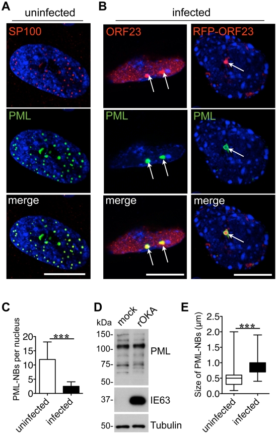
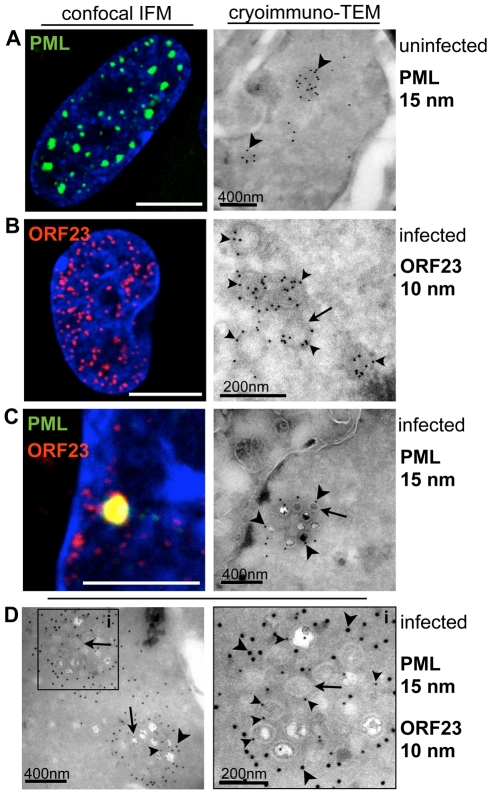
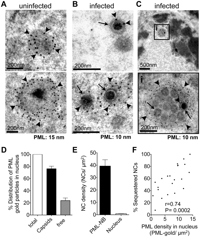
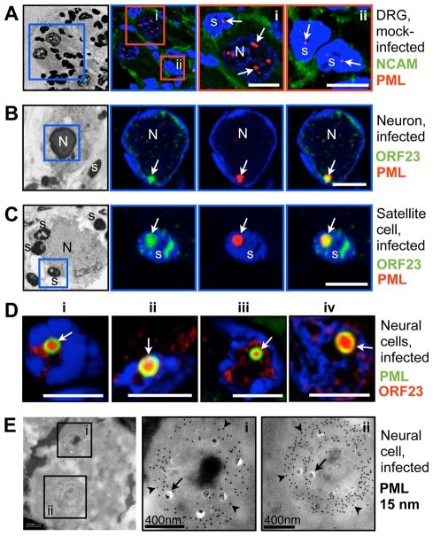
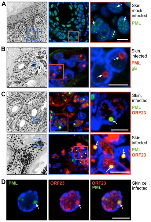
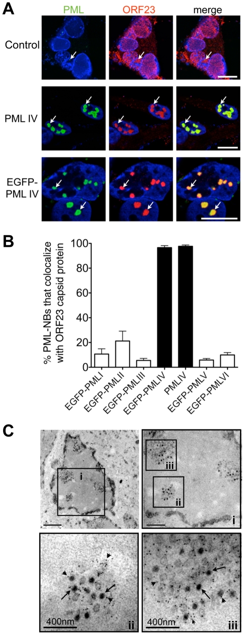
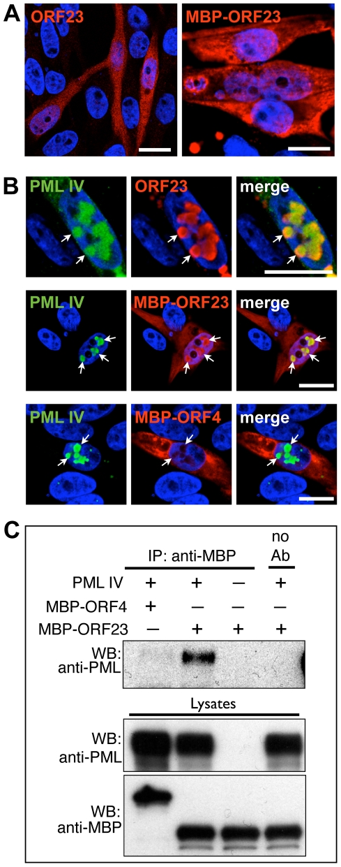
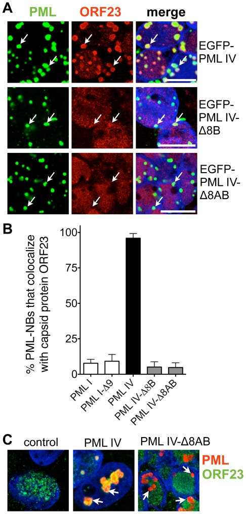
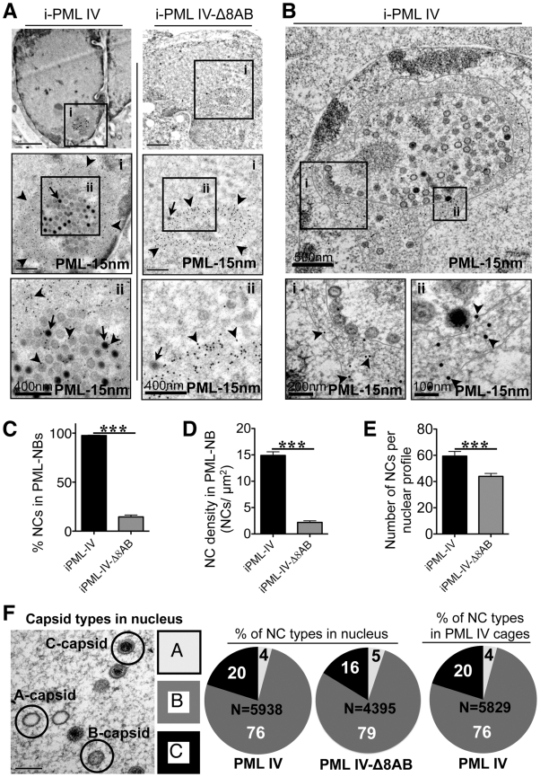
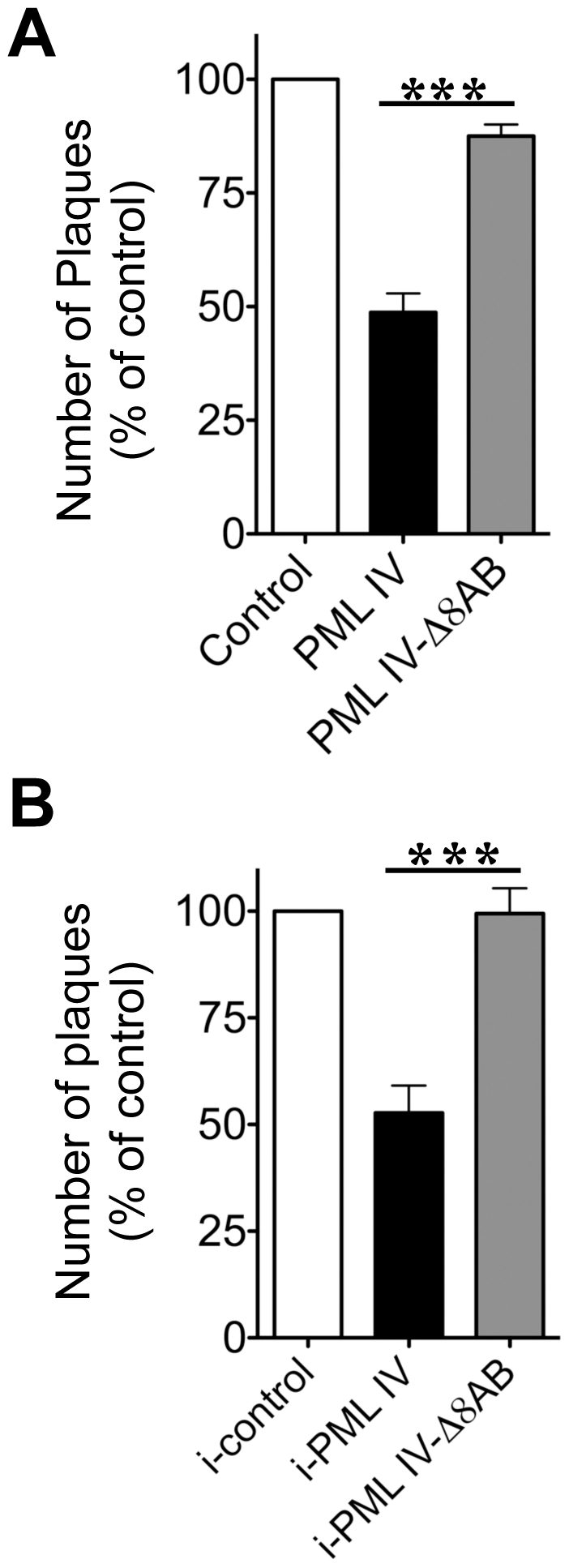
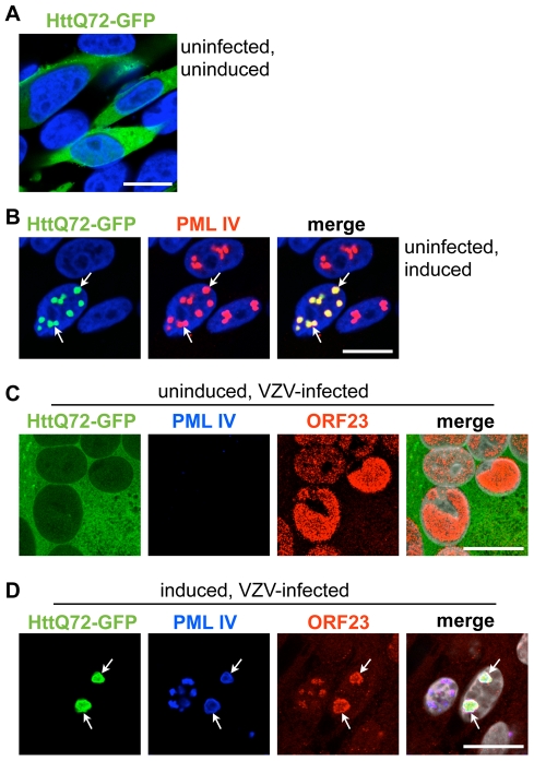
The herpesviruses, like most other DNA viruses, replicate in the host cell nucleus. Subnuclear domains known as promyelocytic leukemia protein nuclear bodies (PML-NBs), or ND10 bodies, have been implicated in restricting early herpesviral gene expression. These viruses have evolved countermeasures to disperse PML-NBs, as shown in cells infected in vitro, but information about the fate of PML-NBs and their functions in herpesvirus infected cells in vivo is limited. Varicella-zoster virus (VZV) is an alphaherpesvirus with tropism for skin, lymphocytes and sensory ganglia, where it establishes latency. Here, we identify large PML-NBs that sequester newly assembled nucleocapsids (NC) in neurons and satellite cells of human dorsal root ganglia (DRG) and skin cells infected with VZV in vivo. Quantitative immuno-electron microscopy revealed that these distinctive nuclear bodies consisted of PML fibers forming spherical cages that enclosed mature and immature VZV NCs. Of six PML isoforms, only PML IV promoted the sequestration of NCs. PML IV significantly inhibited viral infection and interacted with the ORF23 capsid surface protein, which was identified as a target for PML-mediated NC sequestration. The unique PML IV C-terminal domain was required for both capsid entrapment and antiviral activity. Similar large PML-NBs, termed clastosomes, sequester aberrant polyglutamine (polyQ) proteins, such as Huntingtin (Htt), in several neurodegenerative disorders. We found that PML IV cages co-sequester HttQ72 and ORF23 protein in VZV infected cells. Our data show that PML cages contribute to the intrinsic antiviral defense by sensing and entrapping VZV nucleocapsids, thereby preventing their nuclear egress and inhibiting formation of infectious virus particles. The efficient sequestration of virion capsids in PML cages appears to be the outcome of a basic cytoprotective function of this distinctive category of PML-NBs in sensing and safely containing nuclear aggregates of aberrant proteins.


Conflict of interest statement

The authors have declared that no competing interests exist.

Figures

Figure 1
Figure 1. Endogenous PML-NBs colocalize with ORF23 capsid protein in VZV infected cells.
(A) Uninfected human embryonic lung fibroblasts (HELF) were immunostained for SP100 (red) or PML (green) to visualize PML nuclear bodies (PML-NBs). Nuclei were stained with Hoechst (blue). (B) HELF cells were infected with cell-associated VZV for 24 hr and immunostained for PML (green) and ORF23 (red). PML-NBs and ORF23 capsid protein colocalized (white arrows) in HELF infected with either VZV (left panel) or VZV expressing red fluorescent protein (RFP)-tagged ORF23 (right panel). Nuclei were stained with Hoechst (blue). ORF23 capsid protein was identified in 94% (N = 228) of PML-NBs that were present in VZV infected cells at 24 hr. Scale bars in A and B are 5 µm. (C) Quantitation of the mean number of PML-NBs in nuclei of uninfected HELF (N = 180) and in infected HELF cells at 48 hr after infection (N = 180). The graph shows the mean ± SD; asterisks indicate a significant difference (p<0.0001). (D) Immunoblot analysis of PML protein (upper panel), VZV IE63 protein as a marker of VZV infection (middle panel) and tubulin (lower panel) in whole cell lysates of HELF that were mock-infected or infected with VZV (rOka) for 48 hr. (E) Quantitation of the mean size (diameter in µm) of PML-NBs in uninfected HELF and infected HELF. The box and whiskers plot shows the mean, 25 and 75 percentiles and the range of PML-NB sizes from 100 uninfected or infected nuclei, respectively; asterisks indicate a significant difference (p<0.0001).
Figure 2
Figure 2. VZV nucleocapsids are sequestered in endogenous nuclear PML cages.
HELF cells were uninfected (A, left panel) or infected (B, C, left panels) with VZV (rOka) for 48 hr and analyzed by confocal immunofluorescence (IF) microscopy after staining for PML (green) or ORF23 (red). For cryoimmuno-EM, HELF cells were uninfected (A, right panel) or infected (B–C, right panels; D, left and right panels) with VZV (rOka) for 48 hr and analyzed after immunogold labeling of PML (15 nm) or ORF23 (10 nm) by transmission electron microscopy (TEM). (A) PML-NBs visualized by confocal IF and by cryoimmuno-EM. Dense clusters of PML-specific gold particles (15 nm) label PML-NBs, indicated by arrowheads. (B) Tiny fluorescent punctae detected in VZV infected nuclei immunostained for ORF23 (red) by confocal IF may correspond to nucleocapsids (NCs). Cryoimmuno-EM identifies ORF23 (arrowheads) with NCs (arrows) by ORF23 specific gold-labeling of ring-like or hexagonal structures of about 100 nm diameter. (C) PML (green) and ORF23 (red) colocalize in PML-NBs shown by confocal IF microscopy. Cryoimmuno-EM in combination with PML-specific single-immunogold labeling (C, arrowheads) or double-immunogold labeling for PML and ORF23 (D, left panel) identifies clusters of VZV NCs (arrows) surrounded by PML protein. At a higher magnification (D, right panel) of the cluster indicated by the black square, NCs with ORF23-labeling (small arrowheads) and associated PML-labeling (large arrowheads) can be seen. Scale bars in IF images are 5 µm. Scale bars for EM images are as indicated.
Figure 3
Figure 3. PML cages sequester mature and immature VZV nucleocapsids.
(A-C) Representative immunogold-EM images of uninfected (A) or VZV-infected (B and C) HELF cells at 48 hr after infection and processing by high-pressure freezing (HPF) and freeze substitution (FS) to optimally preserve ultra-structural details. PML protein is specifically labeled with (A) 15 nm or (B and C) 10 nm gold particles (arrowheads indicate PML gold particles). Arrows indicate examples of viral NCs. (A) PML-labeled endogenous NBs in uninfected cells; examples are shown in upper and lower panels. (B) Individual VZV NCs (arrows) are associated with varying amounts of PML protein (arrowheads) as shown in the upper and lower panels. (C) Clusters of VZV NCs (arrows) are sequestered in an endogenous fibrous PML cage (arrowheads). The black square in the upper panel demarks the area (i) that is shown at higher magnification on the lower panel. Scale bars are as indicated. (D–F) Quantitative analysis of PML-specific gold particles (N = 1,611) and viral NCs (N = 450) in 30 VZV-infected cell nuclei. (D) Percentage of the total number of PML-specific gold particles associated with VZV capsids (black bar) or found ‘free’ within the nuclear area excluding PML-NBs (grey bar). (E) Density of viral NCs determined as NCs/µm2 (mean ± SD) within PML-NBs (black bar) or within the nuclear area outside PML-NBs (white bar). (F) Correlation between the percentage of NCs sequestered within PML-NBs (y-axis) and the PML labeling density in each nucleus, which is the number of PML-specific gold particles/µm2 of the total nuclear area (x-axis). Since only profiles of infected cell nuclei that contained at least 10 NCs were considered, N = 22 for this analysis. Pearson correlation: r = 0.74, R2 = 0.54, p<0.0002.
Figure 4
Figure 4. PML cages sequester VZV nucleocapsids during infection of human Dorsal Root Ganglia (DRG).
Human DRG xenografts in the SCID mouse model were collected 14 days after mock infection (A) or inoculation with VZV (B–E) and fixed in 4% paraformaldehyde. Semithin (500 nm) cryosections were analyzed by confocal IF microscopy after staining for (A) Neural Cell Adhesion Molecule (NCAM) and PML, or (B–D) ORF23 and PML, as indicated at the right of each row of panels. Nuclei are stained with Hoechst (blue). Images in A-C (left panels) show overviews of the same DRG sections after Hoechst staining using an inverted grey scale. Blue or red squares demark areas shown at higher magnification in the adjacent panels. (A) Uninfected DRG sections show numerous small PML-NBs (red, white arrows) in the nuclei of neurons (N) and satellite cells (s). (B, C) VZV infected neurons and satellite cells with PML-NBs (white arrows) in which PML (red) colocalizes with ORF23 capsid protein (green). Scale bars are 5 µm. (D) Four examples (i–iv) of neural cells in infected DRG with large ring-like PML cages (green, white arrows) that sequester ORF23 protein (red). ORF23 capsid protein was present in 87% (N = 116) of PML-NBs within infected neural cells. Scale bars are 5 µm. (E) PML-specific immunogold-labeling of ultrathin (80 nm) cryosections of infected human DRG shows large ring-like PML cages (PML, 15 nm; arrowheads) that sequester numerous VZV NCs (arrows) in infected neural cells in human DRG. Areas in the black squares (i and ii) are shown at higher magnification in panels at the right. Sizes of scale bars are as indicated.
Figure 5
Figure 5. PML cages sequester VZV nucleocapsids during infection of human skin.
Human skin xenografts in the SCID mouse model were collected 21 days after mock infection (A) or inoculation with VZV (B–D). Thick (5 µm) paraffin sections were analyzed by confocal IF microscopy after staining for PML (A), double-IF staining for PML and the VZV glycoprotein gE (B) or double-IF staining for ORF23 and PML (C and D), as indicated at the right of each row of panels. Nuclei are stained with Hoechst (blue). Images in A-C (left panels) are overviews of the same skin sections, including epidermal and dermal layers and hair follicles, after Hoechst staining (inverted grey scale). Blue and red squares demark areas shown at higher magnification in the panels on the right. Arrows indicate PML-NBs in the nuclei of skin cells. (A) Uninfected skin cell nuclei contain several small PML-NBs (green). (B) Infected cells have fused to form the syncytia (polykaryons). Large ring-like PML-NBs (red) are visible and VZV glycoprotein gE (green) is expressed on plasma membranes. (C) Large ring-like PML-NBs (green) sequester ORF23 protein (red). (D) A single skin cell nucleus showing PML-NBs (green) and ORF23 protein (red). All scale bars are 5 µm.
Figure 6
Figure 6. PML IV promotes the sequestration of VZV nucleocapsids within PML cages.
(A) Representative IF-microscopy images show colocalization of PML IV-NBs (green) with ORF23 capsid protein (red) in VZV-infected melanoma cells at 48 hr after infection. Cells were either untransfected (control, upper panels) or transfected with plasmids expressing PML IV (middle panels) or EGFP-PML IV (lower panels). Nuclei were stained with Hoechst (blue). Arrows depict examples of PML IV bodies. (B) Quantitation of the percentage of PML-NBs that colocalize with ORF23 protein at 48 hr after infection of cells that had been transfected with plasmids expressing the six different PML isoforms, PML I-PML VI as indicated. Data shown for each PML isoform represent the mean percentage ± SD from five different samples with at least 100 PML-NBs each. (C) Representative immunogold-EM images after transfection with EGFP-PML IV and 48 hr after VZV infection. PML protein was identified with a polyclonal (rabbit) anti-PML antibody and Protein-A conjugated with 15 nm gold particles (small arrowheads). Three areas (i-iii, black squares) are shown at higher magnification. Arrows indicate viral NCs within densely labeled PML-NBs. Quantitative EM analysis of 100 infected nuclei showed that more than 90% of VZV NCs (N = 4,900) were sequestered in PML cages. EM scale bars are indicated.
Figure 7
Figure 7. PML IV binds ORF23 capsid protein in the absence of other viral proteins.
(A) Representative IF images show the cellular distribution of either untagged ORF23 (red; left panel) or maltose binding protein (MBP)-tagged ORF23 (red; right panel) at 48 hr after transfection of melanoma cells. (B) IF localization of untagged ORF23 protein (red; upper panels), MBP-tagged ORF23 protein (red; middle panels) or MBP-tagged ORF4 protein (red; lower panels) and PML IV bodies (green, white arrows) at 48 hr after transfection of melanoma cells. Nuclei were stained with Hoechst (blue). All scale bars are 5 µm. (C) Coimmunoprecipitation (Co-IP) of PML IV using an anti-MBP polyclonal (rabbit) antibody in cells co-transfected with PML IV expression plasmid and MBP-ORF23 or the control MBP-ORF4, in the combinations as indicated by ‘+’ and ‘-’. Co-IP of mock-transfected cells (no PML IV overexpression) is shown in the lane at the far right. PML IV was identified by immunoblotting with a polyclonal (rabbit) anti-PML antibody. Western blot of the lysates confirmed the expression of transfected PML IV, MBP-ORF23 and MBP-ORF4 (lower panel).
Figure 8
Figure 8. Sequestration of ORF23 capsid protein requires the C-terminal domain of PML IV.
(A) Representative IF images show that EGFP-PML IV (green; upper panels) colocalized with ORF23 protein (red) but not with the C-terminal deletion mutants, EGFP-PML IV-Δ8B (green; middle panel) or EGFP-PML IV-Δ8AB (green; lower panel) in transfected cells that were infected with VZV for 48 hr. Nuclei were stained with Hoechst (blue). Arrows indicate the location of PML-NBs. Scale bars are 5 µm. (B) Quantitation of the percentages of PML-NBs that colocalized with ORF23 protein in infected cells that expressed the indicated PML isoforms or PML deletion mutants after transfection. The bars show PML-NB colocalization with ORF23 as the mean percentage ± SD for each PML isoform or deletion mutant from seven different areas, each of which contained 60-120 PML-NBs. (C) Doxycycline-inducible melanoma cells were generated that expressed no exogenous PML IV (left), PML IV (middle) or the truncated mutant PML IV-Δ8AB (right). The stable cell lines were induced with 5 µg/ml doxycycline and then infected with VZV for 24 hr. Cells were immunostained for PML (red) and ORF23 capsid protein (green); nuclei were stained with Hoechst (blue). White arrows indicate PML-NBs. Scale bar, 5 µm.
Figure 9
Figure 9. Quantitative ultrastructural analysis of PML IV cages with entrapped VZV nucleocapsids.
(A) Cells induced to express PML IV (left panels) or PML IV-Δ8AB (right panels) and infected with VZV for 48 hr were processed for immunogold-EM. PML-NBs were identified with a polyclonal (rabbit) PML antibody and protein A conjugated to 15 nm gold particles. Areas in black squares (i and ii) are shown at higher magnification. Arrows indicate mature (C-type) capsids. Arrowheads indicate PML-specific gold particles (15 nm). The size of scale bars is indicated. (B) Cells induced to express PML IV and infected with VZV for 48 hr were embedded in Epoxy-resin. Ultrathin sections were surface-etched with hydrogen peroxide before PML-immunogold-labeling (15 nm, arrowheads). A PML IV cage with sequestered VZV NCs is shown. Red lines demark the outer and inner border of the spherical PML cage. PML-specific labeling (15 nm gold particles) is visible when the areas in the black squares (i and ii) are examined at higher magnification. The shell of the PML cage consists of PML-positive fibers (i and ii, lower panels). NCs align along the inner margin of the fibrous PML cage (area i). (C-E) Quantitative ultrastructural analysis of 100 nuclear profiles each in cells expressing PML IV (black bars) or PML IV-Δ8AB (grey bars). The total number of NCs analyzed was N = 5,938 in PML IV cells and N = 4,395 in PML IV-Δ8AB cells. (C) Quantitation of the number of VZV NCs shown as the percentage of the total NCs sequestered by induced PML IV (iPML-IV) and induced PML IV-Δ8AB (iPML IV-Δ8AB); the asterisks indicate a significant difference (p<0.0001). (D) Quantitation of the density of viral NCs within PML IV-NBs or PML IV-Δ8AB PML-NBs (mean ± SEM) within unit areas of PML-NBs (NCs/µm2) from an analysis of 100 PML-NBs (p<0.0001). (E) Comparison of the total number of VZV NCs per nuclear profile in nuclei with PML IV cages and nuclei with PML IV-Δ8AB NBs (mean ± SEM; p = 0.0003). (F) Proportion of the three types of VZV capsids within 100 nuclear profiles of cells induced to express PML IV or PML IV-Δ8AB and within PML IV cages. The left panel illustrates mature infectious (C-type), intermediate (B-type) and abortive (A-type) NCs visualized by TEM; the relative percentage of each type is shown in the pie charts (A-type: light gray; B-type: dark gray; C-type: black). The total number (N) of capsids analyzed is indicated in each pie chart.
Figure 10
Figure 10. PML IV requires the unique C-terminal domain for inhibition VZV replication.
(A) VZV plaque assay with melanoma cells that were transfected either with EGFP control, EGFP-PML IV or EGFP-PML IV-Δ8AB expression constructs and infected for 24 hr with VZV (rOka-RFP-ORF23) that expressed RFP tagged capsid protein. Only cells that were both transfected (green fluorescence) and infected (red fluorescence) were recovered by FACS. Melanoma cell monolayers were inoculated with equal numbers of cells from each of the sorted cell preparations. (A) Plaques were counted after 72 hr. Data are shown as the mean percentage (mean ± SEM) of plaque numbers normalized to the control (EGFP alone) from five independent experiments; plaque numbers were measured in triplicate in each experiment. The asterisks indicate a significant difference (p<0.0001). (B) Infectious virus production 24 hr after VZV infection of doxycycline-induced control, PML IV or PML IV-Δ8AB expressing cell lines, shown as mean plaque numbers (percentage of control ± SEM). The number of plaques formed in cells expressing PML IV was significantly reduced compared to the number in PML IV-Δ8AB expressing cells (p = 0.0018; N = 4).
Figure 11
Figure 11. PML IV cages cosequester ORF23 capsid protein and Huntington's disease protein in VZV infected cells.
PML IV inducible cell lines were transfected to express HttQ72-GFP. (A) HttQ72-GFP (green) expressing cells were fixed in the absence of PML IV induction. (B) HttQ72-GFP (green) expressing cells were induced to express PML IV and fixed after 48 hr. PML IV was detected by PML-specific IF staining (red). Recruitment of HttQ72-GFP to PML IV bodies (white arrows) is visible. (C) HttQ72-GFP transfected cells were infected with VZV for 48 hr without induction of PML IV expression. IF-images show the localization of HttQ72-GFP (green), PML (blue) and ORF23 capsid protein (red) in a syncytium (polykaryon) of fused VZV-infected cells. (D) A HttQ72-GFP transfected cell (the cell on the right) after induction of PML IV and infection with VZV for 48 hr. IF-images show the colocalization (white arrows) of HttQ72-GFP (green), PML (blue) and ORF23 capsid protein (red).

Similar articles

Cited by

References

    1. Ishov AM, Sotnikov AG, Negorev D, Vladimirova OV, Neff N, et al. PML is critical for ND10 formation and recruits the PML-interacting protein daxx to this nuclear structure when modified by SUMO-1. J Cell Biol. 1999;147:221–234. - PMC - PubMed
    1. Zhong S, Muller S, Ronchetti S, Freemont PS, Dejean A, et al. Role of SUMO-1-modified PML in nuclear body formation. Blood. 2000;95:2748–2752. - PubMed
    1. Lallemand-Breitenbach V, Zhu J, Puvion F, Koken M, Honore N, et al. Role of promyelocytic leukemia (PML) sumolation in nuclear body formation, 11S proteasome recruitment, and As2O3-induced PML or PML/retinoic acid receptor alpha degradation. J Exp Med. 2001;193:1361–1371. - PMC - PubMed
    1. Eskiw CH, Dellaire G, Mymryk JS, Bazett-Jones DP. Size, position and dynamic behavior of PML nuclear bodies following cell stress as a paradigm for supramolecular trafficking and assembly. J Cell Sci. 2003;116:4455–4466. - PubMed
    1. Muratani M, Gerlich D, Janicki SM, Gebhard M, Eils R, et al. Metabolic-energy-dependent movement of PML bodies within the mammalian cell nucleus. Nat Cell Biol. 2002;4:106–110. - PubMed

Publication types

MeSH terms