. 2011 Jan 31;6(1):e16571.
doi: 10.1371/journal.pone.0016571.

Intracellular S1P generation is essential for S1P-induced motility of human lung endothelial cells: role of sphingosine kinase 1 and S1P lyase

Affiliations

Intracellular S1P generation is essential for S1P-induced motility of human lung endothelial cells: role of sphingosine kinase 1 and S1P lyase

Evgeny V Berdyshev et al. PLoS One. .

Abstract

Background: Earlier we have shown that extracellular sphingosine-1-phosphate (S1P) induces migration of human pulmonary artery endothelial cells (HPAECs) through the activation of S1P(1) receptor, PKCε, and PLD2-PKCζ-Rac1 signaling cascade. As endothelial cells generate intracellular S1P, here we have investigated the role of sphingosine kinases (SphKs) and S1P lyase (S1PL), that regulate intracellular S1P accumulation, in HPAEC motility.

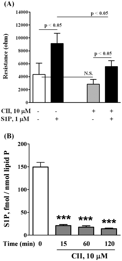
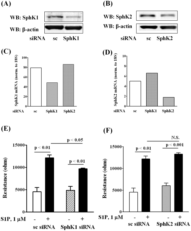
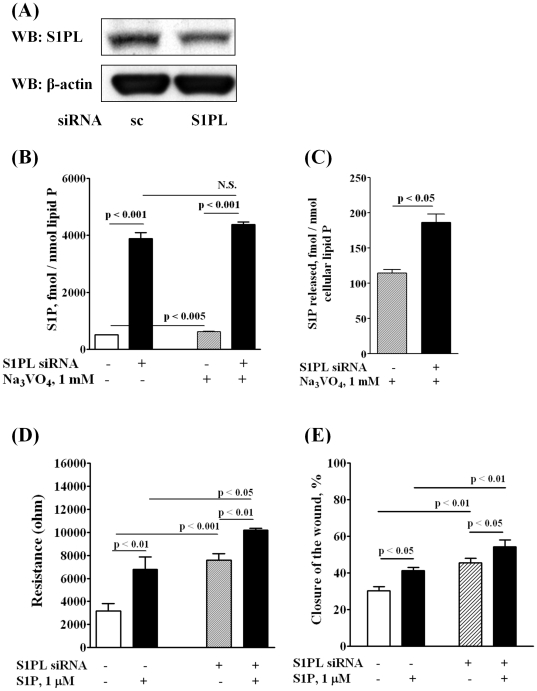
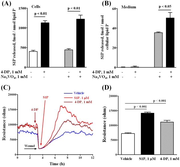
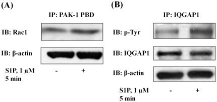
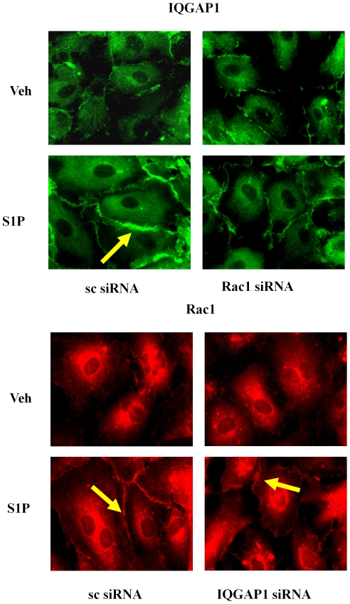
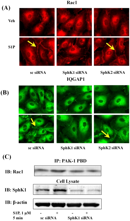
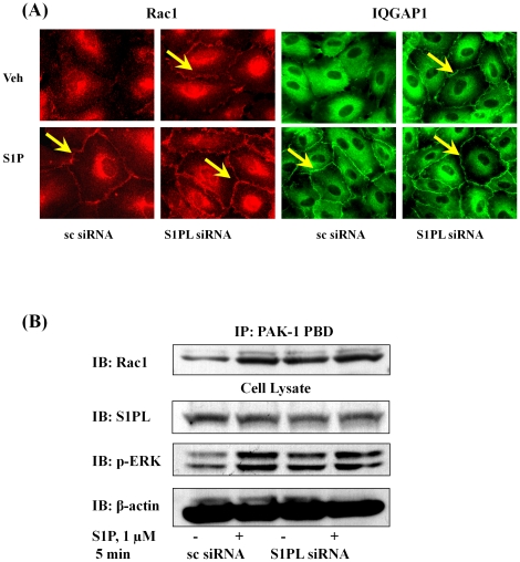
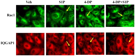
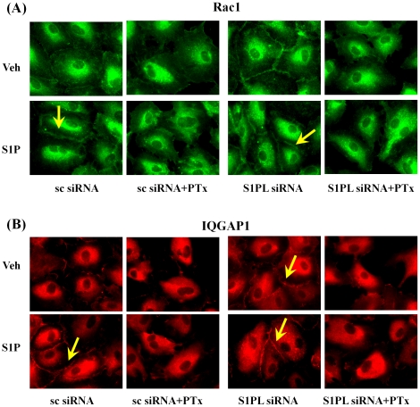
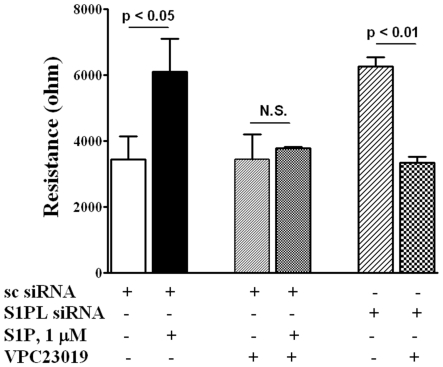
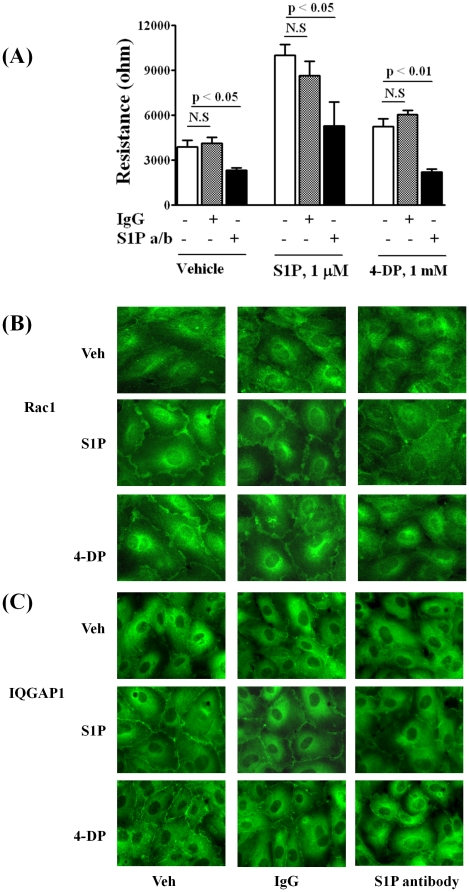
Methodology/principal findings: Inhibition of SphK activity with a SphK inhibitor 2-(p-Hydroxyanilino)-4-(p-Chlorophenyl) Thiazole or down-regulation of Sphk1, but not SphK2, with siRNA decreased S1P(int), and attenuated S1P(ext) or serum-induced motility of HPAECs. On the contrary, inhibition of S1PL with 4-deoxypyridoxine or knockdown of S1PL with siRNA increased S1P(int) and potentiated motility of HPAECs to S1P(ext) or serum. S1P(ext) mediates cell motility through activation of Rac1 and IQGAP1 signal transduction in HPAECs. Silencing of SphK1 by siRNA attenuated Rac1 and IQGAP1 translocation to the cell periphery; however, knockdown of S1PL with siRNA or 4-deoxypyridoxine augmented activated Rac1 and stimulated Rac1 and IQGAP1 translocation to cell periphery. The increased cell motility mediated by down-regulation was S1PL was pertussis toxin sensitive suggesting "inside-out" signaling of intracellularly generated S1P. Although S1P did not accumulate significantly in media under basal or S1PL knockdown conditions, addition of sodium vanadate increased S1P levels in the medium and inside the cells most likely by blocking phosphatases including lipid phosphate phosphatases (LPPs). Furthermore, addition of anti-S1P mAb to the incubation medium blocked S1P(ext) or 4-deoxypyridoxine-dependent endothelial cell motility.

Conclusions/significance: These results suggest S1P(ext) mediated endothelial cell motility is dependent on intracellular S1P production, which is regulated, in part, by SphK1 and S1PL.


Conflict of interest statement

Competing Interests: Dr. Roger A. Sabbadini is an employee of Lpath Inc., which provided anti-s1P antibody specifically for the experiments carried out as reported in Figure 13. They were consulted prior to submission of this manuscript and had no objection for submission nor any financial claims to this manuscript or studies. There are no patents, products in development or marketed products to declare. This does not alter the authors' adherence to all the PLoS ONE policies on sharing data and materials.

Figures

Figure 1
Figure 1. CII, inhibitor of SphK, decreases both the migration of HPAECs in a scratch assay in vitro and the content of S1P in HPAECs.
HPAECs grown to ∼95% confluence in 35 mm dishes were starved for 3 h in 0.1% FBS in EBM-2 without growth factors and treated with 10 µM of CII. Monolayers were scratched, and challenged with medium containing 0.1% BSA or 1.0 µM S1P complexed to 0.1% BSA. A shows the migration of cells into a “wound” that was scratched and exposed to S1P. B shows the decrease of S1P content in cells as measured by LC/MS/MS (see Methods) after lipid extraction from harvested cells. The values are mean ± S.E.M for three independent experiments each performed in triplicate (*** - p<0.001 in comparison to T = 0 min).
Figure 2
Figure 2. Silencing of SphK1, but not SphK2, decreases HPAEC migration by S1P in wound healing ECIS assay.
HPAECs (∼50% confluence) grown on 35-mm dishes or on gold electrodes (10–4 cm2) were transfected with either scrambled siRNA or SphK1 siRNA or SphK2 siRNA (25 nM, 72 h), then starved for 3 h in 0.1% FBS in EBM-2 without growth factors. Cell lysates (20 µg total protein) were subjected to SDS-PAGE on 10% precast Tris-Glycine gels and Western blotted with anti-SphK1 (A) and SphK2 (B) antibody as described under “Experimental procedures”. Western blot is representative of three independent experiments. Total RNA was isolated from control and SphK1 siRNA (C) and SphK2 (D) transfected cells, and Real-time PCR was performed in a Light Cycler using SYBR Green QuantiTect. Control and transfected cells were wounded on the gold electrodes (E and F) as described under “Experimental Procedures” prior to S1P (1.0 µM) challenge. Measurement of transendothelial electrical resistance (TER) using an electrical cell substrate impedance-sensing system (ECIS) for 16 h after wounding the cells on the gold electrode and exposure to 1.0 µM S1P was carried out. Values are the mean ± S.E.M. for three independent experiments in triplicate.
Figure 3
Figure 3. Silencing of S1PL increases intracellular S1P content in HPAECs and stimulates cell migration in an in vitro scratch and in a wound healing ECIS assay.
HPAECs (∼50% confluence) grown on 35-mm dishes or on gold electrodes were transfected with either scrambled siRNA or S1PL siRNA (50 nM, 72 h), then starved for 3 h in 0.1% FBS in EBM-2 without growth factors. A - Cell lysates (20 µg total proteins) were subjected to SDS-PAGE and Western blotted with anti-S1PL antibody as described under “Experimental procedures”. Western blot is representative of three independent experiments. B,C – S1PL was silenced with siRNA (50 nM, 72 h) then intracellular ( B ) and extracellular ( C ) S1P content was determined by LC/MS/MS. Ortho-vanadate (1 mM) was applied 30 min before lipid extraction. S1P level in the medium was normalized per cellular phospholipid content. D – HPAECs (∼50% confluence) were transfected with either scrambled siRNA or S1PL siRNA (50 nM, 72 h) then wounded on the gold electrodes as described under “Experimental Procedures” prior to S1P (1.0 µM) challenge. Transendothelial electrical resistance (TER) was recorded using an electrical cell substrate impedance-sensing system (ECIS) for 16 h. Values are the mean ± S.E.M. for three independent experiments in triplicate. E - HPAECs (∼50% confluence) were transfected with either scrambled siRNA or S1PL siRNA (50 nM, 72 h) prior to scratching the cells for migration assay. Scratched cells were challenged with S1P (1.0 µM) for 16 h. The closure of the wound was evaluated as described under “Experimental Section” 16 h after the wounding of EC monolayer. The values are mean ± S.E.M. for three independent experiments in triplicates.
Figure 4
Figure 4. 4-Deoxypyridoxine increases intracellular content of S1P in HPAECs and stimulates cell migration in a wound healing ECIS assay.
HPAECs (∼90% confluence) grown on 35-mm dishes or on gold electrodes were starved for 3 h in 0.1% FBS in EBM-2 without growth factors and treated with 1 mM 4-DP for 6 h in the same medium. A, B – 4-DP increases intracellular content of S1P ( A ) and S1P release into the medium ( B ). Ortho-vanadate was added 30 min before lipid extraction in a fresh medium. S1P content in cells and medium was determined by LC-MS/MS as described under “Experimental procedures”. ( C, D ) - Control and 4-DP-treated (1 mM for 30 min) cells were wounded on the gold electrodes as described under “Experimental Procedures” prior to S1P (1 µM) challenge. D shows the changes in TER (ohms) in vehicle and 4-DP or S1P-treated cells at 4 h after wounding. Values are the mean ± S.E.M. for three independent experiments.
Figure 5
Figure 5. Silencing of Rac1 and IQGAP1 decreases HPAEC migration in an in vitro scratch assay.
HPAECs (∼50% confluence) grown on 35-mm dishes or on cover slips were transfected with either scrambled siRNA or Rac1 siRNA or IQGAP1 siRNA (50 nM, 72 h), then starved for 3 h in 0.1% FBS in EBM-2 without growth factors. In A, cells on cover slips were stimulated with 0.1% BSA-complexed S1P (1.0 µM) for 5 min, washed, fixed, permeabilized, probed with anti-Rac1 antibody and anti-IQGAP1 antibody, and examined by immunofluorescence microscopy using a ×60 oil objective. In B, HPAECs (∼50% confluence) were transfected with either scrambled siRNA or Rac1 siRNA or IQGAP1 siRNA (50 nM, 72 h) prior to scratching the cells for migration assay. Scratched cells were challenged with S1P (1.0 µM) for 16 h. The values are means ± S.E.M. for three independent experiments each done in triplicates. C - Cell lysates (20 µg total proteins) were subjected to SDS-PAGE and Western blotted with anti-Rac1 and anti-IQGAP1 antibody as described under “Experimental procedures”. Western blot is representative of three independent experiments.
Figure 6
Figure 6. S1P induces Rac1 activation and IQGAP1 tyrosine phosphorylation in HPAECs.
HPAECs (∼90% confluence) grown on 35-mm dishes were starved for 3 h in 0.1% FBS in EBM-2 without growth factors and treated with S1P (1 µM) for 5 min. Activated Rac1 was immunoprecipitated from total cell lysates (500 µg of total protein) from control and S1P (1 µM) treated cells using PAK-1 PBD agarose beads as described under “Experimental Procedures”. A, Rac-1-GTP bound to PAK-1 PBD were separated by SDS-PAGE, transferred to nitrocellulose, and probed with anti-Rac1 antibody. B, IQGAP1 was immunoprecipitated from total cell lysates (500 µg of total protein) from control and S1P (1 µM) treated cells using anti-IQGAP1 antibody. Immunoprecipitates were separated by SDS-PAGE, transferred to nitrocellulose, and probed with anti-p-Tyr antibody. Shown is a representative blot from three independent experiments.
Figure 7
Figure 7. Rac1 siRNA attenuates translocation of IQGAP1 to the cell periphery.
HPAECs (∼50% confluence) grown on coverslips were transfected with either scrambled siRNA or Rac1 siRNA or IQGAP1 siRNA (50 nM, 72 h), starved for 3 h in 0.1% FBS in EBM-2 without growth factors and treated with S1P (1 µM) for 5 min. Cells were washed, fixed, permeabilized, probed with anti-IQGAP1 or anti-Rac1 antibodies, and examined by immunofluorescence microscopy using a ×60 oil objective.
Figure 8
Figure 8. SphK1 siRNA, but not SphK2 siRNA, attenuates Rac1 activation and IQGAP1 translocation to cell periphery of HPAECs.
HPAECs (∼50% confluence) grown on 35-mm dishes or cover slips were transfected with either scrambled siRNA or SphK1 siRNA or SphK2 siRNA (25 nM, 72 h), then starved for 3 h in 0.1% FBS in EBM-2 without growth factors. Cells on cover slips were stimulated with 0.1% BSA-complexed S1P (1.0 µM) for 5 min, washed, fixed, permeabilized, probed with anti-Rac1 antibody (A) and anti-IQGAP1 (B) antibody, and examined by immunofluorescence microscopy using a ×60 oil objective. In C, activated Rac1 was immunoprecipitated from total cell lysates (500 µg of total protein) from control, transfected and S1P (1.0 µM) treated cells using PAK-1 PBD agarose beads as described under “Experimental Procedures”. Rac-1-GTP bound to PAK-1 PBD were separated by SDS-PAGE, transferred to nitrocellulose, and probed with anti-Rac1 antibody. Shown is a representative blot from three independent experiments.
Figure 9
Figure 9. Silencing of S1PL stimulates Rac1 activation and IQGAP1 translocation to cell periphery of HPAECs.
HPAECs (∼50% confluence) grown on 35-mm dishes or cover slips were transfected with either scrambled siRNA or S1PL siRNA (50 nM, 72 h), then starved for 3 h in 0.1% FBS in EBM-2 without growth factors. A - Cells on cover slips were stimulated with 0.1% BSA-complexed S1P (1.0 µM) for 5 min, washed, fixed, permeabilized, probed with anti-Rac1 antibody and anti-IQGAP1 antibody, and examined by immunofluorescence microscopy using a ×60 oil objective. B - Activated Rac1 was immunoprecipitated from total cell lysates (500 µg of total protein) from control, transfected, and S1P (1.0 µM) treated cells using PAK-1 PBD agarose beads as described under “Experimental Procedures”. Rac-1-GTP bound to PAK-1 PBD was separated by SDS-PAGE, transferred to nitrocellulose, and probed with anti-Rac1 antibody. Shown is a representative blot from three independent experiments.
Figure 10
Figure 10. 4-Deoxypyridoxine stimulates translocation of Rac1 and IQGAP1 to the cell periphery.
HPAECs (∼90% confluence) grown on coverslips were starved for 3 h in 0.1% FBS in EBM-2 without growth factors then treated with 4-DP (1 mM) for 6 h, with or without final treatment with S1P (1 µM) for 5 min. Cells were washed, fixed, permeabilized, probed with anti-Rac1 antibody and anti-IQGAP1 antibody, and examined by immunofluorescence microscopy using a ×60 oil objective. Shown is a representative micrograph from three independent experiments.
Figure 11
Figure 11. Pertussis toxin attenuates translocation of Rac1 and IQGAP1 to cell periphery induced by S1P or by silencing of S1PL.
HPAECs (∼50% confluence) grown on cover slips were transfected with either scrambled siRNA or S1PL siRNA (50 nM, 72 h), then treated with Pertussis toxin (100 ng/ml) for 16 h in 2% FBS in EBM-2 without growth factors. Cells were stimulated with 0.1% BSA-complexed S1P (1.0 µM) for 5 min, washed, fixed, permeabilized, probed with anti-Rac1 (A) or anti-IQGAP1 (B) antibodies, and examined by immunofluorescence microscopy using a ×60 oil objective.
Figure 12
Figure 12. Antagonist of S1P1 and S1P3, but not S1P2, blocks HPAEC migration induced by S1P and S1PL silencing in a wound healing ECIS assay.
HPAECs (∼50% confluence) grown on gold electrodes were transfected with either scrambled siRNA or S1PL siRNA (50 nM, 72 h), then starved for 3 h in 0.1% FBS in EBM-2 without growth factors. Control and transfected cells were wounded on the gold electrodes as described under “Experimental Procedures” prior to VPC23019 (10 µM for 15 min) and following S1P (1.0 µM) challenge. Transendothelial electrical resistance (TER) was recorded for 16 h. Values are the mean ± S.E.M. for three independent experiments each performed in triplicates.
Figure 13
Figure 13. S1P antibodies decrease S1P- and 4-Deoxypyridoxine-induced HPAEC migration.
HPAECs (∼90% confluence) grown on gold electrodes or coverslips were starved for 3 h in 0.1% FBS in EBM-2 without growth factors. A -Cells were treated with 4-DP (1 mM for 30 min) then wounded on the gold electrodes as described under “Experimental Procedures” prior to the addition of isotype-matched control IgGk1 or anti-S1P antibody (150 µg/ml) and S1P (1 µM) challenge. Transendothelial electrical resistance was recorded for 16 h. Values are the mean ± S.E.M. for three independent experiments each performed in triplicate. B,C - Cells on cover slips were treated with 4-DP (1 mM) and anti-S1P antibody for 6 h, then stimulated with 0.1% BSA-complexed S1P (1.0 µM for 5 min), washed, fixed, permeabilized, probed with anti-Rac1 antibody (B) or anti-IQGAP1 antibody (C), and examined by immunofluorescence microscopy using a ×60 oil objective. Shown is a representative micrograph from several independent experiments.

Similar articles

Cited by

References

    1. Hammad SM, Pierce JS, Soodavar F, Smith KJ, Al Gadban MM, et al. Blood sphingolipidomics in healthy humans: impact of sample collection methodology. J Lipid Res. 2010;51:3074–3087. - PMC - PubMed
    1. Rosen H, Gonzalez-Cabrera PJ, Sanna MG, Brown S. Sphingosine 1-phosphate receptor signaling. Annu Rev Biochem. 2009;78:743–768. - PubMed
    1. Spiegel S, Milstien S. Exogenous and intracellularly generated sphingosine 1-phosphate can regulate cellular processes by divergent pathways. Biochem Soc Trans. 2003;31:1216–1219. - PubMed
    1. Taha TA, Argraves KM, Obeid LM. Sphingosine-1-phosphate receptors: receptor specificity versus functional redundancy. Biochim Biophys Acta. 2004;1682:48–55. - PubMed
    1. Wang L, Dudek SM. Regulation of vascular permeability by sphingosine 1-phosphate. Microvasc Res. 2009;77:39–45. - PMC - PubMed

Publication types

MeSH terms