. 2011 Jan 31;6(1):e16575.
doi: 10.1371/journal.pone.0016575.

Peripheral delivery of a CNS targeted, metalo-protease reduces aβ toxicity in a mouse model of Alzheimer's disease

Affiliations

Peripheral delivery of a CNS targeted, metalo-protease reduces aβ toxicity in a mouse model of Alzheimer's disease

Brian Spencer et al. PLoS One. .

Erratum in

Abstract

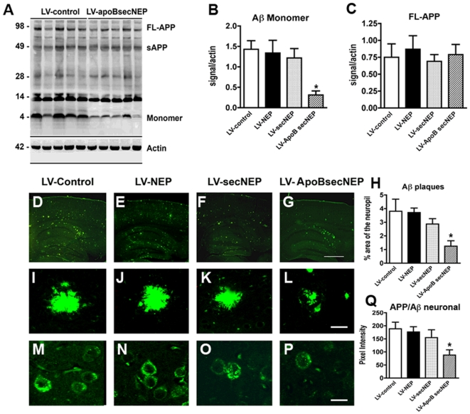
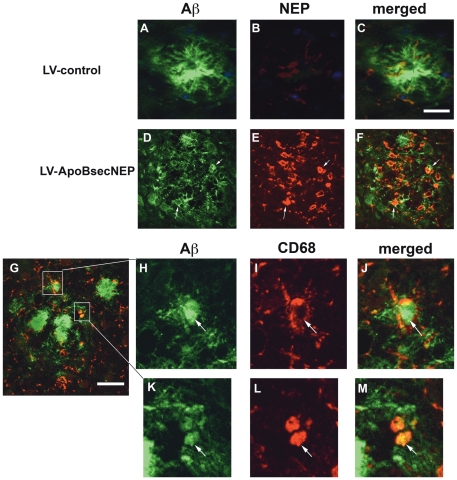
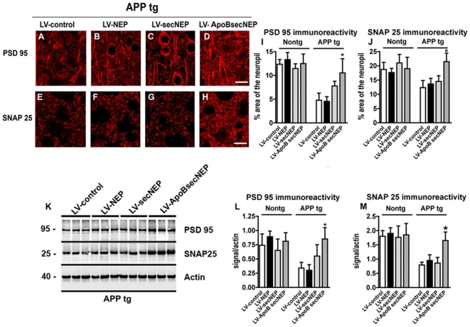
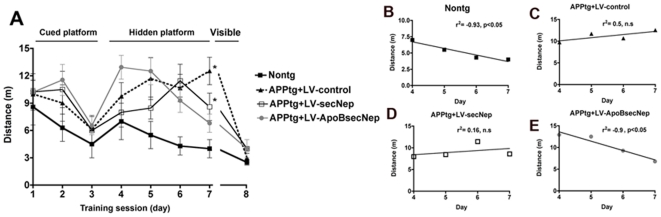
Alzheimer's disease (AD), an incurable, progressive neurodegenerative disorder, is the most common form of dementia. Therapeutic options have been elusive due to the inability to deliver proteins across the blood-brain barrier (BBB). In order to improve the therapeutic potential for AD, we utilized a promising new approach for delivery of proteins across the BBB. We generated a lentivirus vector expressing the amyloid β-degrading enzyme, neprilysin, fused to the ApoB transport domain and delivered this by intra-peritoneal injection to amyloid protein precursor (APP) transgenic model of AD. Treated mice had reduced levels of Aβ, reduced plaques and increased synaptic density in the CNS. Furthermore, mice treated with the neprilysin targeting the CNS had a reversal of memory deficits. Thus, the addition of the ApoB transport domain to the secreted neprilysin generated a non-invasive therapeutic approach that may be a potential treatment in patients with AD.


Conflict of interest statement

Competing Interests: The authors have declared that no competing interests exist.

Figures

Figure 1
Figure 1. Characterization of levels of expression and activity of the ApoBSecNEP fusion protein.
293T cells were infected with LV-NEP, LV-SecNEP or LV-ApoBSecNEP and 48 hours later analyzed. (A) Representative images of immunoblot of cell lysates probed with anti-Neprilysin (upper panel) and anti-Aβ (lower panel) antibodies. The middle panel illustrates the proteolytic activity against a FITC labeled Aβ42. (B) Representative images of immunoblot and proteolysis of FITC labeled Aβ42 with conditioned media. Reactions were collected at 0 or 24 hours in the presence or absence of the neprilysin inhibitor thiorphan and analyzed with the Versadoc gel imaging system (BioRad). (C) Analysis of levels of neprilysin immunoreactive bands and proteolysis of FITC labeled Aβ42 with cell lysates. (D) Levels of neprilysin immunoreactivity and proteolysis of FITC labeled Aβ42 with conditioned media.
Figure 2
Figure 2. Neuroprotective effects of the ApoBSecNEP fusion protein from Aβ toxicity.
Adult rat hippocampal neural progenitor cells were differentiated into neurons, infected with LV-control, LV-NEP, LV-SecNEP or LV-ApoBSecNEP for 48 hours and then challenged with 10 nM Aβ for 24 hours. Neuronal cells were immunostained and analyzed by confocal laser microscopy. (A) Patterns of immunostaining with antibodies against neprilysin (first column). Comparison of effects of Aβ on neuronal morphology and neurite characteristics with an antibody against β-tubulin (second and third columns). Scale bar  = 50 µm. (B) Computer aided image analysis of the levels of β-tubulin immunoreactivity in cells infected with the lentiviruses and treated with vehicle alone or Aβ. (C) Levels of activated caspase-3 immunoreactivity (marker of cell injury via pro-apoptotic pathway) determined by immunocytochemistry and computer aided image analysis in cells infected with the lentiviruses and treated with vehicle alone or Aβ. (D and E) Determinations of levels of neprilysin activity in cell lysates and the conditioned media utilizing an artificial substrate (DAGNPG). * - indicates statistically significant difference by 1-way ANOVA with poshoc Dunnet's when compared to vehicle treated control (p<0.05).
Figure 3
Figure 3. Trafficking of the ApoBSecNEP into the CNS of APP tg mice.
Vibratome sections from nontg and APP tg mice that received a single intra-peritoneal injection of LV-control, LV-NEP, LV-SecNEP or LV-ApoBSecNEP were immunostained with an antibody against NEP and analyzed by laser scanning confocal microscopy. (A, B) Low levels of NEP immunostaining in APP tg mice and (C) nontg mice treated with LV-secNEP (D, E) Accumulation of NEP immunoreactivity (red) in the neocortex and hippocampus of APP tg mice and (F) to a lower extent in nontg mice treated with LV-ApoBsecNEP. Areas of neprilysin immunolabeling (white arrows) were enlarged to show localization within neurons (cutouts E, F). Scale bar  = 100 µm for A, B and 20 µm for C–F. (G) Computer aided image analysis of levels of neprilysin immunoreactivity expressed as pixel intensity. (H) Determinations of levels of neprilysin activity in hippocampal homogenates by measuring an artificial substrate cleavage (DAGNPG). * - indicates statistically significant difference by 1-way ANOVA with poshoc Dunnet's when compared to mice treated with the LV-Control (p<0.05). n = 8 mice per group.
Figure 4
Figure 4. Localization of ApoBSecNEP in hippocampus of APP tg mice.
Sections from APP tg mice that received a single intraperitoneal injection of LV-ApoBSecNEP were double labeled with antibodies against NEP (panels in red) and neuronal markers MAP2 or NeuN (panels in green). (A–C) Co-localization of NEP with MAP2 in the pyramidal neurons in the CA1 regions of the hippocampus; (D–F) co-localization of NEP with MAP2 in granular neurons in the dentate gyrus. (G–I) Co-localization of NEP with NeuN in the pyramidal neurons in the CA1 regions of the hippocampus; (J–L) co-localization of NEP with NeuN in granular neurons in the dentate gyrus. Scale bar  = 50 µm.
Figure 5
Figure 5. ApoBSecNEP reduces Aβ in the CNS of APP tg mice.
Sections from APP tg mice that had received peripheral injections with the lentiviral vectors were homogenized, fractioned and examined for levels of APP and Aβ by western blot. (A) Representative immunoblot probed with the 82E1 monoclonal antibody displaying the reduction in the levels of Aβ in APP tg mice treated with LV-ApoBSecNEP. (B–C) Analysis of immunoblot for levels of Aβ monomers and full length APP. (D–G) Low magnification view of sections from APP tg mice treated with LV-control, LV-NEP, LV-SecNEP or LV-ApoBSecNEP respectively and immunolabeled with a monoclonal antibody against Aβ (82E1) imaged with the laser scanning microscope. (H) Computer aided image analysis of the % area of the neuropil occupied by Aβ immunoreactive deposits. (I–L) Higher magnification view of the Aβ immunoreactive plaques in APP tg mice treated with the various lentiviruses. (M–P) Representative images of the patterns of intraneuronal APP/Aβ immunostaining in the frontal cortex from APP tg mice treated with LV-Control, LV-NEP, LV-SecNEP or LV-ApoBSecNEP respectively immunolabeled with a monoclonal antibody against Aβ (82E1) imaged with the laser scanning microscope. (Q) Image analysis of levels of intracellular APP/Aβ immunostaining. Scale bar  = 15 µm for I-L and 10 µm for M-P. * - indicates statistically significant difference by 1-way ANOVA with poshoc Dunnet's when compared to LV-Control treated animals (p<0.05). n = 8 mice per group.
Figure 6
Figure 6. ApoBSecNEP co-localizes with activated macrophages at the site of plaques.
Sections from APP tg mice that received peripheral injections with (A–C) LV-control or (D–F) LV-ApoBSecNEP were double labeled with antibodies against Aβ (green) and NEP (red) and imaged with the laser scanning confocal microscope. (G–M) Representative plaque from a mouse treated with LV-ApoBSecNEP displaying co-localization of Aβ immunoreactive material in macrophages/microglia around the plaques labeled with CD68 (red). White arrows indicate areas of co-localization of Aβ and neprilysin or CD68. Scale bar  = 20 µm for A–F. Scale bar  = 10 µm for G–M.
Figure 7
Figure 7. Trafficking of ApoBSecNEP to the CNS ameliorates synaptic deficits in APP tg mice.
Brain sections from APP tg mice that had received the lentiviral vectors were immunolabeled with monoclonal antibodies against the post-synaptic marker PSD 95 (red) or the pre-synaptic marker SNAP25 (red) and imaged with the laser scanning confocal microscope. Images are from the frontal-parietal cortex layers 2–3. (A–D) Representative images from APP tg mice treated with LV-control, LV-NEP, LV-SecNEP or LV-ApoBSecNEP respectively and immunolabeled with a monoclonal antibody against PSD 95. (E–H) Images from APP tg mice treated with LV-control, LV-NEP, LV-SecNEP or LV-ApoBSecNEP respectively and immunolabeled with a monoclonal antibody against SNAP25. (I–J) Computer aided image analysis of the % area of the neuropil stained for PSD 95 or SNAP 25 structures. (K) Representative immunoblot probed with antibodies against PSD 95, SNAP25 and actin. For these analysis the posterior aspect of the brain containing the cortex and hippocampus was homogenized and the membrane fraction used for the immunoblot. (L–M) Image analysis of the immunoreactive bands for PSD 95 and SNAP25 expressed as ratio to actin as loading control. Scale bar  = 5 µm for A–H. *  =  indicates statistically significant difference by 1-way ANOVA with poshoc Dunnet's when compared to LV-control treated animals (p<0.05). n = 8 mice per group.
Figure 8
Figure 8. Effects of peripheral treatment with LV-ApoBSecNEP in water maze performance in APP tg mice.
(A) Four weeks after the intra-peritoneal injections with the LV-control, LV-SecNEP or LV-ApoBSecNEP virus, memory and learning were assessed by the Morris water maze. Mice were trained on the cued platform on days 1–3 and then tested for spatial learning on days 4–7 followed by a return of the cued platform on day 8. (B–E) Linear regression analysis showing the slope for the learning curves in the nontg mice and the APP tg mice with LV-control, LV-secNEP and LV-ApoB-sec-NEP respectively. *  =  indicates statistically significant difference by 1-way ANOVA with poshoc Dunnet's when compared to nontg treated animals (p<0.05). n = 8 mice per group.

Similar articles

Cited by

References

    1. Ashford JW. APOE genotype effects on Alzheimer's disease onset and epidemiology. J Mol Neurosci. 2004;23:157–165. - PubMed
    1. Terry R, Hansen L, Masliah E. Structural basis of the cognitive alterations in Alzheimer disease. In: Terry R, Katzman R, editors. Alzheimer disease. New York: Raven Press; 1994. pp. 179–196.
    1. Pardridge WM. Drug and gene delivery to the brain: the vascular route. Neuron. 2002;36:555–558. - PubMed
    1. Howell S, Nalbantoglu J, Crine P. Neutral endopeptidase can hydrolyze beta-amyloid(1-40) but shows no effect on beta-amyloid precursor protein metabolism. Peptides. 1995;16:647–652. - PubMed
    1. Iwata N, Tsubuki S, Takaki Y, Shirotani K, Lu B, et al. Metabolic regulation of brain Abeta by neprilysin. Science. 2001;292:1550–1552. - PubMed

Publication types

MeSH terms