. 2011 Jan 31;6(1):e16386.
doi: 10.1371/journal.pone.0016386.

Light converts endosymbiotic fungus to pathogen, influencing seedling survival and niche-space filling of a common tropical tree, Iriartea deltoidea

Affiliations

Light converts endosymbiotic fungus to pathogen, influencing seedling survival and niche-space filling of a common tropical tree, Iriartea deltoidea

Patricia Álvarez-Loayza et al. PLoS One. .

Abstract

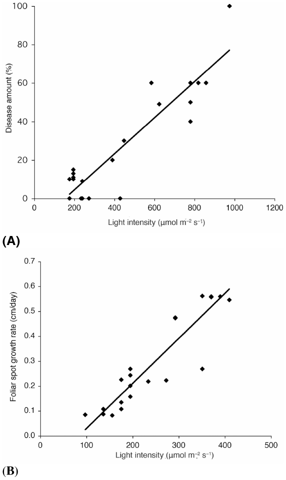
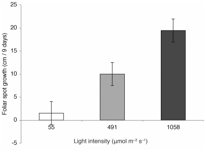
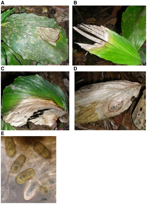
Pathogens are hypothesized to play an important role in the maintenance of tropical forest plant species richness. Notably, species richness may be promoted by incomplete filling of niche space due interactions of host populations with their pathogens. A potentially important group of pathogens are endophytic fungi, which asymptomatically colonize plants and are diverse and abundant in tropical ecosystems. Endophytes may alter competitive abilities of host individuals and improve host fitness under stress, but may also become pathogenic. Little is known of the impacts of endophytes on niche-space filling of their hosts.Here we evaluate how a widespread fungal endophyte infecting a common tropical palm influences its recruitment and survival in natural ecosystems, and whether this impact is modulated by the abiotic environment, potentially constraining host niche-space filling. Iriartea deltoidea dominates many wet lowland Neotropical forests. Diplodia mutila is a common asymptomatic endophyte in mature plants; however, it causes disease in some seedlings. We investigated the effects of light availability on D. mutila disease expression.We found I. deltoidea seedlings to preferentially occur under shady conditions. Correspondingly, we also found that high light triggers endophyte pathogenicity, while low light favors endosymbiotic development, constraining recruitment of endophyte-infested seedlings to shaded understory by reducing seedling survival in direct light. Pathogenicity of D. mutila under high light is proposed to result from light-induced production of H(2)O(2) by the fungus, triggering hypersensitivity, cell death, and tissue necrosis in the palm. This is the first study to demonstrate that endophytes respond to abiotic factors to influence plant distributions in natural ecosystems; and the first to identify light as a factor influencing where an endophyte is placed on the endosymbiont-pathogen continuum. Our findings show that pathogens can indeed constrain niche-space filling of otherwise successful tropical plant species, providing unoccupied niche space for other species.


Conflict of interest statement

Competing Interests: The authors have declared that no competing interests exist.

Figures

Figure 1
Figure 1. Higher light intensities increased disease development produced by Diplodia mutila.
(A) For young seedlings with 2 leaves or less there was a significant interaction between amount of infection (% of D. mutila foliar spots in Iriartea deltoidea leaves) and light level (F 1,22 = 55.4, P = 0.0001**, r 2 = 0.73). (B) The diametric growth rate of the foliar spots produced by D. mutila was higher at higher light conditions (ANOVA, F 1,22 = 93.26, P = 0.0001**, r 2 = 0.816).
Figure 2
Figure 2. Increased light availability switched the endosymbiotic phase of D mutila to its pathogenic phase.
Young seedlings that were colonized with endophytic Diplodia mutila showed faster growth rates of diameter of foliar spots (cm) caused by the pathogenic phase of D. mutila at higher light intensities (∼1058±23 µmol m−2 s−1) than seedlings under shaded conditions (∼55±15 µmol m−2 s−1) (n = 30, t test, P = 0.0001**). There were also significant differences of foliar spot growth rates among plants growing in the greenhouse (∼491±34 µmol m−2 s−1) and plants growing under shaded conditions (n = 30, t test, P = 0.024*). Foliar spot growth rates among plants growing in the greenhouse were lower than plants growing under high light intensities (n = 30, t test, P = 0.013*), (Tukey Kramer HSD test, ANOVA, F 3,30 = 12.62, P = 0.0001**).
Figure 3
Figure 3. Fungal growth variation of Diplodia mutila under different light treatments and two different media.
(A) Mycelial radial growth of Diplodia mutila on Potato Dextrose Agar (PDA) was faster under a 12-hour cycle than the 3-hour cycle: 1.25 (±0.03) cm/day vs. 1.11 (±0.03) cm./day (n = 12, t test, P>0.018*). On Water Agar (WA) the average radial growth rate per day of the colony mycelium was 0.51 cm/day under a 12-hour light cycle; while under a 3-hour light cycle the average growth rate of the colony mycelium was significantly lower, at 0.41 cm/day after 7 days (n = 12, t test, P>0.004*). (B) Colonies grown in PDA under the 12-hour light cycle had a more rapid melanization of the central area of the colony (∼0.71 cm/day) than colonies exposed to 3-hours of light (∼0.5 cm/day) (n = 12, t test, P>0.022*). Similar results were obtained for colonies growing in WA (n = 12, t test, P>0.026*). (C) Melanization of colonies of D. mutila growing in PDA observed on 4 days (from left to right). Rate of melanization was reduced in the 3-hour cycle treatment (above). Faster melanization was observed in cultures maintained in a 12-hour light cycle (below).
Figure 4
Figure 4. In vitro hydrogen peroxide production of Diplodia mutila on Potato Dextrose Agar (PDA).
(A) Hydrogen peroxide (orange pigmentation) accumulation in culture grown under light at 30°C treatment. (B) Culture grown under dark at 30°C treatment, showing no or minimal accumulation of hydrogen peroxide.
Figure 5
Figure 5. Foliar spots in Iriartea deltoidea caused by Diplodia mutila, at different infection stages.
(A) Leaf spot infection for a plant with 2 leaves and one spot covering less than 20% of the leaf (B) A plant with two leaves and with a spot covering ∼40% of one leaf (C) A plant with two leaves and with the two foliar spots covering 50% of both leaves (D) Foliar spots covering the entire plant, representing 100% of infection. These plants died after 15 to 31 days. (E) Diplodia mutila pycnidia produced slowly maturing, non-striate, brown, 1-septate conidia measuring 26–28×15–20 µm. Liquid conidial darkening and septation was recorded to take place after discharge.

Similar articles

Cited by

References

    1. Janzen D. Herbivores and the number of tree species in tropical forests. American Naturalist. 1970;104:501–529.
    1. Connell JH. On the role of natural enemies in preventing competitive exclusion in some marine animals and in rain forest trees. In: Boer PJD, Gradwell GR, editors. Dynamics of numbers in populations. Wageningen, Netherlands: Centre for Agricultural Publication and Documentation; 1971. pp. 298–312.
    1. Peters HA. Neighbour-regulated mortality: the influence of positive and negative density dependence on tree populations in species-rich tropical forests. Ecology Letters, 2003;6:757–765.
    1. Ricklefs RE. Evolutionary diversification, coevolution between populations and their antagonists, and the filling of niche space. Proceedings of the National Academy of Sciences USA. 2010;107:1265–1272. - PMC - PubMed
    1. Bell T, Freckleton RP, Lewis OT. Plant pathogens drive density-dependent seedling mortality in a tropical tree. Ecology Letters. 2006;9:569–574. - PubMed

Publication types