. 2011 Jan 31;6(1):e16414.
doi: 10.1371/journal.pone.0016414.

Immunisation with recombinant PfEMP1 domains elicits functional rosette-inhibiting and phagocytosis-inducing antibodies to Plasmodium falciparum

Affiliations

Immunisation with recombinant PfEMP1 domains elicits functional rosette-inhibiting and phagocytosis-inducing antibodies to Plasmodium falciparum

Ashfaq Ghumra et al. PLoS One. .

Abstract

Background: Rosetting is a Plasmodium falciparum virulence factor implicated in the pathogenesis of life-threatening malaria. Rosetting occurs when parasite-derived P. falciparum Erythrocyte Membrane Protein One (PfEMP1) on the surface of infected erythrocytes binds to human receptors on uninfected erythrocytes. PfEMP1 is a possible target for a vaccine to induce antibodies to inhibit rosetting and prevent severe malaria.

Methodology/findings: We examined the vaccine potential of the six extracellular domains of a rosette-mediating PfEMP1 variant (ITvar9/R29var1 from the R29 parasite strain) by immunizing rabbits with recombinant proteins expressed in E. coli. Antibodies raised to each domain were tested for surface fluorescence with live infected erythrocytes, rosette inhibition and phagocytosis-induction. Antibodies to all PfEMP1 domains recognized the surface of live infected erythrocytes down to low concentrations (0.02-1.56 µg/ml of total IgG). Antibodies to all PfEMP1 domains except for the second Duffy-Binding-Like region inhibited rosetting (50% inhibitory concentration 0.04-4 µg/ml) and were able to opsonize and induce phagocytosis of infected erythrocytes at low concentrations (1.56-6.25 µg/ml). Antibodies to the N-terminal region (NTS-DBL1α) were the most effective in all assays. All antibodies were specific for the R29 parasite strain, and showed no functional activity against five other rosetting strains.

Conclusions/significance: These results are encouraging for vaccine development as they show that potent antibodies can be generated to recombinant PfEMP1 domains that will inhibit rosetting and induce phagocytosis of infected erythrocytes. However, further work is needed on rosetting mechanisms and cross-reactivity in field isolates to define a set of PfEMP1 variants that could induce functional antibodies against a broad range of P. falciparum rosetting parasites.


Conflict of interest statement

Competing Interests: The authors have declared that no competing interests exist.

Figures

Figure 1
Figure 1. Diagram of the ITvar9 PfEMP1 variant showing domains expressed as recombinant proteins in E.coli.
The ITvar9 gene (also known as R29var1 , accession numbers Y13402 and CAA73831) encodes a group A PfEMP1 variant with four Duffy Binding Like (DBL) and two Cysteine-rich InterDomain Region (CIDR) domains. The number of the domain designates its position from the N-terminus and the Greek symbol represents its homology group . PfEMP1 contains a N-Terminal Segment (NTS) and an Acidic Terminal Segment (ATS) proximal to the transmembrane (TM) region. The DBL1α domain is the functional erythrocyte-binding region . The bars represent the E.coli expression constructs and the numbers show the first and last amino acid positions.
Figure 2
Figure 2. SDS-PAGE showing recombinant DBL and CIDR domains from ITvar9 expressed in E. coli.
The purity and quality of the recombinant DBL and CIDR domains were assessed by electrophoresis of reduced and non-reduced pairs of proteins on 10% SDS-polyacrylamide gels. Two µg of protein was used per well and lanes were as follows: 1) NTS-DBL1α, 2) DBL1α, 3) DBL2γ 4) DBL3ε, 5) DBL4δ, 6) CIDR2β and 7) NTS-DBL1α-CIDR1γ. M, molecular weight marker; NR, non-reduced; R, reduced.
Figure 3
Figure 3. Immunofluorescence assay showing that ITvar9 antibodies recognize PfEMP1 on the surface of live infected erythrocytes.
R29 mature infected erythrocytes (pigmented trophozoites and schizonts) were grown to 5% parasitaemia and incubated with rabbit antisera against recombinant ITvar9 DBL and CIDR domains at 1/50 dilution. After washing, the cells were incubated with Alexa Fluor 488-labelled goat anti-rabbit IgG (Invitrogen) at 1/1000 dilution. The example shown here is the binding of anti-DBL2γ antisera, however all antisera to ITvar9 gave similar results. Punctate staining of the membrane of infected erythrocytes (green) was seen with the specific antisera (“immune”) but not with the pre-immune sera. The location of infected erythrocytes is shown by DAPI staining of the parasite (blue). Slides were viewed with a 100× objective using a Leica DM 2000 fluorescent microscope.
Figure 4
Figure 4. Antibodies to ITvar9 domains have anti-rosetting activity against R29 parasites.
Purified IgG raised against NTS-DBL1α disrupted existing rosettes (A) and inhibited the formation of rosettes (B), whereas negative control non-immunized rabbit IgG had no effect on rosetting (A and B). Antibodies raised to all extracellular domains of the ITvar9 PfEMP1 variant inhibited R29 rosetting except for DBL2γ antibodies (C). NTS-DBL1α antibodies showed similar dose-response curves when the R29 parasites were grown in group A or O erythrocytes (D). The results are expressed as percentage of the control value, in which complete binding medium (equivalent volume as that of antibody) was added to the parasite culture. The control always had at least 50% of infected erythrocytes in rosettes. For (A), the mean and standard deviation of three independent experiments is shown. For (B), (C) and (D), the mean and standard deviation of triplicate determinations of rosette frequency at each concentration of antibody within a single experiment is shown.
Figure 5
Figure 5. ELISA to detect binding of PfEMP1 antibodies to recombinant NTS-DBL1α.
Antibodies were added to wells coated with 2 µg/ml of recombinant NTS-DBL1α protein and binding was detected using HRP-conjugated anti-rabbit IgG at 1/10,000 dilution. Antisera raised to recombinant proteins containing DBL1α (i.e. anti-NTS-DBL1α, anti-DBL1α and anti-NTS-DBL1α-CIDR1γ) all recognize the recombinant protein as expected. Antisera to DBL2γ, DBL3ε and CIDR2β do not cross-react with recombinant NTS-DBL1α. However, the antiserum to DBL4δ does shows binding to the recombinant NTS-DBL1α, suggesting that there is cross-reactivity between these two domains.
Figure 6
Figure 6. Rosette inhibition of antibodies depleted by absorption against NTS-DBL1α.
Immunoblotting (A) and rosette inhibition (B) by pairs of antibodies that were either non-absorbed, or absorbed on NTS-DBL1α recombinant protein coupled to sepharose. A) Recombinant NTS-DBL1α protein was spotted onto nitrocellulose membrane at doubling dilutions, starting from 2 µg/ml, and incubated with 1/1000 dilution of absorbed or non-absorbed antibody. 1) non-absorbed anti-NTS-DBL1α, 2) absorbed anti-NTS-DBL1α, 3) non-absorbed anti-NTS-DBL1α-CIDR1γ, 4) absorbed anti-NTS-DBL1α-CIDR1γ, 5) non-absorbed anti-DBL3ε, 6) absorbed anti-DBL3ε, 7) non-absorbed anti-DBL4δ and 8) absorbed anti-DBL4δ. Non-absorbed antibodies to DBLα (lanes 1 and 3) and DBL4δ (lane7) recognized NTS-DBL1α recombinant protein. After absorption, however, this activity was lost (lanes 2, 4 and 8). Antibodies to DBL3ε did not recognize NTS-DBL1α recombinant protein (lanes 4 and 5). B) Rosette inhibition assays showed that the anti-rosetting activity of NTS-DBL1α antibodies was lost after absorption. Antibodies to DBL3ε and DBL4δ retained rosette-inhibitory activity after absorption, showing that their anti-rosetting effects are likely to be independent of DBL1α. Antibodies to NTS-DBL1α-CIDR1γ also retained inhibitory effects after absorption on NTS-DBL1α protein, suggesting that antibodies to the CIDR1γ domain of ITvar9 also have anti-rosetting effects. Data shown are the mean and standard deviation of triplicate determinations of rosette frequency after overnight incubation with absorbed or non-absorbed antibody diluted 1/10 from the 1 mg/ml stock used for absorption. The control (with binding medium only added) had more than 50% of infected erythrocytes in rosettes.
Figure 7
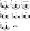
Figure 7. Rosette disruption assays with ITvar9 antisera against six P. falciparum rosetting laboratory strains.
Antisera raised to ITvar9 domains, and paired pre-immune sera, were used at 1/20 dilution in rosette disruption assays with R29, PAR+, Muz12, HB3R+, TM284 and TM180. Antisera were as follows: A) NTS-DBL1α, B) DBL1α, C) NTS-DBL1α-CIDR1γ, D) DBL2γ, E) DBL3ε, F) DBL4δ and G) CIDR2β. Disruption of rosetting was only seen with R29 parasites. Data shown are the mean and standard deviation from three independent experiments. The control (with binding medium only added) had more than 50% of infected erythrocytes in rosettes.
Figure 8
Figure 8. Rosette inhibition assays with ITvar9 antisera against six P. falciparum rosetting laboratory strains.
Total IgG was used at a concentration of 100 µg/ml in rosette inhibition assays with R29, PAR+, Muz12, HB3R+, TM284 and TM180. Antisera were as follows: A) NTS-DBL1α, B) DBL1α, C) NTS-DBL1α-CIDR1γ, D) DBL2γ, E) DBL3ε, F) DBL4δ and G) CIDR2β. Inhibition of rosetting was only seen with R29 parasites. Data shown are the mean and standard deviation of triplicate determinations of rosette frequency within a single experiment. The control (with binding medium only added) had more than 50% of infected erythrocytes in rosettes.
Figure 9
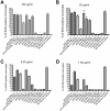
Figure 9. Phagocytosis of R29 infected erythrocytes after opsonization with anti-PfEMP1 antibodies.
Ethidium bromide stained R29-infected erythrocytes were opsonized with antibodies and incubated with the monocytic Thp-1 cell line. The percentage of Thp-1 cells that had phagocytosed one or more infected erythrocytes was assessed by flow cytometry. The positive control was 90 µg/ml rabbit-anti human erythrocyte polyclonal antibody and the negative control was media alone (no serum control). All antibodies to PfEMP1 domains were used at four different concentrations: 100 µg/ml (A), 25 µg/ml (B), 6.25 µg/ml (C) or 1.56 µg/ml (D). Antibodies directed against ITvar9 PfEMP1 domains (first seven bars of each graph) promoted phagocytosis of R29 infected erythrocytes, whereas antibodies to the NTS-DBL1α domains of other PfEMP1 variants (control PAR+, TM284, TM180 and HB3R+) did not. The effect of ITvar9 PfEMP1 antibodies was concentration-dependent, with anti-NTS-DBL1α being the most effective at low concentration (D). Values shown are means and standard deviation from duplicates.

Similar articles

Cited by

References

    1. Snow RW, Guerra CA, Noor AM, Myint HY, Hay SI. The global distribution of clinical episodes of Plasmodium falciparum malaria. Nature. 2005;434:214–217. - PMC - PubMed
    1. Carlson J, Helmby H, Hill AV, Brewster D, Greenwood BM, et al. Human cerebral malaria: association with erythrocyte rosetting and lack of anti-rosetting antibodies. Lancet. 1990;336:1457–1460. - PubMed
    1. Treutiger CJ, Hedlund I, Helmby H, Carlson J, Jepson A, et al. Rosette formation in Plasmodium falciparum isolates and anti-rosette activity of sera from Gambians with cerebral or uncomplicated malaria. Am J Trop Med Hyg. 1992;46:503–510. - PubMed
    1. Ringwald P, Peyron F, Lepers JP, Rabarison P, Rakotomalala C, et al. Parasite virulence factors during falciparum malaria: rosetting, cytoadherence, and modulation of cytoadherence by cytokines. Infect Immun. 1993;61:5198–5204. - PMC - PubMed
    1. Rowe A, Obeiro J, Newbold CI, Marsh K. Plasmodium falciparum rosetting is associated with malaria severity in Kenya. Infect Immun. 1995;63:2323–2326. - PMC - PubMed

Publication types

Substances