

The development and characterization of a human mesothelioma in vitro 3D model to investigate immunotoxin therapy
- PMID: 21305058
- PMCID: PMC3031536
- DOI: 10.1371/journal.pone.0014640
The development and characterization of a human mesothelioma in vitro 3D model to investigate immunotoxin therapy
Abstract
Background: Tumor microenvironments present significant barriers to penetration by antibodies and immunoconjugates. Tumor microenvironments, however, are difficult to study in vitro. Cells cultured as monolayers exhibit less resistance to therapy than those grown in vivo and an alternative research model more representative of the in vivo tumor is more desirable. SS1P is an immunotoxin composed of the Fv portion of a mesothelin-specific antibody fused to a bacterial toxin that is presently undergoing clinical trials in mesothelioma.
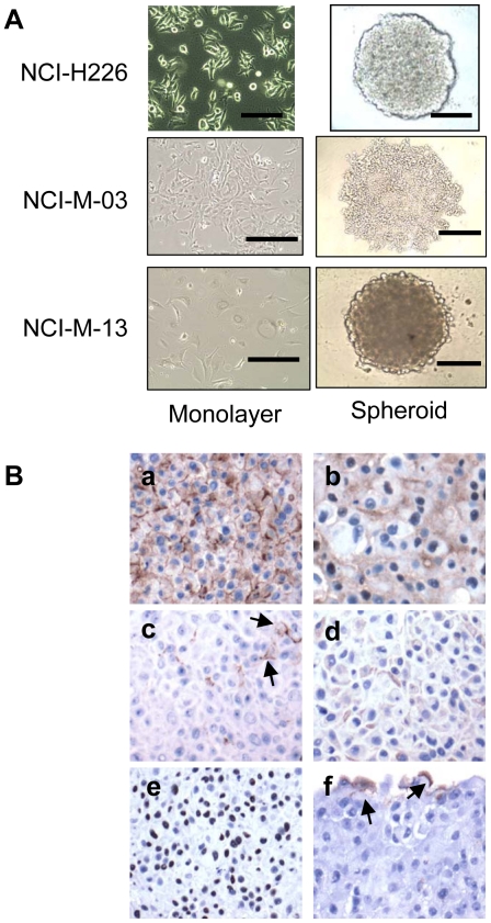
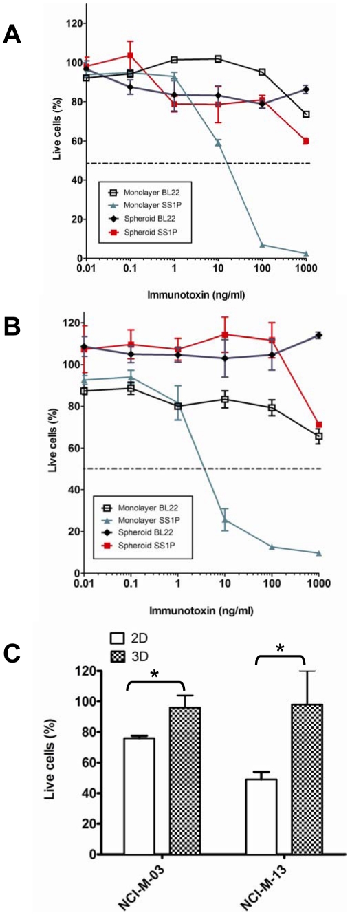
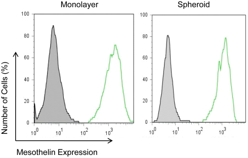
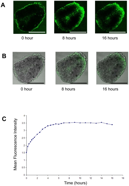
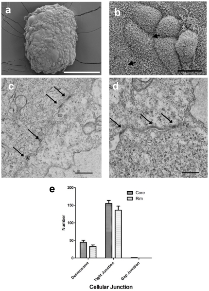
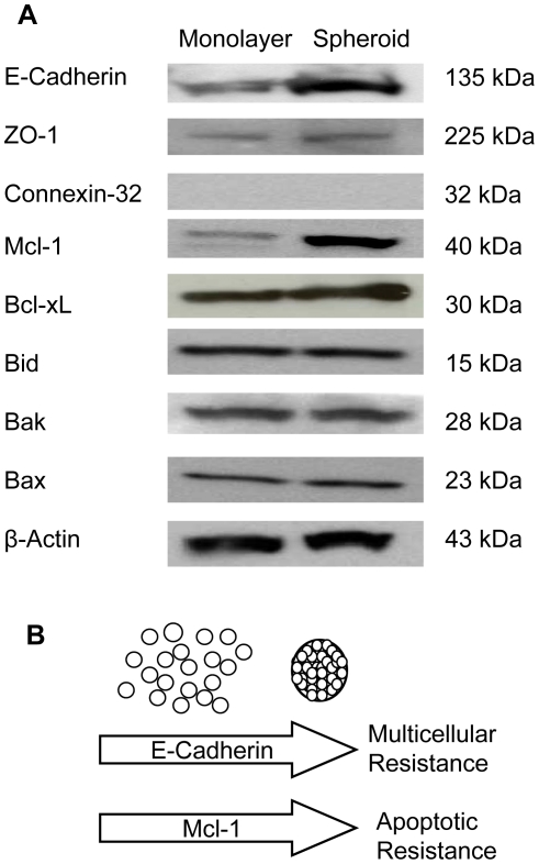
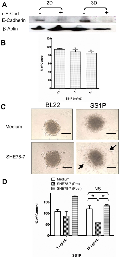
Methodology/principal findings: Here, we examined how the tumor microenvironment affects the penetration and killing activity of SS1P in a new three-dimensional (3D) spheroid model cultured in vitro using the human mesothelioma cell line (NCI-H226) and two primary cell lines isolated from the ascites of malignant mesothelioma patients. Mesothelioma cells grown as monolayers or as spheroids expressed comparable levels of mesothelin; however, spheroids were at least 100 times less affected by SS1P. To understand this disparity in cytotoxicity, we made fluorescence-labeled SS1P molecules and used confocal microscopy to examine the time course of SS1P penetration within spheroids. The penetration was limited after 4 hours. Interestingly, we found a significant increase in the number of tight junctions in the core area of spheroids by electron microscopy. Expression of E-Cadherin, a protein involved in the assembly and sealing of tight junctions and highly expressed in malignant mesothelioma, was found significantly increased in spheroids as compared to monolayers. Moreover, we found that siRNA silencing and antibody inhibition targeting E-Cadherin could enhance SS1P immunotoxin therapy in vitro.
Conclusion/significance: This work is one of the first to investigate immunotoxins in 3D tumor spheroids in vitro. This initial description of an in vitro tumor model may offer a simple and more representative model of in vivo tumors and will allow for further investigations of the microenvironmental effects on drug penetration and tumor cell killing. We believe that the methods developed here may apply to the studies of other tumor-targeting antibodies and immunoconjugates in vitro.
Conflict of interest statement
Figures
Similar articles
-
A flow cytometry method to quantitate internalized immunotoxins shows that taxol synergistically increases cellular immunotoxins uptake.Cancer Res. 2010 Feb 1;70(3):1082-9. doi: 10.1158/0008-5472.CAN-09-2405. Epub 2010 Jan 26. Cancer Res. 2010. PMID: 20103626 Free PMC article.
-
Rapid generation of in vitro multicellular spheroids for the study of monoclonal antibody therapy.J Cancer. 2011;2:507-14. doi: 10.7150/jca.2.507. Epub 2011 Oct 13. J Cancer. 2011. PMID: 22043235 Free PMC article.
-
SS1P Immunotoxin Induces Markers of Immunogenic Cell Death and Enhances the Effect of the CTLA-4 Blockade in AE17M Mouse Mesothelioma Tumors.Toxins (Basel). 2018 Nov 14;10(11):470. doi: 10.3390/toxins10110470. Toxins (Basel). 2018. PMID: 30441807 Free PMC article.
-
Mesothelin-Targeted Recombinant Immunotoxins for Solid Tumors.Biomolecules. 2020 Jun 28;10(7):973. doi: 10.3390/biom10070973. Biomolecules. 2020. PMID: 32605175 Free PMC article. Review.
-
Discovery of mesothelin and exploiting it as a target for immunotherapy.Cancer Res. 2014 Jun 1;74(11):2907-12. doi: 10.1158/0008-5472.CAN-14-0337. Epub 2014 May 13. Cancer Res. 2014. PMID: 24824231 Free PMC article. Review.
Cited by
-
The Biology of Malignant Mesothelioma and the Relevance of Preclinical Models.Front Oncol. 2020 Mar 25;10:388. doi: 10.3389/fonc.2020.00388. eCollection 2020. Front Oncol. 2020. PMID: 32269966 Free PMC article. Review.
-
Antibody or Antibody Fragments: Implications for Molecular Imaging and Targeted Therapy of Solid Tumors.Front Immunol. 2017 Oct 12;8:1287. doi: 10.3389/fimmu.2017.01287. eCollection 2017. Front Immunol. 2017. PMID: 29075266 Free PMC article.
-
Comparative Efficacy of Ribosome-Inactivating Protein-Containing Immunotoxins in 2D and 3D Models of Sarcoma.Toxins (Basel). 2025 Jun 18;17(6):308. doi: 10.3390/toxins17060308. Toxins (Basel). 2025. PMID: 40559886 Free PMC article.
-
Mimicking Tumors: Toward More Predictive In Vitro Models for Peptide- and Protein-Conjugated Drugs.Bioconjug Chem. 2017 Mar 15;28(3):846-856. doi: 10.1021/acs.bioconjchem.6b00699. Epub 2017 Feb 17. Bioconjug Chem. 2017. PMID: 28122451 Free PMC article. Review.
-
In vivo imaging of human malignant mesothelioma grown orthotopically in the peritoneal cavity of nude mice.J Cancer. 2011 Mar 1;2:123-31. doi: 10.7150/jca.2.123. J Cancer. 2011. PMID: 21479131 Free PMC article.
References
-
- Jain RK. Transport of molecules, particles, and cells in solid tumors. Annu Rev Biomed Eng. 1999;1:241–263. - PubMed
-
- Desoize B, Jardillier J. Multicellular resistance: a paradigm for clinical resistance? Crit Rev Oncol Hematol. 2000;36:193–207. - PubMed
-
- Hanahan D, Weinberg RA. The hallmarks of cancer. Cell. 2000;100:57–70. - PubMed
-
- Johnstone RW, Ruefli AA, Lowe SW. Apoptosis – A link between cancer genetics and chemotherapy. Cell. 2002;108:153–164. - PubMed
Publication types
MeSH terms
Substances
Grants and funding
LinkOut - more resources
Full Text Sources
Other Literature Sources
Medical
