

Review
doi: 10.1007/s12105-011-0246-2.
Epub 2011 Feb 9.
Oral verruciform xanthoma associated with chronic graft-versus-host disease: a report of five cases and a review of the literature
Affiliations
- PMID: 21305367
- PMCID: PMC3098333
- DOI: 10.1007/s12105-011-0246-2
Item in Clipboard
Review
Oral verruciform xanthoma associated with chronic graft-versus-host disease: a report of five cases and a review of the literature
Head Neck Pathol.
2011 Jun.
Abstract
Verruciform xanthoma (VX) is an uncommon benign inflammatory mucocutaneous condition that chiefly occurs in the oral cavity. It is often associated with pre-existing epithelial and/or inflammatory disorder and is characterized histopathologically by papillary epithelial hyperplasia and the presence of foamy macrophages in connective tissue papillae. We report of a series of five cases with VX who concurrently had chronic oral graft-versus-host disease following hematopoietic stem cell transplantation.
Figures
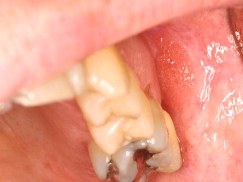
Case 1 showing a well-demarcated papillary reddish plaque of the left buccal mucosa
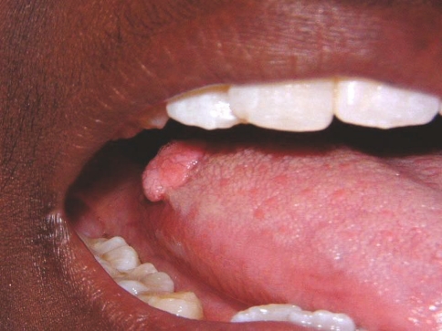
Case 2 showing a pedunculated papillary nodule on the right posterior tongue dorsum

Case 3 showing a well-demarcated yellowish verrucous plaque on the left lateral ventral tongue

Case 4 showing a well-demarcated erythematous papillary plaque on the maxillary labial mucosa

Case 5 showing a well-defined reddish granular plaque (solid arrow) and a separate unrelated area of leukoplakia (dotted arrow) on the right hard palate
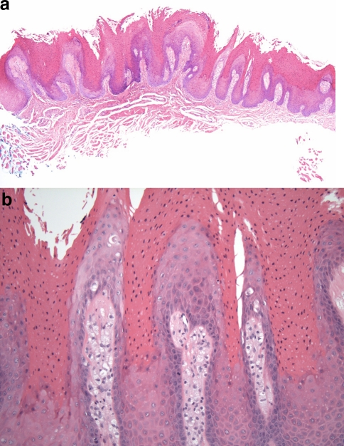
a Photomicrograph of a verruciform xanthoma (case 4) with papillary epithelial hyperplasia, hyperkeratosis, parakeratin plugging, and uniformly elongated rete ridges, (H&E, × 40). b Photomicrograph (case 4) showing numerous large, vacuolated foam cells that fill the connective tissue papillae with overlying parakeratosis (H&E, × 200)
References
Publication types
MeSH terms
LinkOut - more resources
Full Text Sources
Medical
