. 2011 May;63(5):1312-21.
doi: 10.1002/art.30261.

Proteoglycan-induced arthritis and recombinant human proteoglycan aggrecan G1 domain-induced arthritis in BALB/c mice resembling two subtypes of rheumatoid arthritis

Affiliations

Proteoglycan-induced arthritis and recombinant human proteoglycan aggrecan G1 domain-induced arthritis in BALB/c mice resembling two subtypes of rheumatoid arthritis

Tibor T Glant et al. Arthritis Rheum. 2011 May.

Abstract

Objective: To develop a simplified and relatively inexpensive version of cartilage proteoglycan-induced arthritis (PGIA), an autoimmunity model of rheumatoid arthritis (RA), and to evaluate the extent to which this new model replicates the disease parameters of PGIA and RA.

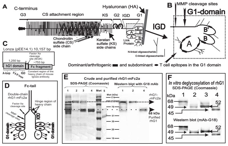
Methods: Recombinant human G1 domain of human cartilage PG containing "arthritogenic" T cell epitopes was generated in a mammalian expression system and used for immunization of BALB/c mice. The development and progression of arthritis in recombinant human PG G1-immunized mice (designated recombinant human PG G1-induced arthritis [GIA]) was monitored, and disease parameters were compared with those in the parent PGIA model.

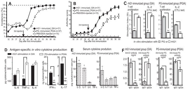
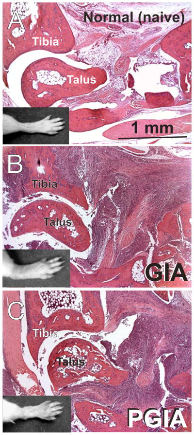
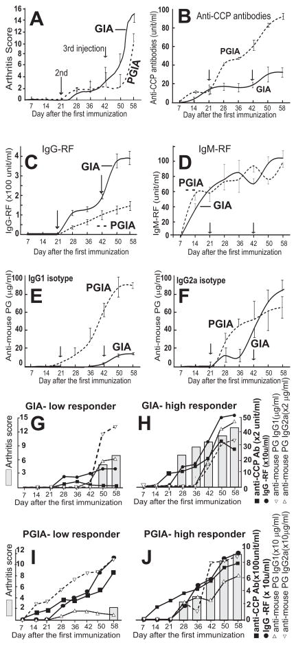
Results: GIA strongly resembled PGIA, although the clinical symptoms and immune responses in mice with GIA were more uniform than in those with PGIA. Mice with GIA showed evidence of stronger Th1 and Th17 polarization than those with PGIA, and anti-mouse PG autoantibodies were produced in different isotype ratios in the 2 models. Rheumatoid factor (RF) and anti-cyclic citrullinated peptide (anti-CCP) antibodies were detected in both models; however, serum levels of IgG-RF and anti-CCP antibodies were different in GIA and PGIA, and both parameters correlated better with disease severity in GIA than in PGIA.

Conclusion: GIA is a novel model of seropositive RA that exhibits all of the characteristics of PGIA. Although the clinical phenotypes are similar, GIA and PGIA are characterized by different autoantibody profiles, and the 2 models may represent 2 subtypes of seropositive RA, in which more than 1 type of autoantibody can be used to monitor disease severity and response to treatment.


Figures

Figure 1
Figure 1
Schematics of the cartilage proteoglycan (PG) aggrecan, G1 domain, the mammalian expression vector containing the rhG1 fusion construct, and the analysis of expressed recombinant proteins. A, The PG molecule consists of a protein core to which hundreds of glycosaminoglycan side chains (chondroitin sulfate [CS] or keratan sulfate [KS] are attached together with O-linked and N-linked oligosaccharides. The B and B’ loops of the G1 domain of the aggrecan PG core protein (PG monomer) interact with hyaluronan (HA) in cartilage; this interaction is stabilized by link protein (LP). Core protein structure: G1, G2 and G3 are the globular domains; IGD is the interglobular domain; KS is the keratan sulfate-rich domain; CS is the chondroitin sulfate attachment region (this figure is an adaptation of figures from references (19) and (5)). B, Detailed structure of the G1 domain, including the major cleavage sites for stromelysin (MMP3) and two aggrecanases (ADAMTS-4 and ADAMTS-5). Three dominant/arthritogenic and four subdominant epitopes are located in the G1 domain (20,21). Thus, less than 0.02% of the molecular mass, or less than 15% of the core protein (i.e., the G1 domain), drives the arthritogenic response to PG in genetically susceptible BALB/c mice. C, The rhG1-Xa-mFc2a construct in a Lonza pEE14.1 mammalian expression vector. D, Schematics of the “double-chain” rhG1-mFc2a fusion protein. The C-terminal end of the heavy chain of mouse IgG2a (Fc tail) is linked, via the hinge region, to the G1 domain of PG. The heavy chains are able to reform the disulfide bridges. A properly folded Fc tail binds to Protein A or Protein G, thus allowing for purification by affinity chromatography. E, Detection of the rhG1-Xa-mFc2a protein, separated by 12% SDS-PAGE and stained with Coomassie Blue G-250 (left-hand panel) and separated by western blot and stained with mAb G18 (right–hand panel). Lane 1: unpurified CHO serum-free medium harvested from rhG1-Xa-mFc2a-transfected and cloned CHO cells (roughly 30 μg protein). Lane 2: Protein G-purified rhG1-Xa-mFc fusion protein from the same CHO-SFM (5 μg protein). Lane 3: purified rhG1-Xa-mFc2a fusion protein after cleavage with factor Xa. Lane 4: rhG1 protein re-purified using Protein G/Sepharose. Lane 5 contains highly purified native human G1 domain (~42 kDa) isolated from human cartilage PG as previously described (19). Molecular weight markers (Mwt) are indicated in kDa. F, In vitro de-glycosylation of rhG1 yields a lower molecular mass protein. Lane 1: purified rhG1 without Fc-tail. Lane 2: rhG1 digested with PNGase F (thus removing all N-linked oligosaccharides). Lane 3: rhG1 digested with keratanases 1 and 2. Lane 4: digested with all enzymes (PNGase F, keratanases, sialidase and O-glycosidase), thus removing both N-linked and O-linked oligosaccharides.
Figure 2
Figure 2
Comparison of disease development and immune responses in mice with GIA and PGIA. A, Incidence and B, severity of arthritis in BALB/c mice immunized with either rhG1-Xa-mFc2a fusion protein (40 μg G1/injection) or cartilage PG (100 μg/injection) isolated from osteoarthritic cartilage (11,17,47). Vertical arrows indicate the third injection administered on day 42. Each animal was scored for arthritis three times a week, and the scores are shown as mean ± SEM. C, T-cell proliferation and IL-2 production in response to stimulation with rhG1 or PG. D, In vitro antigen (rhG1 and human PG)-induced cytokine production by spleen cells isolated from mice with GIA and PGIA, respectively. (E) Serum levels of cytokines and (F) anti-PG antibodies in mice with GIA and PGIA. The results shown in panels C-F are mean values ± SEM. Asterisks indicate statistically significant differences (*p < 0.05 and **p < 0.01).
Figure 3
Figure 3
Macroscopic images of hind limbs (black and white inserts) and histopathology of corresponding ankle joints of a normal (non-immunized) mouse (A) and in mice with GIA (B) or PGIA (C). Sections of decalcified hind paws of mice were stained with hematoxylin and eosin. There were no clinically or histologically detectable differences when arthritic limbs were compared between animals immunized with rhG1-Xa-mFc2a or rhG1(GIA) and those immunized with human cartilage PG (PGIA). In contrast to the normal joint (A), ankles in GIA (B) and PGIA (C) showed histological evidence of inflammation, synovial pannus formation, and cartilage and bone destruction.
Figure 4
Figure 4
Kinetics of autoantibody production in the context of arthritis development in GIA (rhG1-immunized) and PGIA. Arthritis scores (A), and serum levels of anti-cyclic citrullinated peptide (CCP) antibodies (B), rheumatoid factors (IgG- and IgM-type RF) (C, D), and autoantibodies to mouse cartilage PG (E, F) were monitored in rhG1- and PG-immunized mice between days 7 and 58 of immunization. Results from 17 mice with GIA and 14 mice with PGIA are shown. Panels G-J show individually analyzed arthritis scores and corresponding autoantibody titers in low and high responder mice in the GIA and PGIA groups. Although the results for only one low and one high responder mouse from each group are compared here, similar results were obtained in a replicate experiment. Note, the right y-axis scales in panels G, H, I, J are different from those shown in panels B, C, E, F.

References

    1. Gregersen PK. Genetics of rheumatoid arthritis: confronting complexity. Arthritis Res. 1999;1:37–44. - PMC - PubMed
    1. Bevaart L, Vervoordeldonk MJ, Tak PP. Evaluation of therapeutic targets in animal models of arthritis: How does it relate to rheumatoid arthritis? Arthritis Rheum. 2010;62:2192–205. - PubMed
    1. Glant TT, Mikecz K, Arzoumanian A, Poole AR. Proteoglycan-induced arthritis in BALB/c mice. Clinical features and histopathology. Arthritis Rheum. 1987;30:201–12. - PubMed
    1. Mikecz K, Glant TT, Poole AR. Immunity to cartilage proteoglycans in BALB/c mice with progressive polyarthritis and ankylosing spondylitis induced by injection of human cartilage proteoglycan. Arthritis Rheum. 1987;30:306–18. - PubMed
    1. Glant TT, Finnegan A, Mikecz K. Proteoglycan-induced arthritis: immune regulation, cellular mechanisms and genetics. Crit Rev Immunol. 2003;23:199–250. - PubMed

Publication types

MeSH terms