Case Reports
. 2011 Apr;32(4):456-66.
doi: 10.1002/humu.21472.

Human dermal fibroblasts derived from oculodentodigital dysplasia patients suggest that patients may have wound-healing defects

Affiliations
Case Reports

Human dermal fibroblasts derived from oculodentodigital dysplasia patients suggest that patients may have wound-healing defects

Jared M Churko et al. Hum Mutat. 2011 Apr.

Abstract

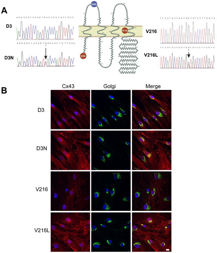
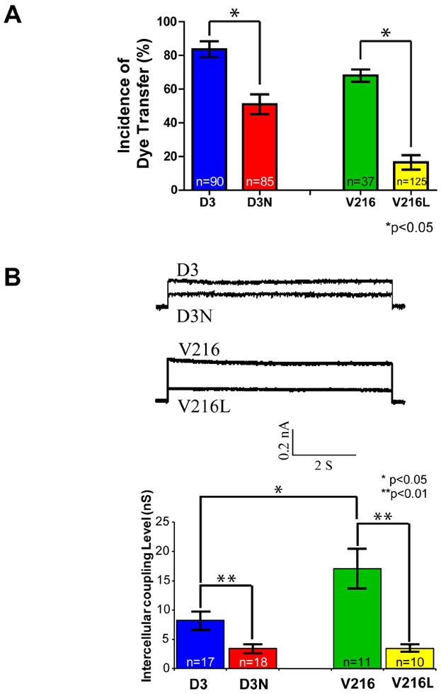
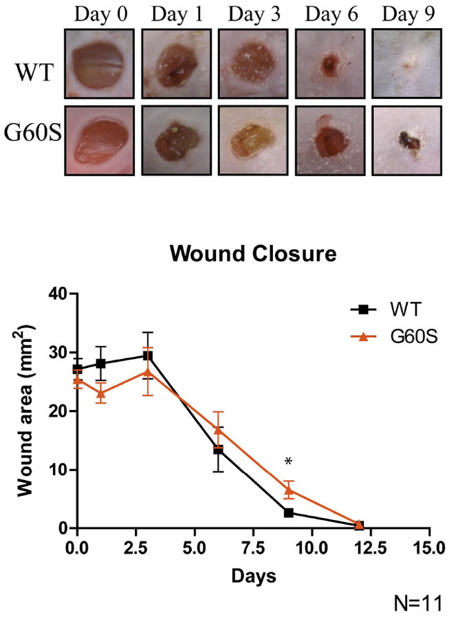
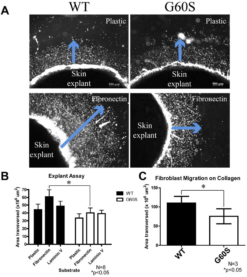
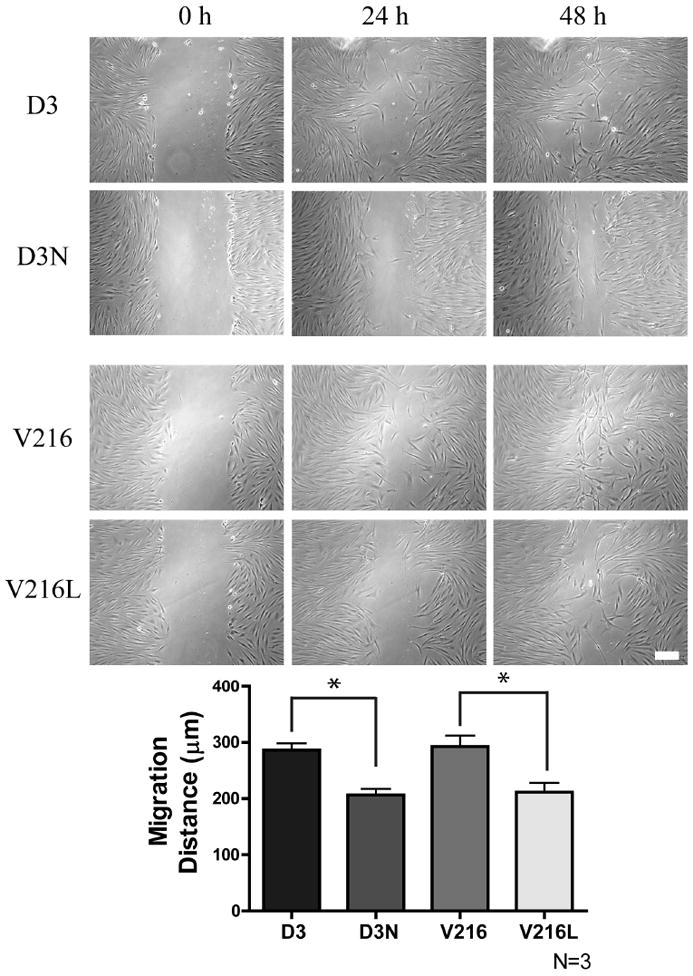
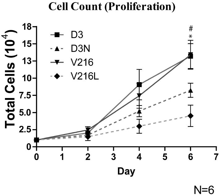
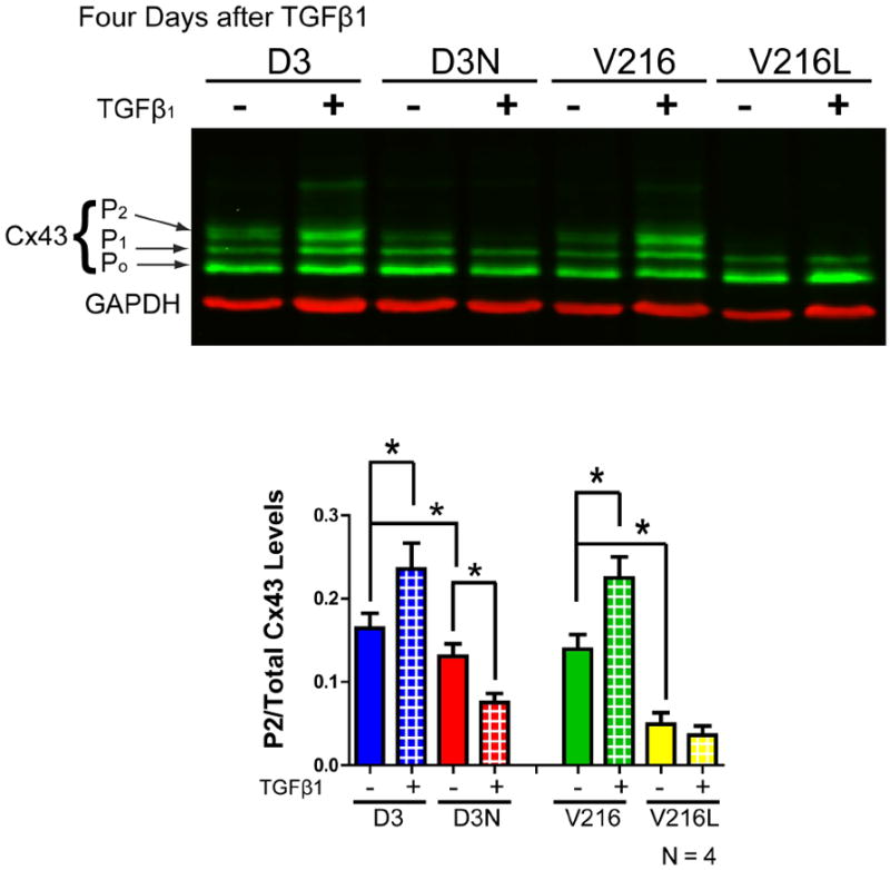
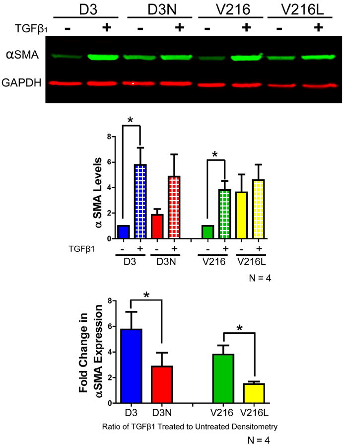
Oculodentodigital dysplasia (ODDD) is primarily an autosomal dominant human disease caused by any one of over 60 mutations in the GJA1 gene encoding the gap junction protein Cx43. In the present study, wound healing was investigated in a G60S ODDD mutant mouse model and by using dermal fibroblasts isolated from two ODDD patients harboring the p.D3N and p.V216L mutants along with dermal fibroblasts isolated from their respective unaffected relatives. Punch biopsies revealed a delay in wound closure in the G60S mutant mice in comparison to wild-type littermates, and this delay appeared to be due to defects in the dermal fibroblasts. Although both the p.D3N and p.V216L mutants reduced gap junctional intercellular communication in human dermal fibroblasts, immunolocalization studies revealed that Cx43 gap junctions were prevalent at the cell surface of p.D3N expressing fibroblasts but greatly reduced in p.V216L expressing fibroblasts. Mutant expressing fibroblasts were further found to have reduced proliferation and migration capabilities. Finally, in response to TGFβ1, mutant expressing fibroblasts expressed significantly less alpha smooth muscle actin suggesting they were inefficient in their ability to differentiate into myofibroblasts. Collectively, our results suggest that ODDD patients may have subclinical defects in wound healing due to impaired function of dermal fibroblasts.


Figures

Figure 1
Figure 1
DNA sequences from two ODDD patients. (A) Genomic DNA was extracted from patient’s blood samples or skin fibroblasts. DNA sequence analysis identified the heterozygous missense mutation in the N-terminal coding region of the GJA1 gene in patient 1 where GAC (Asp, D) changed to AAC (Asn, N) (D3N). The mutation in patient 2 was found in the gene region encoding the fourth transmembrane domain of Cx43 where GTG (Val, V) changed to TTG (Leu, L)(V216L). The G60S mouse mutation is localized within the first extracellular loop domain. (B) Fibroblasts denoted as D3, D3N, V216 and V216L were double-labeled for Cx43 (red) and the resident Golgi apparatus protein GM130 (green). Overlayed images reveal that Cx43 was partially localized to the Golgi apparatus especially in cells expressing the V216L mutant. Bar=10 μm.
Figure 2
Figure 2
Reduced gap junctional intracellular communication in D3N and V216L fibroblasts. (A) D3, D3N, V216 and V216L fibroblasts were preloaded with calcein-AM and DiI, and seeded onto a monolayer of phenotypically-matched fibroblasts for 3 h. Calcein spread from DiI labeled cells was scored as incidences of functional dye transfer. (B) Pairs of D3, D3N, V216 and V216L fibroblasts were double whole cell patch-clamped and assessed for gap junction coupling conductance. Reduced gap junction conductance was found in D3N and V216L fibroblasts when compared to the control D3 and V216 fibroblast counterparts. *p<0.05, **p<0.01
Figure 3
Figure 3
Dermal punch biopsies performed on the backs of WT and G60S mice revealed a delay in wound closure in G60S mutant mice. After nine days of healing, the wounds in the G60S mice were significantly larger than the wounds in the WT mice. *p<0.05
Figure 4
Figure 4
Fibroblasts from mutant mice exhibited reduced migration on fibronectin and collagen. (A) Dermal punch biopsies obtained from the backs of WT and G60S mice were plated in uncoated, fibronectin, laminin, or collagen-coated culture wells. After punch biopsies were cultured for five days, cellular emigration away from the explants could be observed (blue arrow). In these images the cells appear bright due to the nature of image acquisition. (B, C) The area of cellular emigration was quantified around the entire explant and the total cellular emigration area was quantified. There was a significant decrease in the area that fibroblasts derived from the G60S mice migrated out of the skin explants when cultured on fibronectin (B) and collagen type I (C). Bar = 500 μm.
Figure 5
Figure 5
ODDD-linked human fibroblasts exhibit impaired migration. Time-lapse imaging of a scrape wound assay was performed using human fibroblasts from ODDD patients (D3N, V216L) and their matched control fibroblasts (D3, V216). After 24 hours it was evident that ODDD-linked fibroblasts migrated into the wound area slower than control fibroblasts. *p<0.05 Bar = 100μm.
Figure 6
Figure 6
ODDD-linked human fibroblasts exhibited reduced proliferation. D3, D3N, V216 and V216L fibroblasts were plated and counted over a period of 6 days. At day 6, there were significantly fewer cells in the culture wells containing V216L and D3N fibroblasts when compared to V216 and D3 fibroblasts. # D3 vs. D3N p<0.05, * V216 vs. V216L p<0.05
Figure 7
Figure 7
Cx43 levels (green) in untreated and TGFβ1 treated D3, D3N, V216 and V216L fibroblasts as assessed by Western blot. Three species of Cx43 were resolved reflecting various phosphorylation states where the lowest band was denoted as P0, the middle band as P1 and the highest band as P2. Four days after D3, D3N, V216, and V216L fibroblasts were treated with TGFβ1, the levels of the P2 Cx43 phosphorylated species as compared to total Cx43 levels was significantly increased in D3 and V216 fibroblasts but this increase was not observed in D3N and V216L fibroblasts. GAPDH was used as a loading control (red). *p<0.05
Figure 8
Figure 8
The expression of alpha smooth muscle actin was evaluated by Western blot in D3, D3N, V216 and V216L fibroblasts exposed to TGFβ1 for four days. The expression of alpha smooth muscle actin was significantly increased in D3 and V216 fibroblasts after TGFβ1 treatment. The fold change in alpha smooth muscle actin expression after TGFβ1 treatment was also significantly larger in the D3 and V216 fibroblasts when compared to the D3N and V216L fibroblasts. *p<0.05

References

    1. Brenmoehl J, Miller SN, Hofmann C, Vogl D, Falk W, Scholmerich J, Rogler G. Transforming growth factor-beta 1 induces intestinal myofibroblast differentiation and modulates their migration. World J Gastroenterol. 2009;15(12):1431–42. - PMC - PubMed
    1. Churko JM, Langlois S, Pan X, Shao Q, Laird DW. The potency of the fs260 connexin43 mutant to impair keratinocyte differentiation is distinct from other disease-linked connexin43 mutants. Biochem J. 2010;429(3):473–83. - PMC - PubMed
    1. Dobrowolski R, Sasse P, Schrickel JW, Watkins M, Kim JS, Rackauskas M, Troatz C, Ghanem A, Tiemann K, Degen J, et al. The conditional connexin43G138R mouse mutant represents a new model of hereditary oculodentodigital dysplasia in humans. Hum Mol Genet. 2008;17(4):539–54. - PMC - PubMed
    1. Dorsett-Martin WA. Rat models of skin wound healing: a review. Wound Repair Regen. 2004;12(6):591–9. - PubMed
    1. Flenniken AM, Osborne LR, Anderson N, Ciliberti N, Fleming C, Gittens JE, Gong XQ, Kelsey LB, Lounsbury C, Moreno L, et al. A Gja1 missense mutation in a mouse model of oculodentodigital dysplasia. Development. 2005;132(19):4375–86. - PubMed

Publication types