

Seeing touch in the somatosensory cortex: a TMS study of the visual perception of touch
- PMID: 21305659
- PMCID: PMC6870269
- DOI: 10.1002/hbm.21172
Seeing touch in the somatosensory cortex: a TMS study of the visual perception of touch
Abstract
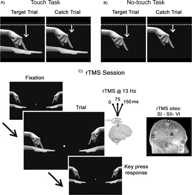
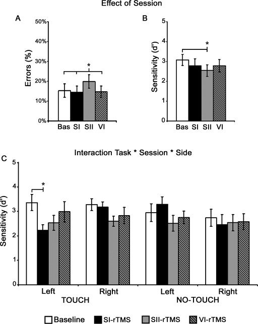
Recent studies suggest the existence of a visuo-tactile mirror system, comprising the primary (SI) and secondary (SII) somatosensory cortices, which matches observed touch with felt touch. Here, repetitive transcranial magnetic stimulation (rTMS) was used to determine whether SI or SII play a functional role in the visual processing of tactile events. Healthy participants performed a visual discrimination task with tactile stimuli (a finger touching a hand) and a control task (a finger moving without touching). During both tasks, rTMS was applied over either SI or SII, and to the occipital cortex. rTMS over SI selectively reduced subject performance for interpreting whether a contralateral visual tactile stimulus contains a tactile event, whereas SII stimulation impaired visual processing regardless of the tactile component. These findings provide evidence for a multimodal sensory-motor system with mirror properties, where somatic and visual properties of action converge. SI, a cortical area traditionally viewed as modality-specific, is selectively implicated in the visual processing of touch. These results are in line with the existence of a sensory mirror system mediating the embodied simulation concept.
Copyright © 2010 Wiley Periodicals, Inc.
Figures
References
-
- Amassian VE, Cracco RQ, Maccabee PJ, Cracco JB, Rudell A, Eberle L ( 1989): Suppression of visual perception by magnetic coil stimulation of human occipital cortex. Electroencephalogr Clin Neurophysiol 74: 458–462. - PubMed
-
- Avenanti A, Bueti D, Galati G, Aglioti SM ( 2005): Transcranial magnetic stimulation highlights the sensorimotor side of empathy for pain. Nat Neurosci 8: 955–960. - PubMed
-
- Avenanti A, Bolognini N, Maravita A, Aglioti SM ( 2007): Somatic and motor components of action simulation. Curr Biol 17: 2129–2135. - PubMed
-
- Avikainen S, Forss N, Hari R ( 2002): Modulated activation of the human SI and SII cortices during observation of hand actions. Neuroimage 15: 640–646. - PubMed
-
- Aziz‐Zadeh L, Maeda F, Zaidel E, Mazziotta J, Iacoboni M ( 2002): Lateralization in motor facilitation during action observation: A TMS study. Exp Brain Res 144: 127–131. - PubMed
Publication types
MeSH terms
LinkOut - more resources
Full Text Sources
