Review
. 2011 Mar-Apr;3(2):231-40.
doi: 10.1002/wsbm.110.

Signaling pathways that control cell migration: models and analysis

Affiliations
Review

Signaling pathways that control cell migration: models and analysis

Erik S Welf et al. Wiley Interdiscip Rev Syst Biol Med. 2011 Mar-Apr.

Abstract

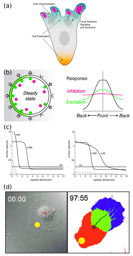
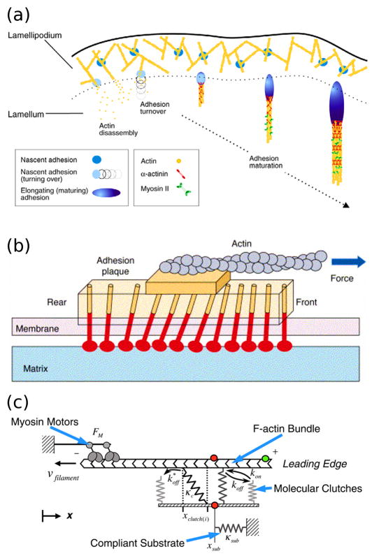
Dissecting the intracellular signaling mechanisms that govern the movement of eukaryotic cells presents a major challenge, not only because of the large number of molecular players involved, but even more so because of the dynamic nature of their regulation by both biochemical and mechanical interactions. Computational modeling and analysis have emerged as useful tools for understanding how the physical properties of cells and their microenvironment are coupled with certain biochemical pathways to actuate and control cell motility. In this focused review, we highlight some of the more recent applications of quantitative modeling and analysis in the field of cell migration. Both in modeling and experiment, it has been prudent to follow a reductionist approach in order to characterize what are arguably the principal modules: spatial polarization of signaling pathways, regulation of the actin cytoskeleton, and dynamics of focal adhesions. While it is important that we 'cut our teeth' on these subsystems, focusing on the details of certain aspects while ignoring or coarse-graining others, it is clear that the challenge ahead will be to characterize the couplings between them in an integrated framework.


Figures

Figure 1
Figure 1. Breaking symmetry of intracellular signaling in migrating cells
(a) Signaling pathways are coordinately localized at the cell front and rear (polarization). They affect, and are in turn affected by, the dynamics of the actin cytoskeleton and cell-matrix adhesion complexes. (b) Local Excitation Global Inhibition (LEGI) models develop intracellular asymmetry in the face of shallow extracellular gradients by amplifying receptor occupancy (gray) via localized activation with positive feedback or cooperativity (green), in tandem with an inhibition mechanism dispersed by fast diffusion (red). (Reprinted with permission from [9]. Copyright 2008 Elsevier Inc.). (c) Polarized signaling responses to two different external gradients, as computed from the “wave pinning” mechanism, are shown by solid lines. The final position of the wavefront (t = 200) does not depend on the stimulus (dotted lines), but rather on the total amount of the signaling protein, which is the same in both cases. (Reprinted with permission from [19]. Copyright 2008 Elsevier Inc.). (d) Guidance of cell migration by focal photo-activation of Rac (yellow spot). (Reprinted with permission from [22]. Copyright 2009 Macmillan Publishers Ltd).
Figure 2
Figure 2. Computational modeling of actin cytoskeletal dynamics
(a) The continuum model assembled by Ditlev et al. encompasses an extensive actin modification network. In the diagram, green circles represent different species or state variables, connected to the reactions (yellow) by solid lines, and the dashed lines represent catalytic interactions. (Reprinted with permission from [24]. Copyright 2009 Elsevier Inc.). (b) Implementation of the model by Ditlev et al. in three dimensions following activation of dendritic nucleation at the cell edge. The top row shows how the protrusion velocity (calculated as a function of actin branching) is localized to the cell front and decays rapidly behind the leading edge. The bottom row shows how actin filament length depends on the rapid nucleation and capping of actin filaments at the cell front that results from depletion of G-actin. (Reprinted with permission from [24]. Copyright 2009 Elsevier Inc.). (c) The model of actin filament dynamics by Lacayo et al. shows how excessive filament capping at the leading edge can produce rough cell morphologies. In cells with high VASP activity at the leading edge (top row), actin filaments are protected from capping and are subject to lateral flows that smooth heterogeneities in protrusive force at the leading edge. (Reprinted with permission from [26]).
Figure 3
Figure 3. Focal adhesion signaling and remodeling
(a) Formation, maturation, and turnover of adhesions at the leading edge occur in different locations and in response to different mechanical and biochemical cues. (Reprinted with permission from [52]. Copyright 2009 The Company of Biologists). (b) Conceptual model of focal adhesion remodeling in response to mechanical strain. The mechanosensitive lower layer of the adhesion complex experiences strain induced by the coupling of actin filaments to the upper layer, inducing growth of the focal adhesion in the direction of force. (Reprinted with permission from [33]. Copyright 2006 Elsevier Inc.). (c) The computational model proposed by Chan and Odde recapitulates the mechanical “clutching” observed between moving actin filaments and adhesion receptors bound to a compliant substrate. (Reprinted with permission from [44]. Copyright 2008 The American Association for the Advancement of Science).

References

    1. Ridley AJ, Schwartz MA, Burridge K, Firtel RA, Ginsberg MH, Borisy G, Parsons TJ, Horwitz AR. Cell migration: Integrating signals from front to back. Science. 2003;302:1704–1709. - PubMed
    1. Friedl P, Wolf K. Plasticity of cell migration: A multiscale tuning model. J Cell Biol. 2010;188:11–19. - PMC - PubMed
    1. Choi CK, Vicente-Manzanares M, Zareno J, Whitmore LA, Mogilner A, Horwitz AR. Actin and α-actinin orchestrate the assembly and maturation of nascent adhesions in a myosin ii motor-independent manner. Nat Cell Biol. 2008;10:1039–1050. - PMC - PubMed
    1. Brandman O, Meyer T. Feedback loops shape cellular signals in space and time. Science. 2008;322:390–395. - PMC - PubMed
    1. Bosgraaf L, Van Haastert PJM. The ordered extension of pseudopodia by amoeboid cells in the absence of external cues. PLoS ONE. 2009;4 - PMC - PubMed

Further Reading

    1. Pollard TD, Borisy GG. Cellular motility driven by assembly and disassembly of actin filaments. Cell. 2003;112:453–465. - PubMed
    1. Mogilner A. Mathematics of cell motility: Have we got its number? J Math Biol. 2009;58:105–134. - PMC - PubMed
    1. Dorn JF, Danuser G, Yang G. Computational processing and analysis of dynamic fluorescence image data. Methods Cell Biol. 2008;85:497–538. - PubMed
    1. Wang Y. Flux at focal adhesions: Slippage clutch, mechanical gauge, or signal depot. Sci STKE. 2007;377:pe10. - PubMed
    1. Orr AW, Helmke BP, Blackman BR, Schwartz MA. Mechanisms of mechanotransduction. Dev Cell. 2006;10:11–20. - PubMed

Publication types