. 2011 Jul;163(5):912-26.
doi: 10.1111/j.1476-5381.2011.01254.x.

Colchicine inhibits cationic dye uptake induced by ATP in P2X2 and P2X7 receptor-expressing cells: implications for its therapeutic action

Affiliations

Colchicine inhibits cationic dye uptake induced by ATP in P2X2 and P2X7 receptor-expressing cells: implications for its therapeutic action

C Marques-da-Silva et al. Br J Pharmacol. 2011 Jul.

Abstract

Background and purpose: The two longest C-termini of the purinergic P2X receptors occur in the P2X2 and P2X7 receptors and are thought to interact with multiple cytoplasmic proteins, among which are members of the cytoskeleton, including microtubules. In this work we asked whether disrupting the microtubule cytoskeleton might affect the functions of these receptors.

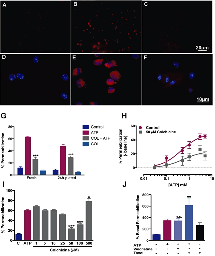
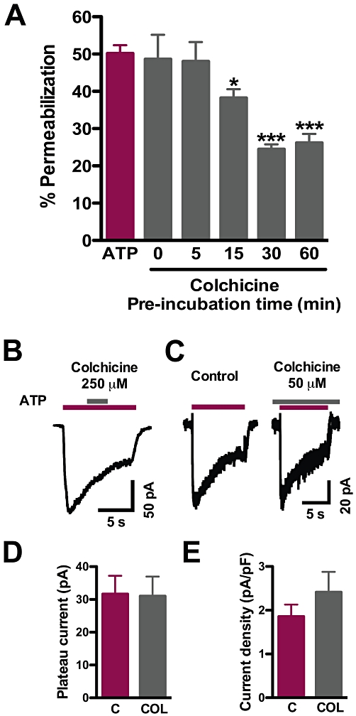
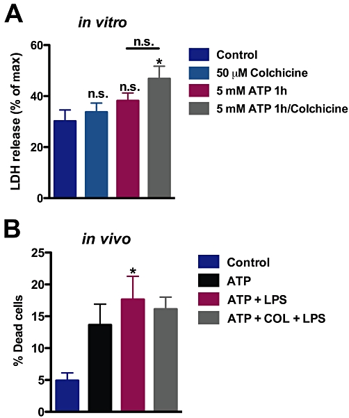
Experimental approach: Functions of heterologously expressed P2X2 and P2X7 receptors were evaluated with electrophysiology and dye uptake following ATP application. Permeabilization and secretion of pro-inflammatory agents were quantified from fresh or cultured peritoneal mouse macrophages, treated in vitro or in vivo with colchicine.

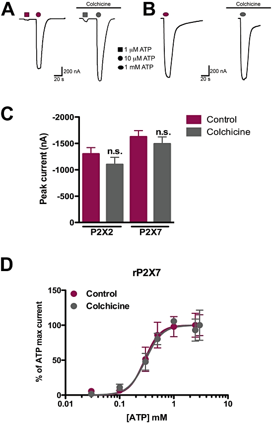
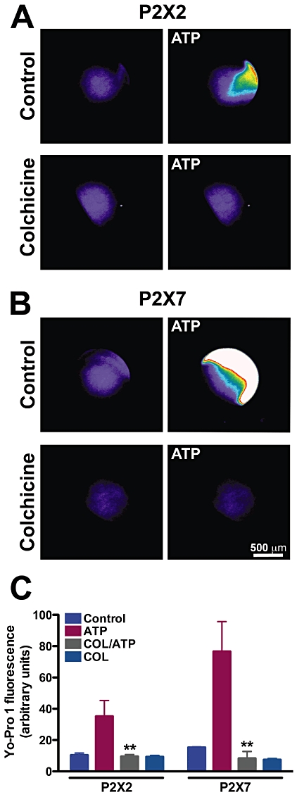
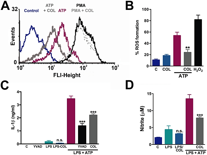
Key results: Disrupting the microtubule network with colchicine did not affect currents generated by ATP in P2X2 and P2X7 receptor-expressing cells but inhibited uptake of the dye Yo-Pro-1 in Xenopus oocytes and HEK293 cells expressing these channels. Peritoneal mouse macrophages showed less ATP-induced permeabilization to ethidium bromide in the presence of colchicine, and less reactive oxygen species (ROS) formation, nitric oxide (NO) and interleukin (IL)-1β release. Colchicine treatment did not affect ATP-evoked currents in macrophages. Finally, in vivo assays with mice inoculated with lipopolysaccharide and ATP showed diminished ROS, IL-1β, interferon-γ and NO production after colchicine treatment.

Conclusions and implications: Colchicine has known anti-inflammatory actions and is used to treat several conditions involving innate immunity, including gout and familial Mediterranean fever. Here we propose a new mechanism of action - inhibition of pore formation induced by activation of P2X receptors - which could explain some of the anti-inflammatory effects of colchicine.


Figures

Figure 1
Figure 1
Colchicine does not affect currents evoked by ATP in P2X2 and P2X7 receptors in Xenopus oocytes. Oocytes were injected with cRNAs encoding rat P2X2 and rat P2X7 receptors and subjected to electrophysiological recordings following 1 h 50 µM colchicine or ND-96 solution (control) treatment. (A) Representative recordings of P2X2 receptor-expressing oocytes in control (left) and following colchicine treatment (right). (B) Typical traces of P2X7 receptor-expressing oocytes, following incubation with colchicine (right) or not (left). (C) Quantification of ATP-evoked current amplitudes in P2X2 and P2X7 receptor-expressing oocytes, with or without colchicine. The differences were not statistically significant (n.s.) as tested by one-way anova followed by Bonferroni's post hoc test (P = 0.29, n = 8 and P = 0.46, n = 6 for P2X2 and P2X7 receptors respectively). (D) Concentration-response curves of ATP-evoked currents in oocytes expressing P2X7 receptors, after treatment with colchicine or with control solution. Estimated EC50s were 0.30 mM (control) and 0.29 mM (colchicine); n = 3–8 oocytes per concentration/group.
Figure 2
Figure 2
Colchicine inhibits Yo-Pro-1 uptake elicited by ATP in P2X2 and P2X7 receptor-expressing oocytes. Oocytes were treated with 50 µM colchicine for 1 h prior to Yo-Pro-1 exposure and the presence of ATP. (A) Pseudo-colour photographs of oocytes expressing P2X2 (A) or P2X7 (B) before and after 100 µM or 1 mM ATP respectively. Lower micrographs are of colchicine-treated oocytes in panels A and B. Scale bar is 500 µm. (C) Quantification of Yo-Pro-1 uptake in P2X2 and P2X7 receptor-expressing oocytes. **P < 0.01, one-way anova followed by Bonferroni's post hoc test; n = 8 (P2X2) and n = 3 (P2X7).
Figure 3
Figure 3
Colchicine effects on the permeabilization of HEK293 cells transfected with rat P2X2, rat P2X7, human P2X4 and human P2X7 receptors. (A) Time course of Yo-Pro-1 uptake following ATP administration (300 µM) in P2X2 receptor-transfected HEK293 cells. Cells were previously treated with or without 50 µM colchicine for 1 h and then used in a plate-reader assay. P < 0.001 when comparing ATP plus colchicine to ATP alone, two-way anova followed by Bonferroni's post hoc test, n = 3 experiments each in triplicate. (B) Permeabilization to 10 µM ethidium bromide (EB) after 5 mM ATP in cells transfected with different P2X receptor subunits, as indicated. The percentage of cells taking up EB was measured with FACS. *P < 0.05 and ***P < 0.001 when comparing ATP plus colchicine to ATP alone, two-way anova followed by Bonferroni's post hoc test, n = 3 experiments each in triplicate.
Figure 4
Figure 4
Colchicine inhibits cationic dye uptake in fresh and plated macrophages. Fresh or plated macrophages obtained by peritoneal wash were exposed or not exposed to 50 µM colchicine for 30 min. Then cells were treated with 5 mM ATP and 10 µM ethidium bromide (EB) or 3 mM sulphorhodamine for additional 15 min at same conditions. Cells were then washed and counted by optical microscopy (G and H), images acquired (A–F) or quantified by flow cytometry (I and J). (A–C) Micrographs of ATP-induced permeabilization to EB (A: control; B: ATP; C: colchicine pretreatment and then ATP). Micrographs of ATP-induced sulforhodamine-B permeabilization (D: control; E: ATP; F: colchicine pretreatment and then ATP). (G) Quantification of EB uptake induced by ATP in freshly isolated and 24 h plated macrophages (P < 0.001, two-way anova followed by Bonferroni's post hoc test, n = 5 experiments each in triplicate). (H) Concentration-response curves of ATP-induced permeabilization following colchicine or control incubation. Estimated EC50s were 0.45 and 0.54 mM for control and colchicine-treated macrophages (n = 4 experiments each in triplicate). (I) Quantification of permeabilization to EB induced by ATP in the presence of different colchicine concentrations in freshly isolated macrophages (*P < 0.05 and ***P < 0.001, relative to ATP only, one-way anova followed by Bonferroni'spost hoc test, n = 3 experiments ieach in triplicate). (J) Quantification of permeabilization to EB induced by ATP in the presence of vincristine or taxol in freshly isolated macrophages (**P < 0.01, one-way anova followed by Bonferroni's post hoc test, n = 3 experiments each in triplicate).
Figure 5
Figure 5
ROS, NO and IL-1β production induced by ATP are inhibited by colchicine. Macrophages obtained by peritoneal wash were pretreated with or without 50 µM colchicine for 30 min, followed by incubation with H2CFDA for additional 30 min. ATP (5 mM) was applied and 5–10 min later cells were analysed by FACScan. As a positive control, we exposed cells to 50 nM PMA (A) or 500 µM H2O2 (B) in substitution to ATP (**P < 0.01, relative to ATP only, one-way anova followed by Bonferroni's post hoc test, n = 2 experiments each in triplicate). (C and D) Peritoneal macrophages were plated for 24 h and stimulated with LPS for additional 24 h, then treated with 50 µM colchicine or Z-YVAD (1:1000) for 30 min, then exposed for 60 min to 5 mM ATP. The supernatants were collected to measure IL-1β (C) and NO (D). The concentrations of IL-1β and NO were determined in ng·mL−1 and µM, respectively, for each experimental group. ***P < 0.001, relative to LPS plus ATP, one-way anova followed by Bonferroni's post hoc test, n = 3 experiments each in triplicate. Differences between LPS only and LPS with colchicine were not significant (P = 0.26 and P = 0.36 for IL-1β and NO, respectively). NO, nitric oxide; LPS, lipopolysaccharide; PMA, phorbol 12-myristate 13-acetate; ROS, reactive oxygen species.
Figure 6
Figure 6
Time course of colchicine action and lack of acute and long-term modulatory effect on ATP-gated currents in macrophages. (A) Time course of colchicine inhibition of ATP-evoked permeabilization. Macrophages were incubated with 50 µM colchicine for the indicated times and then used in permeabilization assays (*P < 0.05 and ***P < 0.001, one-way anova followed by Bonferroni's post hoc test, n = 3 experiments each in triplicate for each time). (B) Whole-cell patch-clamp recording of an acute application of 250 µM colchicine during a 1 mM ATP pulse. (C) Representative recordings of ATP-evoked currents in control (left) and 50 µM colchicine-treated (right) cultured macrophages. (D) Quantification of plateau currents from cells in C (P = 0.47) and of current density (E) (P = 0.35, Student's t-test, n = 8–9 cells).
Figure 7
Figure 7
In vivo treatment with colchicine alters cytokine and other pro-inflammatory agents in mice. (A) Diagram depicting the experimental groups and respective treatments in B and C. At time point I, mice were inoculated with LPS or PBS (i.p.), then 5 h later inoculated with colchicine or PBS (II), following inoculation with ATP or PBS after 1 h (III), and measurements were made at time IV. (B) Cells obtained by peritoneal wash were incubated with DCFH2DA for 30 min, and ROS production was analyzed by FACScan, collecting 10 000 events. The supernatants obtained by peritoneal wash were assayed with Griess reagent or cytokine kits. The concentrations of ROS, cytokines and nitrite were normalized to the production induced by LPS + ATP (100%). Representative measurements of IL-1 [in pg·mL−1 were 860 ± 15 (control), 1460 ± 24 (LPS + ATP), 721 ± 21 (LPS + COL + ATP), 933 ± 40 (LPS only) and 708 ± 22 (LPS + COL)]. Typical nitrite levels were (in µM; mean ± standard deviation): 0.49 ± 0.18 (control), 3.62 ± 0.86, (LPS + ATP), 1.45 ± 0.43 (LPS + COL + ATP), 0.94 ± 0.18 (LPS only) and 0.52 ± 0.14 (LPS + COL). Representative measurements of IFN- (in pg·mL−1) were: 81 ± 5.2 (control), 1077.1 ± 34.3 (LPS + ATP), 156.8 ± 31.2 (LPS + COL + ATP), 108.3 ± 11 (LPS only) and 95.5 ± 22.4 (LPS + COL).***P < 0.001, one-way anova followed by Bonferroni's post hoc test, at least three experiments with two animals were performed for each experimental group. The difference between LPS only and LPS + colchicine was non-significant (n.s.) in all measurements (ROS: P = 0.155; IL-1β: P = 0.160; nitrite: P = 0.33; and IFNγ: P = 0.51). (C) Time course of variation in body temperature (Δ) of mice, relative to values before treatments. Temperature was measured soon after each treatment with a rectal thermometer. Arrows indicate when each agent was administered. Some error bars are shown only in one direction for clarity. ***P < 0.001 two-way anova followed by Bonferroni's post hoc test, n = 3 animals in each group. DCFH2DA, dichlorodihydrofluorescein diacetate; LPS, lipopolysaccharide; ROS, reactive oxygen.
Figure 8
Figure 8
Cell death was not significantly affected by colchicine. (A) Following treatment in vitro, with 5 mM ATP for 1 h, LDH release was measured, relative to maximum release induced by Triton. In the colchicine group, cells were incubated with this agent for 1 h before addition of ATP. *P < 0.05, significantly different from control levels, one-way anova followed by Bonferroni's post hoc test, n = 3 experiments each in triplicate. (B) Animals were treated as in Figure 7; cells were obtained by peritoneal wash and used in Trypan blue exclusion assays. *P < 0.05, significantly different from control levels, one-way anova followed by Bonferroni's post hoc test, n = 2 experiments each in triplicate.

Similar articles

Cited by

References

    1. Alexander SPH, Mathie A, Peters JA. Guide to receptors and channels (GRAC), 4th edn. Br J Pharmacol. 2009;158(Suppl 1):S1–S254. - PMC - PubMed
    1. Bezalel Y, Gershoni-Baruch R, Dagan E, Lidar M, Livneh A. The 3435T polymorphism in the ABCB1 gene and colchicine unresponsiveness in familial Mediterranean fever. Clin Exp Rheumatol. 2009;27:S103–S104. - PubMed
    1. Brone B, Moechars D, Marrannes R, Mercken M, Meert T. P2X currents in peritoneal macrophages of wild type and P2X4 -/- mice. Immunol Lett. 2007;113:83–89. - PubMed
    1. Brough D, Rothwell NJ. Caspase-1-dependent processing of pro-interleukin-1beta is cytosolic and precedes cell death. J Cell Sci. 2007;120:772–781. - PubMed
    1. Castro NG, de Mello MC, de Mello FG, Aracava Y. Direct inhibition of the N-methyl-D-aspartate receptor channel by dopamine and (+)-SKF38393. Br J Pharmacol. 1999;126:1847–1855. - PMC - PubMed

Publication types

MeSH terms

LinkOut - more resources