

Foot kinematics in patients with two patterns of pathological plantar hyperkeratosis
- PMID: 21306644
- PMCID: PMC3045305
- DOI: 10.1186/1757-1146-4-7
Foot kinematics in patients with two patterns of pathological plantar hyperkeratosis
Abstract
Background: The Root paradigm of foot function continues to underpin the majority of clinical foot biomechanics practice and foot orthotic therapy. There are great number of assumptions in this popular paradigm, most of which have not been thoroughly tested. One component supposes that patterns of plantar pressure and associated hyperkeratosis lesions should be associated with distinct rearfoot, mid foot, first metatarsal and hallux kinematic patterns. Our aim was to investigate the extent to which this was true.
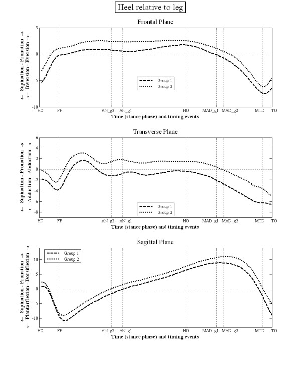
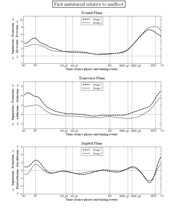
Methods: Twenty-seven subjects with planter pathological hyperkeratosis were recruited into one of two groups. Group 1 displayed pathological plantar hyperkeratosis only under metatarsal heads 2, 3 and 4 (n = 14). Group 2 displayed pathological plantar hyperkeratosis only under the 1st and 5th metatarsal heads (n = 13). Foot kinematics were measured using reflective markers on the leg, heel, midfoot, first metatarsal and hallux.
Results: The kinematic data failed to identify distinct differences between these two groups of subjects, however there were several subtle (generally <3°) differences in kinematic data between these groups. Group 1 displayed a less everted heel, a less abducted heel and a more plantarflexed heel compared to group 2, which is contrary to the Root paradigm.
Conclusions: There was some evidence of small differences between planter pathological hyperkeratosis groups. Nevertheless, there was too much similarity between the kinematic data displayed in each group to classify them as distinct foot types as the current clinical paradigm proposes.
Figures


References
-
- Root ML, Weed JH, Sgarlato TE, Bluth DR. Axis of motion of the subtalar joint. J Am Podiatry Assoc. 1966;56:149–155.
-
- Root ML, Orien WP, Weed JH. Normal and Abnormal function of the Foot. Los Angeles: Clinical Biomechanics Corp; 1977.
-
- Nester C, Jones R, Liu A, Howard D, Lundberg A, Arndt T, Lundgren P, Stacoff A, Wolf P. Invasive study of rearfoot, midfoot and forefoot kinematics during walking. J Biomech. 2006;39:S77–S77. doi: 10.1016/S0021-9290(06)83195-7. - DOI
-
- Sage RA, Webster JK, Fisher SG. Outpatient Care and Morbidity Reduction in Diabetic Foot Ulcers Associated with Chronic Pressure Callus. J Am Podiatry Assoc. 2001;91:275–279. - PubMed
LinkOut - more resources
Full Text Sources
