Review
. 2011 Apr;23(2):84-91.
doi: 10.1016/j.smim.2011.01.007.

T cell receptor recognition of self and foreign antigens in the induction of autoimmunity

Affiliations
Review

T cell receptor recognition of self and foreign antigens in the induction of autoimmunity

Kai W Wucherpfennig et al. Semin Immunol. 2011 Apr.

Abstract

The major histocompatibility complex (MHC) on human chromosome 6 represents the most important genetic locus for a number of common human autoimmune diseases. Specific alleles that differ from closely related alleles by only one or a few amino acids in the peptide binding groove are frequently strongly associated with disease susceptibility, raising the important question of which peptide presentation events are critical in disease initiation and progression. This review will cover a number of topics pertinent to this fundamental question, including MHC linked disease susceptibility to autoimmune diseases, molecular mechanisms for the role of MHC molecules in autoimmune diseases as well as the recognition of self and microbial peptides by self-reactive T cell receptors (TCRs).


Figures

Figure 1
Figure 1. Structural features of MHC class II molecules associated with susceptibility to autoimmune diseases
A. One of the characteristic features of the MS associated HLA-DR2 (DRA, DRB1*1501) molecule is the large hydrophobic P4 pocket that accommodates a phenylalanine residue (P4 Phe) of the bound MBP (85–99) peptide in this structure. A key polymorphic residue, DRβ 71, is a small amino acid (alanine) which provides the room for the large peptide side chain in this pocket (the neighboring amino acid Q70 is highlighted). The bound peptide is shown as a stick model. A positive charge is indicated by a blue surface on the MHC molecule, a negative charge by a red surface. B. The rheumatoid arthritis associated DR4 (DRB1*0401) molecule has a P4 pocket with very different characteristics. A key polymorphic amino acid, DRβ71, is a lysine (K71), which makes the pocket smaller than in DR2 and gives it a positive charge. As a consequence, this pocket has a preference for negatively charged amino acids, like the aspartic acid residue (P4 Asp) of the bound type II collagen peptide. C. The type 1 diabetes associated DQ8 molecule has a positively charged P9 pocket because the key polymorphic residue, DQβ 57, lacks a charge (an alanine, A57). This pocket therefore has a positive charge, indicated by the blue color.
Figure 2
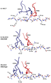
Figure 2. Ob.1A12 TCR with MBP and engA peptides
A. Recognition of the influenza hemagglutinin (306–318) peptide by the human HA1.7 TCR. Shown are the TCR CDR3 loops, CDR3α in blue and CDR3β in red, as well as the peptide as a stick model. Peptide residues representing key TCR contacts are labeled, as well as peptide residues occupying key pocket of the MHC molecule (HLA-DR1). B. Recognition of the self-peptide MBP (85–99) by the Ob.1A12 TCR isolated from a patient with relapsing-remitting MS. The CDR3 loops of this TCR are focused over the P2 peptide position (P2 His), rather than the P5 position as for HA1.7 TCR. Key TCR contacts of Ob.1A12 TCR are P2 His, P3 Phe and P5 Lys (ENPVVHFFKNIVTPR), key HLA-DR2 binding residues are P1 Val and P4 Phe. C. Recognition of a cross-reactive microbial peptide by Ob.1A12 TCR. This TCR cross-reacts with the engA peptide of E. coli and Hemophilus influenzae (DFARVHFISALHGSG) which shares the P2 His and P3 Phe residues with the MBP peptide (bold, underlined). The peptide also has hydrophobic anchor residues for the P1 and P4 pockets (P1 Val and P4 Ile), but is otherwise quite distinct from the MBP peptide. Nevertheless, the binding of Ob.1A12 TCR to the two peptides is very similar (compare B and C).

Similar articles

Cited by

References

    1. Hafler DA, Compston A, Sawcer S, Lander ES, Daly MJ, De Jager PL, de Bakker PI, Gabriel SB, Mirel DB, Ivinson AJ, Pericak-Vance MA, Gregory SG, Rioux JD, McCauley JL, Haines JL, Barcellos LF, Cree B, Oksenberg JR, Hauser SL. Risk alleles for multiple sclerosis identified by a genomewide study. N Engl J Med. 2007;357:851–862. - PubMed
    1. Barrett JC, Clayton DG, Concannon P, Akolkar B, Cooper JD, Erlich HA, Julier C, Morahan G, Nerup J, Nierras C, Plagnol V, Pociot F, Schuilenburg H, Smyth DJ, Stevens H, Todd JA, Walker NM, Rich SS. Genome-wide association study and meta-analysis find that over 40 loci affect risk of type 1 diabetes. Nat Genet. 2009 - PMC - PubMed
    1. MacKay K, Eyre S, Myerscough A, Milicic A, Barton A, Laval S, Barrett J, Lee D, White S, John S, Brown MA, Bell J, Silman A, Ollier W, Wordsworth P, Worthington J. Whole-genome linkage analysis of rheumatoid arthritis susceptibility loci in 252 affected sibling pairs in the United Kingdom. Arthritis Rheum. 2002;46:632–639. - PubMed
    1. Oksenberg JR, Barcellos LF, Cree BA, Baranzini SE, Bugawan TL, Khan O, Lincoln RR, Swerdlin A, Mignot E, Lin L, Goodin D, Erlich HA, Schmidt S, Thomson G, Reich DE, Pericak-Vance MA, Haines JL, Hauser SL. Mapping multiple sclerosis susceptibility to the HLA-DR locus in African Americans. Am J Hum Genet. 2004;74:160–167. - PMC - PubMed
    1. Lincoln MR, Ramagopalan SV, Chao MJ, Herrera BM, Deluca GC, Orton SM, Dyment DA, Sadovnick AD, Ebers GC. Epistasis among HLA-DRB1, HLA-DQA1, and HLA-DQB1 loci determines multiple sclerosis susceptibility. Proc Natl Acad Sci U S A. 2009;106:7542–7547. - PMC - PubMed

Publication types