. 2011 Mar;37(2):238-43.
doi: 10.1093/schbul/sbq165. Epub 2011 Feb 9.

Dysfunctional thalamus-related networks in schizophrenia

Affiliations

Dysfunctional thalamus-related networks in schizophrenia

Didier Pinault. Schizophr Bull. 2011 Mar.

Abstract

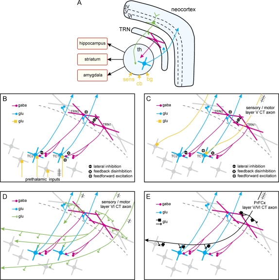
Thalamus abnormalities are common in neurological and psychiatric illnesses. Therefore, it is essential to understand the properties of the thalamus-related networks. The thalamic reticular nucleus (TRN) is a thin GABAergic layer interface strategically located between the thalamus and the neocortex. It is, at the very beginning of life, an essential neurodevelopmental guide for the accurate build up of reciprocal anatomical glutamatergic connections between the thalamus and neocortex. It is more than the mediator of selective attention. It appears as a combinatorial matrix because it holds and can combine multiple functional modalities. TRN cells work like integrators, thanks to their extraordinary intrinsic electrophysiological properties, under the contextual and leading influence of corticothalamic inputs. The TRN and thalamus principally form 2-neuron open-loop circuits (no reciprocal connection). The major functioning principle of such GABAergic-glutamatergic circuits is lateral inhibition, which is a gold standard device to set up, via differential amplifications, coherent structured thalamocortical activity patterns. Thereby, it selects relevant streams of information and deletes distractors during action, resting states, and information integration, including during consciousness, cognition, emotion, and thought. Disruption of thalamic lateral inhibition may contribute to a lack of coordination in activity between brain regions, as observed in psychiatric disorders like schizophrenia.


Figures

Fig. 1.
Fig. 1.
Thalamic Lateral Inhibition And Corticothalamic (CT) Influence. (A) The 3 principal elements of the thalamocortical (TC)/CT systems. The CT (of layer VI, in green) and TC (blue) axons are glutamatergic and cross the thalamic reticular nucleus (TRN), where they give off axon collaterals. The principal prethalamic inputs (deep yellow) originate from sensory receptors (sens), the cerebellum (cb), and basal ganglia (bg). Parts of the thalamus innervate the hippocampus, striatum, and amygdala. (B–E) The 4 drawings illustrate likely synaptic interactions between single TC, CT neurons, which are glutamatergic, and TRN neurons, which are GABAergic (in red). TC and TRN neurons form open-loop circuits. Some of the terminal axonal boutons of 1 TC neuron contact 1 TRN cell; similarly, some terminal boutons of 1 TRN cell make synaptic contacts with 1 TC neuron. The oversimplified drawings do not include thalamic interneurons and do not respect the anatomical scale in dimension and in number. (B) In a first-order (eg, specific sensory or motor) thalamic nucleus, activation (feedforward excitation) starts with neuron TC1, for instance, following an afferent discharge on a specific prethalamic, sensory or motor, glutamatergic axonal input (yellow). TC1 cell, via its axon collaterals, excites TRN2 cell, which then inhibits TC2 cell (lateral inhibition). TRN2 neuron, via dendrodendritic GABAergic synapses, inhibits TRN1 cell, which then disinhibits TC1 neuron (feedback disinhibition). (C) The same scenario might occur in higher-order (nonspecific, associative, cognitive) thalamic nuclei with layer V CT axons (yellow), which are thought to be drivers like prethalamic sensory inputs. In this scenario, the active CT input is indicated by the train of action potentials. (D) Layer VI axons (green), in contrast to layer V CT axons, innervate both the TRN and the thalamus; in addition, they reciprocally innervate larger territories in first-order and higher-order thalamic nuclei. CT axons are thought to supply contextual information to the TC-TRN circuits. (E) The prefrontal cortex (PrFCx) contains layer V and VI CT neurons (black), which innervate TRN and TC neurons in sensory, motor, and limbic systems. Because prefrontal CT axons have a dual mode of terminals (small and large), they may be seen as differential drivers. The functional scenario may roughly be a combination of those suggested in (C) and (D). For clarity, the prethalamic inputs shown in (B) (yellow) are not shown in (C), (D), and (E). The functional role of the distinct CT pathways and how they work together remain a grand mystery.

References

    1. Ross CA, Margolis RL, Reading SA, Pletnikov M, Coyle JT. Neurobiology of schizophrenia. Neuron. 2006;52:139–153. - PubMed
    1. Friston KJ. Dysfunctional connectivity in schizophrenia. World Psychiatry. 2002;1:66–71. - PMC - PubMed
    1. Stephan KE, Baldeweg T, Friston KJ. Synaptic plasticity and dysconnection in schizophrenia. Biol Psychiatry. 2006;59:929–939. - PubMed
    1. Tononi G, Edelman GM. Schizophrenia and the mechanisms of conscious integration. Brain Res Rev. 2000;31:391–400. - PubMed
    1. Zhang N, Zhu XH, Zhang Y, Chen W. An fMRI study of neural interaction in large-scale cortico-thalamic visual network. Neuroimage. 2008;42:1110–1117. - PMC - PubMed

Publication types

MeSH terms

Substances