. 2011 Mar;138(6):1131-42.
doi: 10.1242/dev.058149. Epub 2011 Feb 9.

Pkd1l1 establishes left-right asymmetry and physically interacts with Pkd2

Affiliations

Pkd1l1 establishes left-right asymmetry and physically interacts with Pkd2

Sarah Field et al. Development. 2011 Mar.

Abstract

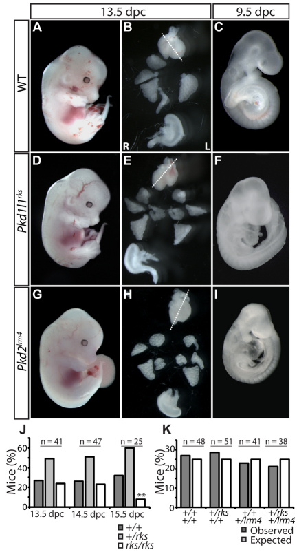
In mammals, left-right (L-R) asymmetry is established by posteriorly oriented cilia driving a leftwards laminar flow in the embryonic node, thereby activating asymmetric gene expression. The two-cilia hypothesis argues that immotile cilia detect and respond to this flow through a Pkd2-mediated mechanism; a putative sensory partner protein has, however, remained unidentified. We have identified the Pkd1-related locus Pkd1l1 as a crucial component of L-R patterning in mouse. Systematic comparison of Pkd1l1 and Pkd2 point mutants reveals strong phenocopying, evidenced by both morphological and molecular markers of sidedness; both mutants fail to activate asymmetric gene expression at the node or in the lateral plate and exhibit right isomerism of the lungs. Node and cilia morphology were normal in mutants and cilia demonstrated typical motility, consistent with Pkd1l1 and Pkd2 activity downstream of nodal flow. Cell biological analysis reveals that Pkd1l1 and Pkd2 localise to the cilium and biochemical experiments demonstrate that they can physically interact. Together with co-expression in the node, these data argue that Pkd1l1 is the elusive Pkd2 binding partner required for L-R patterning and support the two-cilia hypothesis.


Figures

Fig. 1.
Fig. 1.
rks disrupts a PKD domain of Pkd1l1. (A) The rks mutation maps to a ~200 kb region on mouse chromosome 11 containing the Pkd1l1 locus and two non-coding pseudogenes. Recombination events at flanking markers were recorded. Sequencing of the critical region revealed an A-to-G transition at nucleotide 1232 in exon 8 of Pkd1l1 (box). (B) Multiple sequence alignment of orthologous PKD domains with the Pkd1l1rks point mutation in the conserved WDFDGDS motif highlighted (red). (C) Domain structure and membrane topology of mouse Pkd2 and Pkd1l1 with the Pkd2lrm4 and Pkd1l1rks amino acid substitutions indicated (red arrows). (D) Wild-type (WT) and Pkd1l1rks mutant PKD domains modelled using nFOLD3. A schematic of the domains is given beneath. In the mutant, the C′ and much of the E β-sheets are destabilised (asterisks).
Fig. 2.
Fig. 2.
Gross situs abnormalities in Pkd1l1rks and Pkd2lrm4 embryos. (A-C) WT mouse embryo (A) showing left-sided heart apex and stomach and normal lung situs at 13.5 dpc (B), and normal embryonic turning at 9.5 dpc (C). (D-I) Pkd1l1rks (D-F) and Pkd2lrm4 (G-I) embryos demonstrate incidences of reversed heart apex and stomach, right pulmonary isomerism and reversed embryonic turning. Dotted lines indicate primary axis of the heart. (J) Time-of-death analysis reveals that Pkd1l1rks embryos arrest between 14.5 and 15.5 dpc. Two mutant 15.5 dpc embryos were close to death when examined (**). (K) Pkd1l1rks × Pkd2lrm4 intercrosses resulted in no departure from Mendelian ratios when genotyped at weaning.
Fig. 3.
Fig. 3.
Failure to activate left-side loci in Pkd1l1rks and Pkd2lrm4 embryos. (A-C) Nodal mRNA was expressed around the node and in the left lateral plate mesoderm (LPM) in WT mouse embryos, but was absent from the left LPM in Pkd1l1rks and Pkd2lrm4 mutants. Node Nodal expression remained symmetrical in mutants (B′,C′) but became more strongly expressed on the left in WT (A′). (D-I) Lefty1/2 (D-F) and Pitx2 (G-I) expression was absent in both Pkd1l1rks and Pkd2lrm4 mutants. (J-L) Cerl2 mRNA was expressed more strongly on the right side of the node in 3-somite stage WT embryos (J); expression remained symmetrical in mutants (K,L). In all panels, left (L) and right (R) are as indicated in A.
Fig. 4.
Fig. 4.
Node morphology and cilia motility are normal in Pkd1l1rks and Pkd2lrm4 embryos. (A-F) Scanning electron micrographs of WT (A,B), Pkd1l1rks/rks (C,D) and Pkd2lrm4/lrm4 (E,F) 8.25 dpc mouse embryos. Scale bar: 5 μm for A,C,E; 1 μm for B,D,F. (G) Average cilia length does not vary between WT and mutant embryos. (H) Rotational speed of cilia is equivalent in WT and mutant embryos. Error bars indicate s.d.
Fig. 5.
Fig. 5.
Pkd1l1 is strongly enriched in the embryonic node. (A-F) Pkd1l1 mRNA expression visualised by WISH in 7.5 dpc (A-C) and 8.5 dpc (D-F) WT mouse embryos. Highly enriched expression evident in the forming embryonic node (A,B) is clearly localised to the ventral node when viewed in section (C,C′). Higher-magnification views reveal low-level Pkd1l1 expression in the visceral endoderm (C′, arrows). Enriched Pkd1l1 expression in the node and midline (D,E) is particularly obvious in sections (F,F′), revealing node expression solely in the ventral layer. n, node; nc, notochord. Planes of section are indicated by dashed lines.
Fig. 6.
Fig. 6.
Pkd1l1 and PKD2 interact via the intracellular coiled coil domain of Pkd1l1. (A) Pkd1l1 and the Pkd1l1-GFP, Pkd1l1_CC-GFP and Pkd1l1_CCL2554D-GFP constructs. The cleavage site producing Pkd1l1_53 is indicated (blue arrow). (B-D) Immunoprecipitation experiments show that Pkd1l1 and PKD2 interact. (B) A 53 kDa C-terminal Pkd1l1 cleavage product (80 kDa) co-purifies with Myc-PKD2 (140 kDa). (C) The C-terminal coiled coil (CC) domain of Pkd1l1 is sufficient for interaction with PKD2, whereas mutation of the CC prevents the interaction (D). E indicates empty line.
Fig. 7.
Fig. 7.
Pkd1l1 and Pkd2 co-localise to primary cilia. (A-D) IMCD3 cells transiently transfected with Pkd1l1-GFP (B), Myc-PKD2 (C), Pkd1l1-GFP and Myc-PKD2 (D), or with no DNA as control (A). Anti-acetylated tubulin staining (blue) marks cilia. Anti-GFP antibody identifies Pkd1l1-GFP (green) and anti-Myc staining marks Myc-PKD2 (red). Merged images (left column) show that, individually, Pkd1l1-GFP and Myc-PKD2 are within the cell body, but localise to the cilia when co-expressed. (E) Following transfection of Pkd1l1-GFP, 250 cilia were visualised and the number of cilia showing Pkd1l1 with and without Pkd2 was counted. (F,G) IMCD3 cells transfected with Pkd1l1-GFP and Myc-PKD2 (F) or Pkd1l1_CC-GFP and Myc-PKD2 (G) and visualised for acetylated tubulin (red) and GFP (green).

References

    1. Afzelius B. A. (2004). Cilia-related diseases. J. Pathol. 204, 470-477 - PMC - PubMed
    1. Bisgrove B. W., Snarr B. S., Emrazian A., Yost H. J. (2005). Polaris and Polycystin-2 in dorsal forerunner cells and Kupffer's vesicle are required for specification of the zebrafish left-right axis. Dev. Biol. 287, 274-288 - PubMed
    1. Bloodgood R. A. (2010). Sensory reception is an attribute of both primary cilia and motile cilia. J. Cell Sci. 123, 505-509 - PubMed
    1. Blum M., Weber T., Beyer T., Vick P. (2008). Evolution of leftward flow. Semin. Cell Dev. Biol. 20, 464-471 - PubMed
    1. Bogani D., Siggers P., Brixey R., Warr N., Beddow S., Edwards J., Williams D., Wilhelm D., Koopman P., Flavell R. A., et al. (2009). Loss of mitogen-activated protein kinase kinase kinase 4 (MAP3K4) reveals a requirement for MAPK signalling in mouse sex determination. PLoS Biol. 7, e1000196 - PMC - PubMed

Publication types

MeSH terms