. 2011 Mar;138(6):1045-55.
doi: 10.1242/dev.056671. Epub 2011 Feb 9.

EGF signaling regulates the proliferation of intestinal stem cells in Drosophila

Affiliations

EGF signaling regulates the proliferation of intestinal stem cells in Drosophila

Benoît Biteau et al. Development. 2011 Mar.

Abstract

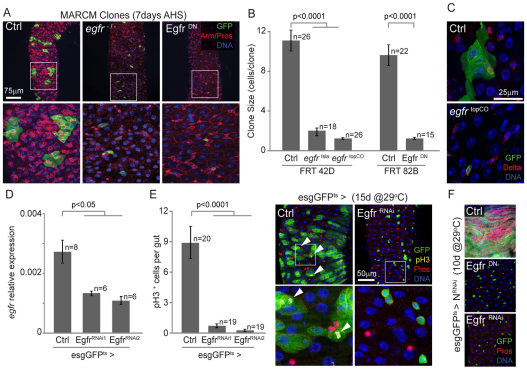
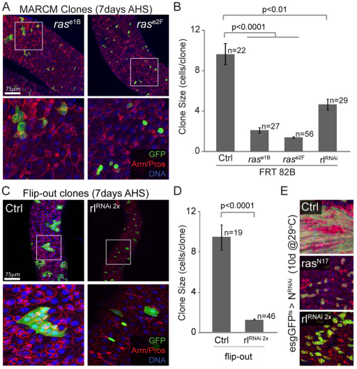
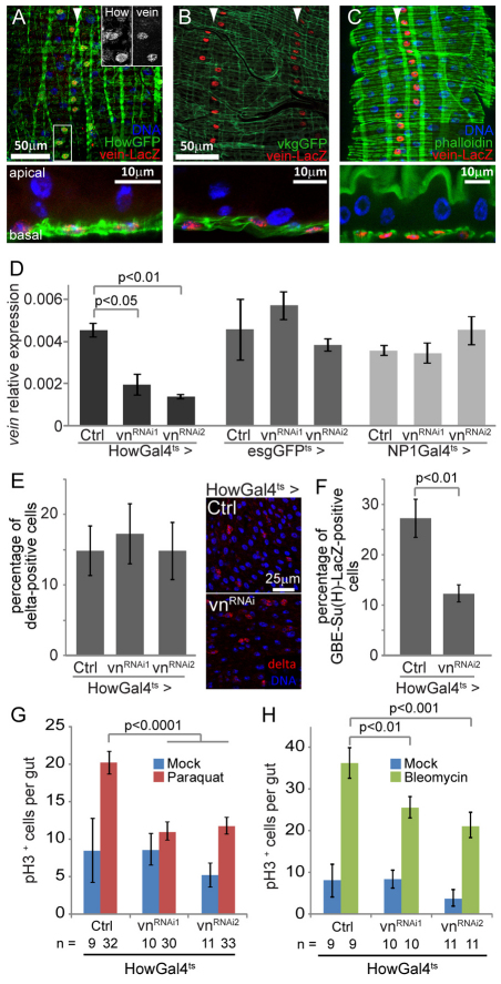
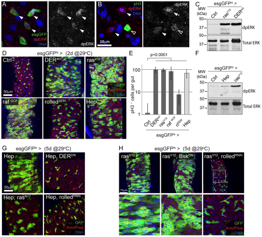
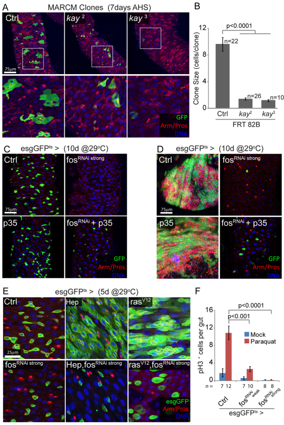
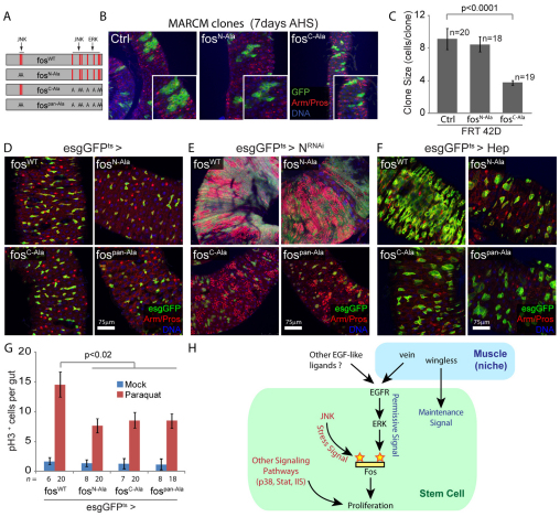
Precise control of somatic stem cell proliferation is crucial to ensure maintenance of tissue homeostasis in high-turnover tissues. In Drosophila, intestinal stem cells (ISCs) are essential for homeostatic turnover of the intestinal epithelium and ensure epithelial regeneration after tissue damage. To accommodate these functions, ISC proliferation is regulated dynamically by various growth factors and stress signaling pathways. How these signals are integrated is poorly understood. Here, we show that EGF receptor signaling is required to maintain the proliferative capacity of ISCs. The EGF ligand Vein is expressed in the muscle surrounding the intestinal epithelium, providing a permissive signal for ISC proliferation. We find that the AP-1 transcription factor FOS serves as a convergence point for this signal and for the Jun N-terminal kinase (JNK) pathway, which promotes ISC proliferation in response to stress. Our results support the notion that the visceral muscle serves as a functional 'niche' for ISCs, and identify FOS as a central integrator of a niche-derived permissive signal with stress-induced instructive signals, adjusting ISC proliferation to environmental conditions.


Figures

Fig. 1.
Fig. 1.
EGF receptor activity is essential for ISC proliferation. (A-C) MARCM clones overexpressing a dominant-negative form of EGFR (DERDN) and homozygous mutant clones for Egfr (egfr−/−) fail to grow, demonstrating that components of the EGFR activity is required for ISC proliferation. Boxed areas are magnified in the lower panels. Quantification of the clone size, measured by the number of cells per clone, 7 days after clone induction [7d after heat shock (AHS)] is shown in B. Error bars represent s.e.m. FRT 42D, MARCM using Flip recombination target at 42D; FRT 82B, MARCM using Flip recombination target at 82B. Staining for the ISC-specific marker Delta (red), indicates that Egfr-null single cell clones (egfrtopCO) are non-dividing ISCs (C). (D) Egfr expression in the intestine, 5 days after induction of expression of dsRNA constructs against Egfr, using the esgGal4 driver. Expression of the two distinct transgenes significantly alters Egfr expression compared with controls. (E) Knockdown of Egfr in ISCs and EBs using the temperature-sensitive driver esgGFPts (esgGal4;tubGal80ts) is sufficient to prevent age-related induction of proliferation and intestinal dysplasia. Proliferation was quantified by counting the number of pH3+ cells per gut after immunostaining, 15 days after transgenes induction at 29°C. Representative confocal images are shown to illustrate the reduced number of pH3+ cells (indicated by arrowheads) and limited intestinal dysplasia observed in esgGFPts>EgfrRNAi flies. Boxed areas are magnified in the lower panels. (F) Reduction of Egfr activity prevents NotchRNAi-induced tumor formation. EgfrDN or EgfrRNAi transgenes were expressed together with NRNAi using esgGFPts. Ten days after induction, tumors composed of esg+ and PROS+ cells accumulate in the posterior midgut of control flies (NRNAi alone), whereas the intestinal epithelium architecture is preserved when EGFR activity is inhibited. In A, C, E and F, GFP expression is shown in green, cell boundaries are stained using β-Catenin/Armadillo (red, membrane), EEs are marked by the expression of Prospero (red nuclei) and DNA is labeled using Hoechst (blue). EB, enteroblast; EE, enteroendocrine cells; ISC, intestinal stem cell; MARCM, mosaic analysis with a repressible cell marker.
Fig. 2.
Fig. 2.
Components of the MAPK signaling pathway are required for ISC proliferation. (A) MARCM clones homozygous for Ras loss-of-function alleles fail to grow compared with control clones (see Fig. 1A). Boxed areas are magnified in the lower panels. (B) Quantification of clone size 7 days after induction, including MARCM clones expressing one copy of the rolledRNAi construct. Error bars represent s.e.m. (C,D) Flip-out clones overexpressing two copies of the rolledRNAi construct mostly remain as single stem cells, confirming the essential role of ERK in ISC proliferation. The clone size, measured by the number of cells per clone, 7 days after clone induction, is quantified in D. (E) Inhibition of Ras and ERK prevents NotchRNAi-induced tumor formation. Confocal images of posterior midguts co-expressing a dominant form of Ras (rasN17) or two copies of the rolledRNAi construct with NRNAi in ISCs and EBs, 10 days after induction at 29°C. In A, C and E, GFP expression is shown in green, cell boundaries are stained using β-Catenin/Armadillo (red, membrane), EEs are marked by the expression of Prospero (red nuclei) and DNA is labeled using Hoechst (blue). EB, enteroblast; EE, enteroendocrine cells; ISC, intestinal stem cell; MARCM, mosaic analysis with a repressible cell marker.
Fig. 3.
Fig. 3.
The EGFR ligand vein is expressed in the intestinal muscle and is required for ISC proliferation. (A-C) The Vein-lacZ transcriptional reporter is expressed in muscle. Nuclear β-galactosidase expression is detected by immunostaining (red), in longitudinal rows of cells located basally, along the entire posterior midgut (indicated by arrowheads). Cells positive for the Vein-lacZ reporter also express GFP when driven with the muscle-specific HowGal4 driver (A, inserts show single channel images of the boxed area), demonstrating that Vein-lacZ is expressed in muscle. Co-localization with vkgGFP (a GFP fusion with the basement membrane component collagen IV) and phalloidin (staining F-actin) further demonstrates the basal position of β-galactosidase+ cells in the epithelium (B,C). (D) vein mRNA can be detected in the intestine and is expressed in muscle. Expression of dsRNA constructs against Vein in the muscle (HowGal4ts driver) is sufficient to significantly reduce mRNA level in the intestine, whereas expression of vnRNAi constructs in ISCs or EBs (esgGFPts driver) or in enterocytes (NP1Gal4ts driver) has no effect. The expression of vein is measured by real-time RT-PCR, relative to the expression of Actin5c. (E) vein knockdown in the muscle does not affect ISC maintenance. The proportion of Delta-positive cells is similar in the epithelium of control and HowGal4ts>vnRNAi flies 5 days after transgenes induction. Representative images are shown to illustrate the maintenance of small Delta-positive cells in the epithelium of HowGal4ts>vnRNAi flies (shown in red). (F) vein knockdown reduces the number of EBs. The proportion of cells positive for the EB marker GBE-Su(H)-lacZ is significantly lower in the intestine of HowGal4ts>vnRNAi flies compared with wild-type controls. (G,H) Knocking down vein expression in the muscle limits stress-induced proliferation. The number of pH3-positive cells, in mock-treated flies or after exposure to paraquat or bleomycin (for 24 and 48 hours, respectively) was measured in the epithelium of control and HowGal4ts>vnRNAi flies. EB, enteroblast; ISC, intestinal stem cell.
Fig. 4.
Fig. 4.
The MAPK signaling pathway is active in ISCs and its activation is sufficient to promote ISC proliferation. (A) The active, double-phosphorylated (dp) form of ERK can be detected by immunostaining in the ISCs under normal conditions (red, left-hand panel; monochrome, right-hand panel). (B) Similar dpERK staining is observed in dividing (pH3-positive; open arrowhead) and non-dividing ISCs (solid arrowhead). (C) Expression of activated forms of the EGFR (DERact) or RAS (rasV12), using the esgGFPts driver, dramatically increases the level of activated ERK in the intestine. dpERK is detected by Western blot from dissected midguts 2 days after transgene induction at 29°C. Levels of total ERK protein serve as loading control. (D,E) Activation of the EGFR/MAPK and JNK signaling pathways is sufficient to induce ISC proliferation, resulting in intestinal dysplasia. Activated forms of DER (DERact), RAS (rasV12), RAF (rafGOF) and ERK (rolledSEM; rlSEM) or HEP (Hep) were expressed for 2 days in ISCs using the temperature-sensitive driver (esg-Gal4,UAS-GFP;tub-Gal80ts). Proliferation rates in the midgut were quantified (E) by counting the number of pH3+ cells per gut in the same genetic conditions. (F) Activation of JNK in ISCs and EBs does not significantly elevate dpERK levels. dpERK is detected by Western blot from dissected midguts 2 days after transgene induction at 29°C. Levels of total ERK protein serve as loading control. (G) Overexpression of HEP in ISCs and EBs results in dyplastic phenotype in the intestinal epithelium (esgGFPts>Hep). Co-expression of dominant forms of EGFR (DERDN) and ras (rasN17), or a dsRNA construct directed against ERK (rolledRNAi), is sufficient to prevent HEP-induced dysplasia. (H) RAS-induced dysplasia specifically requires ERK activity. Inhibiting ERK (rolledRNAi) prevents the expansion of esg+ cells observed when RasV12 is expressed under the control of the esgGFPts driver. Co-expression of a JNK dominant form (BskDN) does not affect this phenotype. Boxed areas are enlarged in lower panels. EB, enteroblast; ISC, intestinal stem cell.
Fig. 5.
Fig. 5.
FOS is required for stem cell survival and proliferation, downstream of JNK and RAS. (A,B) FOS is required in ISCs for clone formation. Posterior midguts showing MARCM clones homozygous for fos (kay) loss-of-function alleles, 7 days after induction. Mutant clones remain smaller than controls, often limited to single stem cells, and are recovered less frequently for the kay3 allele. Boxed areas are enlarged in lower panels. Clone size (number of cells per clone) is quantified in B. FRT 82B, MARCM using Flip recombination target at 82B. (C) Prolonged inhibition of FOS results in ISC death by apoptosis. Expression of the fosRNAi construct that causes strong knockdown of FOS expression, using the esgGFPts driver, leads to the disappearance of esg+ cells. This phenotype is rescued when the anti-apoptotic protein p35 is co-expressed, whereas expression of p35 alone has no effect. (D) A similar experiment performed in N loss-of-function background demonstrates that p35 expression rescues cell death but is unable to restore NRNAi-induced proliferation and tumor formation, suggesting that FOS affects both survival and proliferative capacity of ISCs. (E) FOS is required for HEP- and RAS-induced ISC proliferation. Intestines 5 days after induction of HEP or rasV12 together with fosRNAi, using the temperature-sensitive esgGal4 driver. The HEP- and rasV12-induced expansion of esg+ cells is entirely blocked by fosRNAi, demonstrating that FOS is required downstream of JNK and RAS. Note that knocking down FOS does not block RAS-induced cell growth. (F) Paraquat-induced ISC proliferation requires FOS. Expression of two different fosRNAi constructs, using esgGFPts (for 2 days at 29°C prior to treatment), significantly reduces Paraquat-induced proliferation in the intestinal epithelium, as shown by the limited number of pH3+ cells per gut 48 hours after paraquat exposure. In A, C and D GFP is shown in green, armadillo (Arm) outlines cell boundaries (red), prospero (Pros) identifies EEs (nuclear red), DNA is shown in blue. AHS, after heat shock; EE, enteroendocrine cells; ISC, intestinal stem cell; MARCM, mosaic analysis with a repressible cell marker.
Fig. 6.
Fig. 6.
FOS integrates JNK and ERK signaling pathways through distinct phosphorylation sites. (A) Schematic representation of the FOS protein and the different mutants carrying substitution of JNK and/or ERK phosphorylation sites. Red lines indicate phosphorylation sites. (B,C) Posterior midguts showing MARCM clones overexpressing fosC-Ala and fosN-Ala, 7 days after induction. Expression of FOS carrying substitution ERK phosphorylation sites (fosC-Ala) prevents the formation of large clones. Insets show enlarged cells. Clone size (number of cells per clone) is quantified in C. (D) Expression of FOS mutant forms in ISCs and EBs does not affect the architecture of the posterior midgut. (E) ERK phosphorylation sites are required for N loss-of-function tumor formation. Intestines 10 days after induction of NRNAi and fos mutants carrying substitution of JNK and/or ERK phosphorylation sites. Expression of the mutant forms lacking ERK phosphorylation prevents NRNAi-induced ISC overproliferation. (F) ERK and JNK phosphorylation sites are required for HEP-induced proliferation. Representative confocal images of intestines 5 days after induction of JNK and HEP together with the wild-type or mutant forms of FOS in ISCs/EBs. The HEP-induced expansion of esg+ cells is blocked by all the FOS mutant forms. (G) Overexpression of FOS mutant forms partially prevents paraquat-induced stem cell proliferation, as shown by reduced number of pH3+ cells, 48 hours after paraquat exposure. In B, D and E, GFP is shown in green, armadillo (Arm) outlines cell boundaries (red), prospero (Pros) identifies EEs (nuclear red), DNA is shown in blue. (H) Model representing the role of the EGFR signaling pathway and circular muscle acting as a niche for ICS and the integration of EGFR and JNK signaling by FOS in ISCs to regulate proliferation. EB, enteroblast; EE, enteroendocrine cells; ISC, intestinal stem cell; MARCM, mosaic analysis with a repressible cell marker.

Similar articles

Cited by

References

    1. Amcheslavsky A., Jiang J., Ip Y. T. (2009). Tissue damage-induced intestinal stem cell division in Drosophila. Cell Stem Cell 4, 49-61 - PMC - PubMed
    1. Apidianakis Y., Pitsouli C., Perrimon N., Rahme L. (2009). Synergy between bacterial infection and genetic predisposition in intestinal dysplasia. Proc. Natl. Acad. Sci. USA 106, 20883-20888 - PMC - PubMed
    1. Baker N. E., Rubin G. M. (1989). Effect on eye development of dominant mutations in Drosophila homologue of the EGF receptor. Nature 340, 150-153 - PubMed
    1. Barker N., van de Wetering M., Clevers H. (2008). The intestinal stem cell. Genes Dev. 22, 1856-1864 - PMC - PubMed
    1. Barker N., Ridgway R. A., van Es J. H., van de Wetering M., Begthel H., van den Born M., Danenberg E., Clarke A. R., Sansom O. J., Clevers H. (2009). Crypt stem cells as the cells-of-origin of intestinal cancer. Nature 457, 608-611 - PubMed

Publication types

MeSH terms

Substances