

Bovine plasmacytoid dendritic cells are the major source of type I interferon in response to foot-and-mouth disease virus in vitro and in vivo
- PMID: 21307187
- PMCID: PMC3126242
- DOI: 10.1128/JVI.02495-10
Bovine plasmacytoid dendritic cells are the major source of type I interferon in response to foot-and-mouth disease virus in vitro and in vivo
Abstract
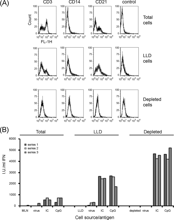
Type I interferons (alpha/beta interferons [IFN-α/β]) are the main innate cytokines that are able to induce a cellular antiviral state, thereby limiting viral replication and disease pathology. Plasmacytoid dendritic cells (pDCs) play a crucial role in the control of viral infections, especially in response to viruses that have evolved mechanisms to block the type I IFN signal transduction pathway. Using density gradient separation and cell sorting, we have highly enriched a population of bovine cells capable of producing high levels of biologically active type I IFN. These cells represented less than 0.1% of the total lymphocyte population in blood, pseudoafferent lymph, and lymph nodes. Phenotypic analysis identified these cells as bovine pDCs (CD3(-) CD14(-) CD21(-) CD11c(-) NK(-) TCRδ(-) CD4(+) MHC II(+) CD45RB(+) CD172a(+) CD32(+)). High levels of type I IFN were generated by these cells in vitro in response to Toll-like receptor 9 (TLR-9) agonist CpG and foot-and-mouth disease virus (FMDV) immune complexes. In contrast, immune complexes formed with UV-inactivated FMDV or FMDV empty capsids failed to elicit a type I IFN response. Depletion of CD4 cells in vivo resulted in levels of type I IFN in serum early during FMDV infection that were significantly lower than those for control animals. In conclusion, pDCs interacting with immune-complexed virus are the major source of type I interferon production during acute FMDV infection in cattle.
Figures


Similar articles
-
Loss of plasmacytoid dendritic cell function coincides with lymphopenia and viremia during foot-and-mouth disease virus infection.Viral Immunol. 2010 Feb;23(1):29-41. doi: 10.1089/vim.2009.0078. Viral Immunol. 2010. PMID: 20121400
-
Foot-and-mouth disease virus exhibits an altered tropism in the presence of specific immunoglobulins, enabling productive infection and killing of dendritic cells.J Virol. 2011 Mar;85(5):2212-23. doi: 10.1128/JVI.02180-10. Epub 2010 Dec 22. J Virol. 2011. PMID: 21177807 Free PMC article.
-
Infection with foot-and-mouth disease virus (FMDV) induces a natural killer (NK) cell response in cattle that is lacking following vaccination.Comp Immunol Microbiol Infect Dis. 2014 Sep;37(4):249-57. doi: 10.1016/j.cimid.2014.07.004. Epub 2014 Aug 12. Comp Immunol Microbiol Infect Dis. 2014. PMID: 25150134
-
Innate immune responses against foot-and-mouth disease virus: current understanding and future directions.Vet Immunol Immunopathol. 2009 Mar 15;128(1-3):205-10. doi: 10.1016/j.vetimm.2008.10.296. Epub 2008 Oct 17. Vet Immunol Immunopathol. 2009. PMID: 19026453 Review.
-
Molecular Mechanisms of Foot-and-Mouth Disease Virus Targeting the Host Antiviral Response.Front Cell Infect Microbiol. 2017 Jun 13;7:252. doi: 10.3389/fcimb.2017.00252. eCollection 2017. Front Cell Infect Microbiol. 2017. PMID: 28660175 Free PMC article. Review.
Cited by
-
Phenotypic, ultra-structural, and functional characterization of bovine peripheral blood dendritic cell subsets.PLoS One. 2014 Oct 8;9(10):e109273. doi: 10.1371/journal.pone.0109273. eCollection 2014. PLoS One. 2014. PMID: 25295753 Free PMC article.
-
Alphaherpesvirus-induced activation of plasmacytoid dendritic cells depends on the viral glycoprotein gD and is inhibited by non-infectious light particles.PLoS Pathog. 2021 Nov 29;17(11):e1010117. doi: 10.1371/journal.ppat.1010117. eCollection 2021 Nov. PLoS Pathog. 2021. PMID: 34843605 Free PMC article.
-
The Cell-Mediated Immune Response against Bovine alphaherpesvirus 1 (BoHV-1) Infection and Vaccination.Vaccines (Basel). 2023 Apr 2;11(4):785. doi: 10.3390/vaccines11040785. Vaccines (Basel). 2023. PMID: 37112697 Free PMC article. Review.
-
Cell mediated innate responses of cattle and swine are diverse during foot-and-mouth disease virus (FMDV) infection: a unique landscape of innate immunity.Immunol Lett. 2013 May;152(2):135-43. doi: 10.1016/j.imlet.2013.05.007. Epub 2013 May 30. Immunol Lett. 2013. PMID: 23727070 Free PMC article. Review.
-
Regulation of porcine plasmacytoid dendritic cells by cytokines.PLoS One. 2013;8(4):e60893. doi: 10.1371/journal.pone.0060893. Epub 2013 Apr 8. PLoS One. 2013. PMID: 23577175 Free PMC article.
References
-
- Abe M., Zahorchak A. F., Logar A. J., Thomson A. W. 2004. Migratory responses of murine liver myeloid, lymphoid-related, and plasmacytoid dendritic cells to CC chemokines. Am. J. Transplant. 4:180 - PubMed
-
- Alexandersen S., Zhang Z. D., Donaldson A. I. 2002. Aspects of the persistence of foot-and-mouth disease virus in animals—the carrier problem. Microbes Infect. 4:1099–1110 - PubMed
-
- Ankel H., Westra D. F., Welling-Wester S., Lebon P. 1998. Induction of interferon-alpha by glycoprotein D of herpes simplex virus: a possible role of chemokine receptors. Virology 251:317–326 - PubMed
-
- Asselin-Paturel C., et al. 2001. Mouse type I IFN-producing cells are immature APCs with plasmacytoid morphology. Nat. Immunol. 2:1144–1150 - PubMed
-
- Asselin-Paturel C., Brizard G., Pin J. J., Briere F., Trinchieri G. 2003. Mouse strain differences in plasmacytoid dendritic cell frequency and function revealed by a novel monoclonal antibody. J. Immunol. 171:6466–6477 - PubMed
Publication types
MeSH terms
Substances
Grants and funding
LinkOut - more resources
Full Text Sources
Research Materials
Miscellaneous
