. 2011 Apr;85(8):3708-16.
doi: 10.1128/JVI.02133-10. Epub 2011 Feb 9.

The IRF-3/Bax-mediated apoptotic pathway, activated by viral cytoplasmic RNA and DNA, inhibits virus replication

Affiliations

The IRF-3/Bax-mediated apoptotic pathway, activated by viral cytoplasmic RNA and DNA, inhibits virus replication

Saurabh Chattopadhyay et al. J Virol. 2011 Apr.

Abstract

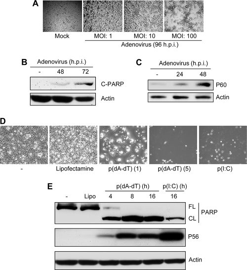
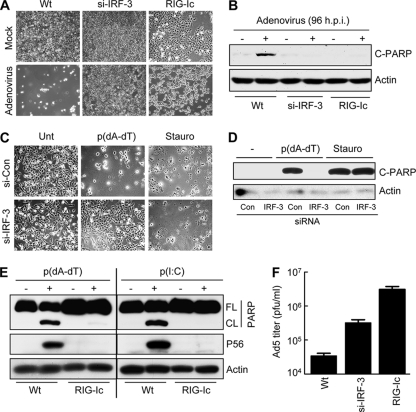
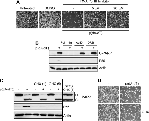
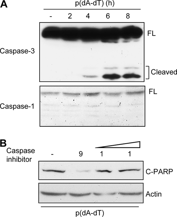
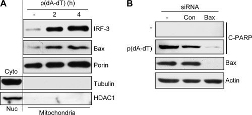
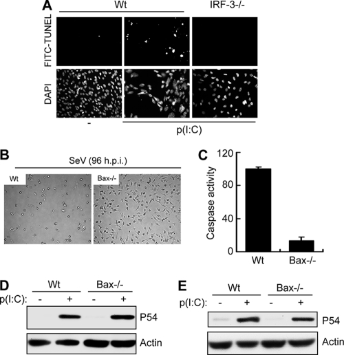
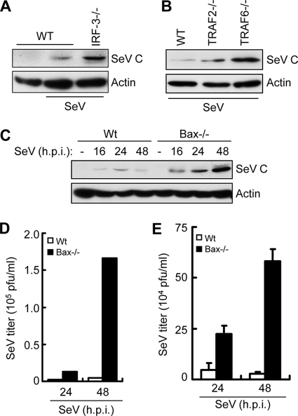
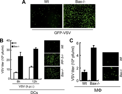
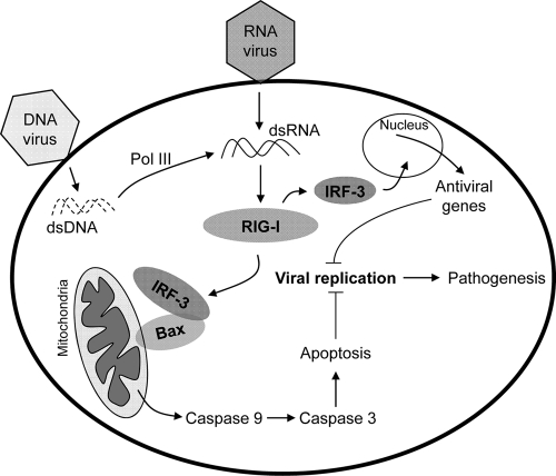
Induction of apoptosis in cells infected by Sendai virus (SeV), which triggers the cytosolic RIG-I pathway, requires the presence of interferon regulatory factor 3 (IRF-3). Independent of IRF-3's transcriptional role, a novel IRF-3 activation pathway causes its interaction with the proapoptotic protein Bax and its mitochondrial translocation to induce apoptosis. Here we report that two other RNA viruses, vesicular stomatitis virus (VSV) and encephalomyocarditis virus (EMCV), may also activate the same pathway. Moreover, cytosolic DNA, produced by adenovirus or introduced by transfection, activated the pathway in an RNA polymerase III-dependent fashion. To evaluate the contribution of this newly discovered apoptotic pathway to the host's overall antiviral response, we measured the efficiencies of replication of various viruses in vitro and viral pathogenesis in vivo, using cells and mice that are selectively deficient in components required for the apoptotic pathway of IRF-3. Our results clearly demonstrate that the IRF-3/Bax-mediated apoptotic signaling branch contributes significantly to the host's protection from viral infection and consequent pathogenesis.


Figures

Fig. 1.
Fig. 1.
Induction of apoptosis by adenovirus infection and cytosolic DNA. (A) HT1080 cells were infected with adenovirus (Ad5) at the indicated MOIs. Representative culture fields are shown for 96 h postinfection. (B) HT1080 cells were infected with adenovirus (Ad5) (at an MOI of 10) for the indicated times, when the cell lysates were analyzed for cleaved PARP (C-PARP). (C) HT1080 cells were infected with adenovirus (at an MOI of 10) for the indicated times, when the cell lysates were analyzed for induction of P60, an IRF-3-dependent gene product. (D) HT1080 cells were transfected with poly(dA-dT) [p(dA-dT)] at 1 and 5 μg/ml, as indicated, or with poly(I:C) [p(I:C)] at 2 μg/ml. Representative culture fields are shown for 24 h posttransfection. (E) HT1080 cells were transfected with poly(dA-dT) (1 μg/ml) or poly(I:C) (2 μg/ml) by use of Lipofectamine 2000 (Lipo), and at the indicated times, cell lysates were analyzed for PARP cleavage (FL, full length; CL, cleaved) and induction of P56, an IRF-3-dependent gene product, by Western blotting.
Fig. 2.
Fig. 2.
Adenovirus- and cytosolic DNA-induced apoptosis is dependent on RIG-I and IRF-3. (A) HT1080 (wt), HT1080/si-IRF-3 (IRF-3 knockdown), or HT1080/RIG-Ic (expressing a dominant-negative mutant of RIG-I) cells were either mock infected or infected with adenovirus (at an MOI of 10). Pictures of the culture fields were taken at 96 h postinfection. (B) HT1080, HT1080/si-IRF-3, or HT1080/RIG-Ic cells were infected with adenovirus (at an MOI of 10) for 96 h, when the cell lysates were analyzed for cleaved PARP (C-PARP) by Western blotting. (C) HT1080/si-IRF-3 cells or HT1080/si-con cells (cells expressing a nontargeting short hairpin RNA [Sigma] by lentiviral transduction) were either left untreated (Unt), transfected with poly(dA-dT), or treated with staurosporine (1 μM) (Stauro). Representative culture fields are shown for 18 h posttreatment. (D) HT1080/si-IRF-3 or HT1080/si-con cells were either left untreated, transfected with poly(dA-dT), or treated with staurosporine (1 μM). Cell lysates from 12 h posttreatment were analyzed for PARP cleavage by Western blotting. (E) HT1080 (wt) or HT1080/RIG-Ic cells were transfected with poly(dA-dT), and at 8 h posttransfection, the cell lysates were analyzed for PARP cleavage and induction of P56 by Western blotting. (F) HT1080 (wt), HT1080/si-IRF-3, or HT1080-RIG-Ic cells were infected with adenovirus (at an MOI of 10), and at 4 days postinfection, the cells and the culture medium were analyzed for infectious virus particles.
Fig. 3.
Fig. 3.
dsDNA-induced apoptosis is mediated by RNA polymerase III but independent of de novo protein synthesis. (A) HT1080 cells were pretreated with an inhibitor of RNA polymerase III (ML-60218) at the indicated dose or with a vehicle control (dimethyl sulfoxide [DMSO]) overnight and then transfected with poly(dA-dT). Representative culture fields are shown for 24 h posttransfection. (B) HT1080 cells were pretreated with an inhibitor of RNA polymerase III (Pol III-inh; 20 μM), actinomycin D (ActD; 0.5 μg/ml), or DRB (50 μM) and then transfected with poly(dA-dT) for 8 h, when the cell lysates were analyzed for cleaved PARP and induction of P56 by Western blotting. (C) HT1080 cells were transfected with poly(dA-dT) or poly(I:C) in the absence or presence of CHX (1 and 5 μg/ml, as indicated), and 8 h posttransfection, the cell lysates were analyzed for cleaved PARP and induction of P56 by Western blotting. (D) HT1080 cells were transfected with poly(dA-dT) in the absence or presence of CHX (1 μg/ml, as in panel C). Representative culture fields are shown for 18 h posttransfection.
Fig. 4.
Fig. 4.
Apoptosis is triggered by activation of caspase-9 and caspase-3 but not caspase-1. (A) HT1080 cells were transfected with poly(dA-dT) (1 μg/ml) for the indicated times, and the cell lysates were analyzed for the cleavage of caspase-3 and caspase-1 by Western blotting. (B) HT1080 cells were transfected with poly(dA-dT) in the absence or presence of an inhibitor of caspase-9 (10 μg/ml) or caspase-1 (10 and 25 μg/ml, as shown by increasing dose), and at 8 h posttransfection, cell lysates were analyzed for cleaved PARP by Western blotting.
Fig. 5.
Fig. 5.
dsDNA-induced apoptosis requires translocation of IRF-3 and Bax to mitochondria. (A) HT1080/si-IRF-3 cells reconstituted with wt IRF-3 were transfected with poly(dA-dT) for the indicated times, when the mitochondrial fractions were isolated and analyzed for IRF-3, Bax, porin (a mitochondrial marker protein), tubulin (a cytosolic marker protein), and HDAC1 (a nuclear marker protein) by Western blotting. (B) RNA interference-mediated knockdown of Bax was carried out by transfection of HT1080 cells with an siRNA against Bax (Dharmacon) or a nontargeting siRNA (Con) (Dharmacon), using DharmaFECT 4 reagent (Dharmacon). Forty-eight hours later, these cells were transfected with poly(dA-dT) for 8 h, and the cell extracts were analyzed for cleaved PARP by Western blotting.
Fig. 6.
Fig. 6.
Bax is required for apoptosis, but not for gene induction, in mouse primary cells. (A) wt or IRF-3−/− MEF cells were grown on coverslips and either left untreated or transfected with poly(I:C) for 16 h, after which the cells were stained with fluorescein isothiocyanate (FITC)-conjugated TUNEL reagent and analyzed microscopically. Representative culture fields are shown. Average TUNEL positivity rates for poly(I:C)-transfected cells in multiple fields were 36.1% ± 3.6% for wt cells and 1.8% ± 0.6% for IRF-3−/− cells. DAPI, 4′,6-diamidino-2-phenylindole. (B) Primary bone marrow-derived dendritic cells from wt or Bax−/− mice were infected with SeV (at an MOI of 10), and culture fields were photographed after 96 h. (C) Primary peritoneal macrophages from wt or Bax−/− mice were transfected with poly(I:C) for 16 h, and caspase activity was measured. (D) Primary bone marrow-derived dendritic cells from wt or Bax−/− mice were transfected with poly(I:C) for 8 h, and then induction of P54 was analyzed by Western blotting. (E) Primary peritoneal macrophages from wt or Bax−/− mice were transfected with poly(I:C), and induction of P54 was analyzed by Western blotting after 8 h.
Fig. 7.
Fig. 7.
Deficiency of apoptotic components leads to enhanced SeV replication. (A) wt or IRF-3−/− MEF cells were infected with SeV for 16 h, after which the cell lysates were analyzed for SeV C protein expression by Western blotting. (B) wt, TRAF2−/−, or TRAF6−/− MEF cells were infected with SeV for 16 h, after which the cell lysates were analyzed for SeV C protein expression by Western blotting. (C) wt or Bax−/− MEF cells were infected with SeV for the indicated times, after which the cell lysates were analyzed for SeV C protein expression by Western blotting. (D) wt or Bax−/− MEF cells were infected with SeV (at an MOI of 10) for the indicated times. The cells and the culture medium were analyzed together to determine the titer of infectious virus particles by plaque assay. (E) Primary bone marrow-derived dendritic cells from wt or Bax−/− mice were infected with SeV (at an MOI of 10). The cells and the culture medium were analyzed to determine the titer of infectious virus particles.
Fig. 8.
Fig. 8.
Deficiency of apoptotic components leads to enhanced VSV replication. (A) wt or Bax−/− MEF cells were infected with GFP-VSV (at an MOI of 10). Representative GFP-positive culture fields are shown for 12 h postinfection. (B) Primary bone marrow-derived dendritic cells (DCs) from wt or Bax−/− mice were infected with GFP-VSV (at an MOI of 10) for the indicated times, after which the cells and culture medium were analyzed to determine the titer of infectious virus particles by plaque assay. Representative GFP-positive culture fields for wt, IRF-3−/−, and Bax−/− DCs infected with GFP-VSV are shown for 12 h postinfection. (C) Primary peritoneal macrophages (Mφ) derived from wt or Bax−/− mice were infected with GFP-VSV (at an MOI of 10), and the cells and culture medium were analyzed at 16 h postinfection to determine the viral titer. Representative GFP-positive culture fields are shown for 16 h postinfection.
Fig. 9.
Fig. 9.
Bax deficiency leads to enhanced EMCV replication and pathogenesis in mice. (A) IRF-3−/− MEF cells expressing GFP-IRF-3 were either mock infected or infected with EMCV (at an MOI of 1), and at 8 h postinfection (Mitotracker Red was used only for the final hour of infection), cells were fixed and analyzed by confocal microscopy. The EMCV-infected cell shows clear nuclear localization of IRF-3 and strong colocalization of GFP with Mitotracker Red (yellow staining), indicating mitochondrial localization. (B) wt or Bax−/− MEF cells were infected with EMCV (at an MOI of 0.01), and the cell lysates were analyzed for caspase activity at 6 h postinfection. (C) wt or Bax−/− MEF cells were infected with EMCV (at an MOI of 0.01), and the cells and culture medium were analyzed to determine the titer of infectious virus particles at 6 h postinfection. (D) Six- to 8-week-old wt (n = 11) or Bax−/− (n = 8) mice were infected with EMCV (500 PFU) intraperitoneally and assessed for survival (P < 0.005). (E) Six- to 8-week-old wt or Bax−/− mice (five mice for each genotype) were infected with EMCV as described above, and brain tissues were collected at 48 h postinfection, homogenized in PBS, and analyzed to determine the titer of infectious virus particles by plaque assay (viral titers are presented as PFU/g of brain tissue).
Fig. 10.
Fig. 10.
IRF-3- and Bax-mediated apoptosis acts as a host antiviral response. Infection of cells by DNA or RNA viruses triggers the cellular apoptotic pathway by RIG-I signaling-mediated activation of IRF-3 and Bax to cause their translocation to mitochondria, which in turn activates the apoptotic pathway. Cytosolic DNA, generated upon infection by DNA viruses or other microbes, activates RIG-I signaling via an intermediate dsRNA generated by RNA polymerase III. The IRF-3-mediated apoptotic pathway, in addition to the gene induction pathway, significantly contributes to the host antiviral response.

References

    1. Ablasser A., et al. 2009. RIG-I-dependent sensing of poly(dA:dT) through the induction of an RNA polymerase III-transcribed RNA intermediate. Nat. Immunol. 10:1065–1072 - PMC - PubMed
    1. Alexopoulou L., Holt A. C., Medzhitov R., Flavell R. A. 2001. Recognition of double-stranded RNA and activation of NF-kappaB by Toll-like receptor 3. Nature 413:732–738 - PubMed
    1. Andersen J., VanScoy S., Cheng T. F., Gomez D., Reich N. C. 2008. IRF-3-dependent and augmented target genes during viral infection. Genes Immun. 9:168–175 - PubMed
    1. Burckstummer T., et al. 2009. An orthogonal proteomic-genomic screen identifies AIM2 as a cytoplasmic DNA sensor for the inflammasome. Nat. Immunol. 10:266–272 - PubMed
    1. Chattopadhyay S., et al. 2010. Viral apoptosis is induced by IRF-3-mediated activation of Bax. EMBO J. 29:1762–1773 - PMC - PubMed

Publication types

MeSH terms