. 2011 Feb 9;31(6):2216-24.
doi: 10.1523/JNEUROSCI.2653-10.2011.

The Caenorhabditis elegans JIP3 protein UNC-16 functions as an adaptor to link kinesin-1 with cytoplasmic dynein

Affiliations

The Caenorhabditis elegans JIP3 protein UNC-16 functions as an adaptor to link kinesin-1 with cytoplasmic dynein

Makoto Arimoto et al. J Neurosci. .

Abstract

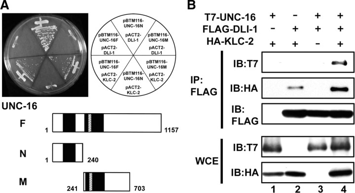
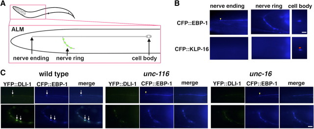
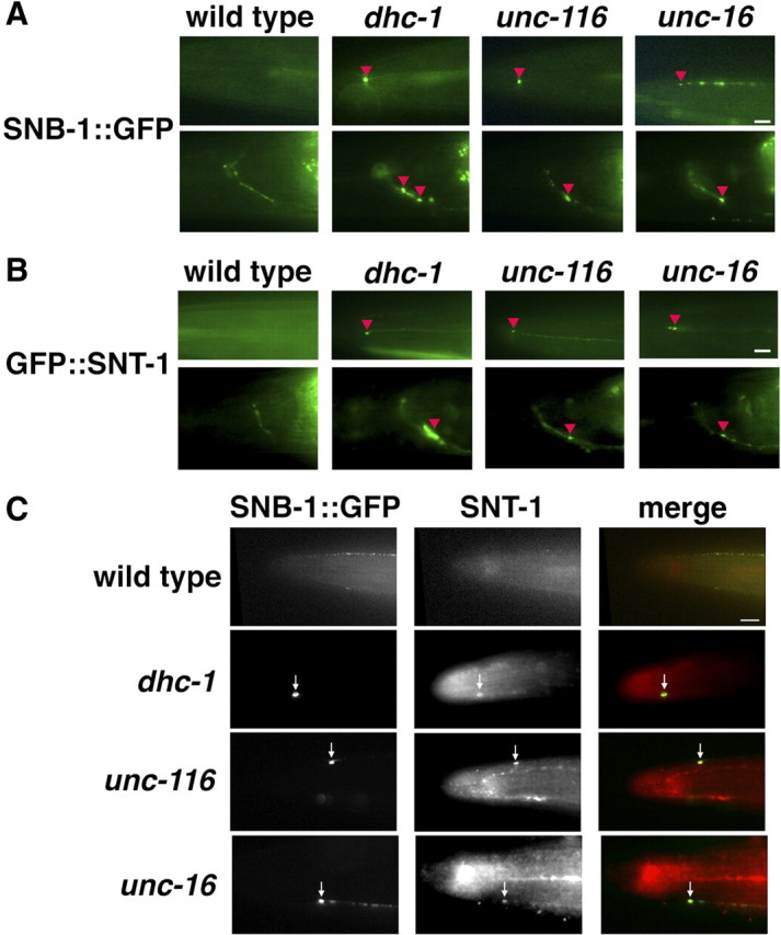
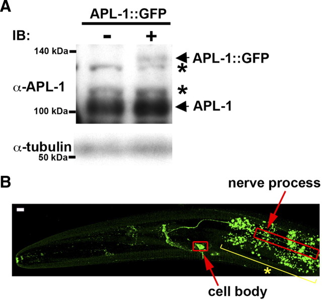
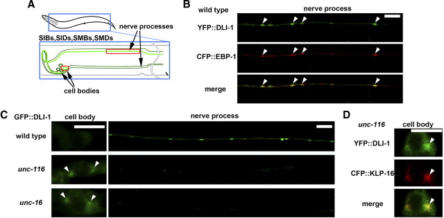
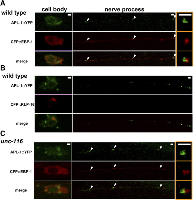
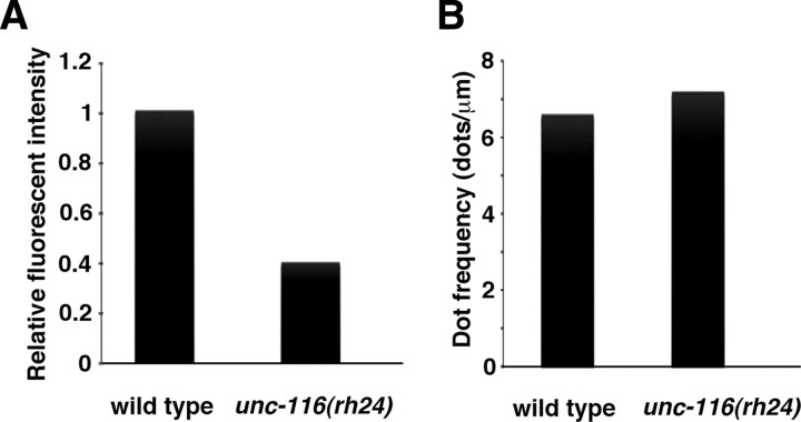
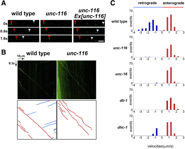
Kinesin-1 is a microtubule plus-end-directed motor that transports various cargos along the axon. Previous studies have elucidated the physical and genetic interactions between kinesin-1 and cytoplasmic dynein, a microtubule minus-end-directed motor, in neuronal cells. However, the physiological importance of kinesin-1 in the dynein-dependent retrograde transport of cargo molecules remains obscure. Here, we show that Caenorhabditis elegans kinesin-1 forms a complex with dynein via its interaction with UNC-16, which binds to the dynein light intermediate (DLI) chain. Both kinesin-1 and UNC-16 are required for localization of DLI-1 at the plus ends of nerve process microtubules. In addition, retrograde transport of APL-1 depends on kinesin-1, UNC-16, and dynein. These results suggest that kinesin-1 mediates the anterograde transport of dynein using UNC-16 as a scaffold and that dynein in turn mediates the retrograde transport of cargo molecules in vivo. Thus, UNC-16 functions as an adaptor for kinesin-1-mediated transport of dynein.


Figures

Figure 1.
Figure 1.
Interaction among UNC-16, DLI-1, and KLC-2. A, Interactions by yeast two-hybrid assay. Yeasts carrying the indicated plasmids were grown on a selective plate lacking histidine for 5 d. UNC-16 constructs used for this assay are shown below. The black and hatched boxes represent coiled-coil and leucine zipper domains, respectively. B, Interactions by coimmunoprecipitation in mammalian cells. COS-7 cells were transfected with control vector, T7-UNC-16, FLAG-DLI-1, and/or HA-KLC-2 as indicated. Cell lysates were immunoprecipitated (IP) with anti-FLAG antibodies. Immunoprecipitates were immunoblotted (IB) with anti-T7, anti-HA, and anti-FLAG antibodies. Whole-cell extracts (WCE) were immunoblotted with anti-T7 and anti-HA antibodies.
Figure 2.
Figure 2.
Localization of DLI-1 in ALM touch neurons. A, Schematic image of ALM touch neuron. Presynapses are indicated by green. B, Localization of CFP::EBP-1 and CFP::KLP-16 in ALM neurons. The yellow arrowhead indicates the location of CFP::EBP-1 at the nerve ending of ALM neuron. The red arrowhead indicates the accumulation of CFP::KLP-16 in the cell body. C, Localization of YFP::DLI-1 and CFP::EBP-1 in ALM neurons in wild-type, unc-116(e2281), and unc-16(e109) mutant worms. The top and bottom panels show the processes and nerve rings of ALM neurons, respectively. The arrows indicate the positions of colocalized YFP::DLI-1 and CFP::EBP-1 at the ends of ALM neuron of processes and nerve rings. The yellow arrowheads indicate the locations at the nerve endings. The merged images are shown in right panels. Scale bars, 10 μm.
Figure 3.
Figure 3.
Localization of SV proteins in ALM touch neurons. A, B, Localization of SNB-1::GFP (A) and GFP::SNT-1 (B) in the head region of ALM neurons. The top and bottom panels show the processes and nerve rings of ALM neurons, respectively. The red arrowheads indicate the positions of accumulated SNB-1::GFP (A) and GFP::SNT-1 (B) proteins at the ends of ALM neuronal processes and near nerve ring synapses. C, Colocalization of SNB-1::GFP and endogenous SNT-1 in the head region of ALM neurons. The arrows indicate the positions of accumulated SNB-1::GFP and SNT-1 proteins at the tip of ALM neurons. The merged images are shown in the bottom panels. Scale bars, 10 μm.
Figure 4.
Figure 4.
Expression and localization of APL-1::GFP. A, Immunoblotting of APL-1 proteins. Whole extracts were prepared from worms at mixed stages. The top panels show immunoblotting with anti-APL-1 antibody. Positions of APL-1::GFP and endogenous APL-1 are indicated by arrows. The asterisks indicate nonspecific bands. The bottom panels show immunoblotting with anti-tubulin antibody as a loading control. B, Image of fluorescent APL-1::GFP protein in the heads of L4 larva. The red open boxes indicate the cell body and nerve processes of the sublateral motor neurons. The yellow line with an asterisk indicates gut fluorescent granules. Images are from a confocal Z series projected onto a single plane. Scale bar, 5 μm.
Figure 5.
Figure 5.
Localization of DLI-1 in sublateral motor neurons. A, Schematic image of sublateral motor neurons. SIB, SID, SMB, and SMD neurons are indicated by green lines. B, Localization of YFP::DLI-1 and CFP::EBP-1 in the nerve processes of sublateral neurons. The merged image is also indicated. The arrowheads indicate the merged sites. Scale bar, 5 μm. C, Localization of GFP::DLI-1 in the sublateral neurons. The left and right panels show cell bodies and nerve processes, respectively. The arrowheads indicate the accumulated positions of GFP::DLI-1 in cell bodies. Scale bar, 5 μm. D, Localization of YFP::DLI-1 and CFP::KLP-16 in the cell bodies of sublateral neurons in unc-116(e2281) mutants. The arrowheads indicate the merged sites.
Figure 6.
Figure 6.
Localization of APL-1 in sublateral motor neurons. A, C, Localization of APL-1::YFP and CFP::EBP-1 in the sublateral neurons of wild-type (A) and unc-116(e2281) (C) worms. The merged images in each animal are also indicated. The arrowheads indicate the merged sites. The right panels show magnified images of merged sites in nerve processes. Anterior is to the left in panels. Scale bars, 1 μm. B, Localization of APL-1::YFP and CFP::KLP-16 in sublateral neurons of wild-type worms.
Figure 7.
Figure 7.
Effects of the unc-116 mutation on APL-1. A, Relative fluorescent intensities of APL-1::GFP per micrometer along the lateral processes in wild-type and unc-116(rh24) mutant worms. B, Numbers of accumulated spots of APL-1::GFP per micrometer along the lateral processes in wild-type and unc-116(rh24) mutant worms.
Figure 8.
Figure 8.
Movement of APL-1 in sublateral motor neurons. A, Time-lapse images of APL-1::GFP fluorescence along the sublateral neurons of worms. Each photograph was taken at 0.8 s intervals. The red and pale blue arrowheads indicate APL-1::GFP vesicles moving toward neurite endings (anterograde) and cell bodies (retrograde), respectively. Scale bar, 2.5 μm. B, Kymographs for APL-1::GFP movements along the lateral processes. The horizontal arrow represents 10 μm. The vertical arrow represents 8.3 s. Schematic diagrams are shown below. The red and blue lines represent anterograde and retrograde movements, respectively. C, Velocity profiles of moving vesicles containing APL-1::GFP in wild-type, unc-116(e2281), unc-16(e109), dli-1(ku266), and dhc-1(or195ts) worms. Moving structures were tracked, and the velocities were calculated as described in Materials and Methods.

References

    1. Araki Y, Kawano T, Taru H, Saito Y, Wada S, Miyamoto K, Kobayashi H, Ishikawa HO, Ohsugi Y, Yamamoto T, Matsuno K, Kinjo M, Suzuki T. The novel cargo Alcadein induces vesicle association of kinesin-1 motor components and activates axonal transport. EMBO J. 2007;26:1475–1486. - PMC - PubMed
    1. Bowman AB, Kamal A, Ritchings BW, Philp AV, McGrail M, Gindhart JG, Goldstein LS. Kinesin-dependent axonal transport is mediated by the sunday driver (SYD) protein. Cell. 2000;103:583–594. - PubMed
    1. Byrd DT, Kawasaki M, Walcoff M, Hisamoto N, Matsumoto K, Jin Y. UNC-16, a JNK-signaling scaffold protein, regulates vesicle transport in C. elegans. Neuron. 2001;32:787–800. - PubMed
    1. Cavalli V, Kujala P, Klumperman J, Goldstein LS. Sunday Driver links axonal transport to damage signaling. J Cell Biol. 2005;168:775–787. - PMC - PubMed
    1. Caviston JP, Holzbaur EL. Microtubule motors at the intersection of trafficking and transport. Trends Cell Biol. 2006;16:530–537. - PubMed

Publication types

MeSH terms

LinkOut - more resources