. 2011 Feb 9;31(6):2225-37.
doi: 10.1523/JNEUROSCI.3730-10.2011.

LRRK2 controls synaptic vesicle storage and mobilization within the recycling pool

Affiliations

LRRK2 controls synaptic vesicle storage and mobilization within the recycling pool

Giovanni Piccoli et al. J Neurosci. .

Abstract

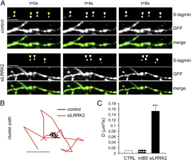
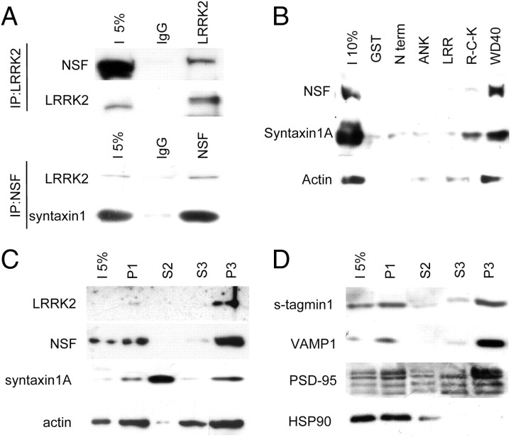
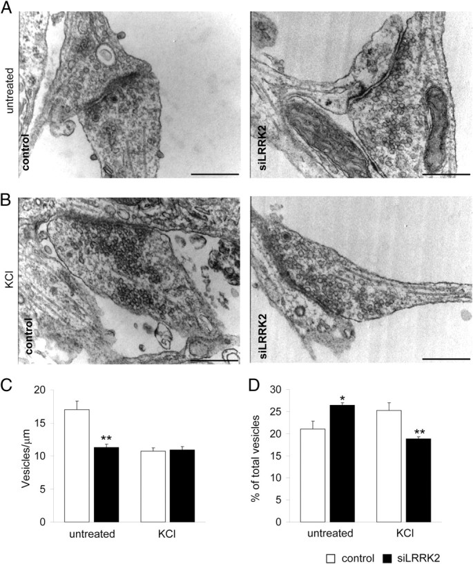
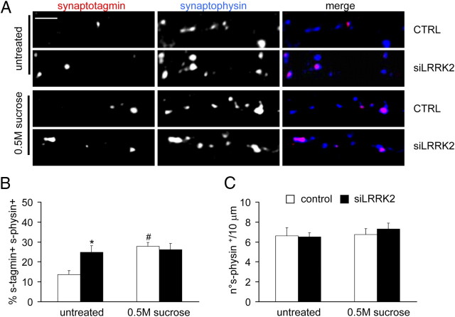
Mutations in leucine-rich repeat kinase 2 (LRRK2) are the single most common cause of inherited Parkinson's disease. Little is known about its involvement in the pathogenesis of Parkinson's disease mainly because of the lack of knowledge about the physiological role of LRRK2. To determine the function of LRRK2, we studied the impact of short hairpin RNA-mediated silencing of LRRK2 expression in cortical neurons. Paired recording indicated that LRRK2 silencing affects evoked postsynaptic currents. Furthermore, LRRK2 silencing induces at the presynaptic site a redistribution of vesicles within the bouton, altered recycling dynamics, and increased vesicle kinetics. Accordingly, LRRK2 protein is present in the synaptosomal compartment of cortical neurons in which it interacts with several proteins involved in vesicular recycling. Our results suggest that LRRK2 modulates synaptic vesicle trafficking and distribution in neurons and in consequence participates in regulating the dynamics between vesicle pools inside the presynaptic bouton.


Figures

Figure 1.
Figure 1.
LRRK2 expression increases during in vitro neuron development. A, Neurons were cultured and solubilized at the indicated DIV. Equal amounts of protein were loaded on SDS-PAGE gel and stained with the indicated antibodies. LRRK2 expression increases along in vitro synapse maturation, as demonstrated by the parallel increment of PSD-95 and synaptophysin amount. Two representative independent experiments are shown. B, Mean ± SE protein levels expressed as optical density (in arbitrary units) normalized against total protein amount. n = 5, *p < 0.05 versus DIV4. C, Cortical neurons were left untreated (mock) or infected at DIV10 with control (LVTH) or two LRRK2-silencing (miB3 and miB4) viruses, solubilized at DIV18 and analyzed for the expression of the indicated protein. D, LRRK2 protein levels expressed as percentage over control; mean ± SE; **p < 0.01, n = 5, ANOVA followed by Tukey's post hoc test. s-physin, Synaptophysin.
Figure 2.
Figure 2.
LRRK2 silencing modifies synaptic transmission. Paired recordings were performed between a control or siLRRK2-transfected neuron (presynaptic) and an adjacent nontransfected neuron. A, Representative EPSC traces from control and siLRRK2 pairs after a single depolarizing stimulus (100 mV, 1 ms). B, Representative traces from control and siLRRK2 pairs after a paired-pulse stimulation protocol (100 mV, 1 ms, 50 ms interpulse interval). C, LRRK2 silencing significantly increased evoked EPSC amplitude after a single-pulse stimulation but enhanced paired-pulse depression. D, LRRK2 silencing increased the latency of the second EPSC. Data are expressed as mean ± SE. **p < 0.01, ***p < 0.001, Student's t test; n = 5.
Figure 3.
Figure 3.
LRRK2 silencing modifies synaptic vesicle recycling. The synaptotagmin uptake assay was performed on cortical neurons at DIV18, infected at DIV10 with control or siLRRK2 virus. A, Synaptotagmin (s-tagmin)-positive spots colocalized with synaptophysin (s-physin) clusters along neuron processes. B, Neurons were left untreated or treated with KCl (50 mm, 5 min) or TTX (30 min before labeling, 2 μm) and then assayed for exo-endocytosis. Scale bar, 5 μm. CTRL, Control. C, The percentage of synaptotagmin and synaptophysin-positive clusters within the totality of synaptophysin-positive clusters reflects the pool of recycling synapses. LRRK2-silenced neurons showed an increased ratio of recycling synapses in basal condition but a reduced activity after KCl stimulation. Instead, LRRK2 knockdown did not modify the number of recycling synapses during TTX repression. NT, Nontreated. D, Active zone number, monitored as synaptophysin-positive dots along neuronal processes, was not modified by LRRK2 silencing. Data are expressed as mean ± SE. *p < 0.05, **p < 0.01, same treatment, Student's t test, #p < 0.01 same infection, ANOVA, Tukey's post hoc test, n = 25.
Figure 4.
Figure 4.
LRRK2 silencing affects Ca2+ sensitivity. A, The ratio of recycling synapses was assayed in neurons in presence of increasing external Ca2+ concentration. Scale bar, 5 μm. CTRL, Control. B, The ratio of recycling synapses rose to a higher extent in siLRRK2 neurons in response to increasing Ca2+ concentration. C, Active zone number was not modified by LRRK2 silencing or Ca2+ concentration. Data are expressed as mean ± SE. *p < 0.05 versus control, Student's t test, n = 14.
Figure 5.
Figure 5.
LRRK2 silencing increases vesicle motility. A, Control and siLRRK2-infected neurons were exposed to anti-synaptotagmin antibody coupled to a fluorochrome. Synaptotagmin (s-tagmin)-positive clusters within GFP-positive processes were tracked in basal condition under laser confocal microscopy. Clusters in siLRRK2 neurons showed an increased motility. Yellow arrows indicate the position of cluster at t = 0 s. White arrows report instead the position of the clusters relative to the previous time point. Scale bar, 5 μm. B, The diagram reports representative path of single cluster from control or siLRRK2 neuron. Scale bar, 1 μm. C, Quantification of diffusion coefficient D, where MSD(Δt) = 4Dt; mean ± SE; ***p < 0.001, Student's t test, n = 9. CTRL, Control.
Figure 6.
Figure 6.
LRRK2 interacts with presynaptic protein. A, Immunoprecipitation (IP) of endogenous LRRK2 (top) and NSF (bottom) from adult brain lysate shows that LRRK2 and NSF interact physiologically. NSF antibody precipitates equally syntaxin1 and LRRK2. B, LRRK2 interacts with presynaptic proteins. GST–LRRK2 domains have been used to pull down putative LRRK2 interactors from adult mouse brain. LRRK2 WD40 domain precipitates presynaptic proteins as NSF and syntaxin 1A and actin. C, D, LRRK2 is present in a subcellular fraction (P3) in which its putative interactors and presynaptic and postsynaptic markers are found. s-tagmin, Synaptotagmin.
Figure 7.
Figure 7.
LRRK2 silencing perturbs vesicle distribution inside the presynaptic bouton. Neurons were infected at DIV10 as described and processed for electron microscopy at DIV18. A, B, High-magnification (40,000×) images from control or LRRK2-silenced neurons kept in basal condition (A) or depolarized with 50 mm KCl 5 min (B). C, The number of docked vesicle (vesicle/μm presynaptic membrane) was reduced in siLRRK2 neurons kept in basal condition. D, LRRK2 silencing affected the fraction of total vesicle within the 75–150 nm range from the presynaptic membrane in untreated and depolarized neurons. Data are expressed as mean ± SE. *p < 0.05, **p < 0.01, Student's t test; n = 35. Scale bar, 500 nm.
Figure 8.
Figure 8.
LRRK2 silencing does not affect RRP size. A, The ratio of recycling synapses was assayed in neurons in presence of a hypertonic sucrose solution (0.5 m sucrose, 45 s). Scale bar, 5 μm. B, The ratio of recycling synapses was not significantly different between control and siLRRK2 neurons after sucrose application. C, Active zone number was not modified by LRRK2 silencing or sucrose application. Data are expressed as mean ± SE. *p < 0.05 versus control, #p < 0.05 versus untreated, ANOVA, Tukey's post hoc test; n = 15. s-physin, Synaptophysin; s-tagmin, synaptotagmin.

Comment in

References

    1. Aasly JO, Toft M, Fernandez-Mata I, Kachergus J, Hulihan M, White LR, Farrer M. Clinical features of LRRK2-associated Parkinson's disease in central Norway. Ann Neurol. 2005;57:762–765. - PubMed
    1. Andres-Mateos E, Mejias R, Sasaki M, Li X, Lin BM, Biskup S, Zhang L, Banerjee R, Thomas B, Yang L, Liu G, Beal MF, Huso DL, Dawson TM, Dawson VL. Unexpected lack of hypersensitivity in LRRK2 knock-out mice to MPTP (1-methyl-4-phenyl-1,2,3,6-tetrahydropyridine) J Neurosci. 2009;29:15846–15850. - PMC - PubMed
    1. Augustin I, Rosenmund C, Südhof TC, Brose N. Munc13-1 is essential for fusion competence of glutamatergic synaptic vesicles. Nature. 1999;400:457–461. - PubMed
    1. Bacci A, Coco S, Pravettoni E, Schenk U, Armano S, Frassoni C, Verderio C, De Camilli P, Matteoli M. Chronic blockade of glutamate receptors enhances presynaptic release and downregulates the interaction between synaptophysin-synaptobrevin-vesicle-associated membrane protein 2. J Neurosci. 2001;21:6588–6596. - PMC - PubMed
    1. Bardoni R, Torsney C, Tong CK, Prandini M, MacDermott AB. Presynaptic NMDA receptors modulate glutamate release from primary sensory neurons in rat spinal cord dorsal horn. J Neurosci. 2004;24:2774–2781. - PMC - PubMed

Publication types

MeSH terms