. 2011 Feb 9;31(6):2248-57.
doi: 10.1523/JNEUROSCI.3153-09.2011.

Membrane penetration by synaptotagmin is required for coupling calcium binding to vesicle fusion in vivo

Affiliations

Membrane penetration by synaptotagmin is required for coupling calcium binding to vesicle fusion in vivo

Brie E Paddock et al. J Neurosci. .

Abstract

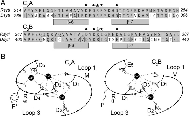
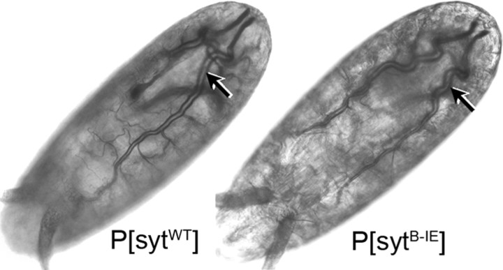
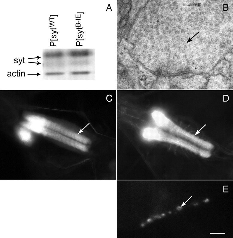
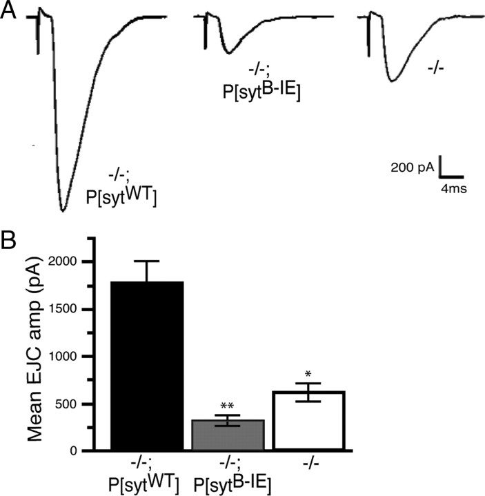
The vesicle protein synaptotagmin I is the Ca(2+) sensor that triggers fast, synchronous release of neurotransmitter. Specifically, Ca(2+) binding by the C(2)B domain of synaptotagmin is required at intact synapses, yet the mechanism whereby Ca(2+) binding results in vesicle fusion remains controversial. Ca(2+)-dependent interactions between synaptotagmin and SNARE (soluble N-ethylmaleimide-sensitive fusion protein attachment receptor) complexes and/or anionic membranes are possible effector interactions. However, no effector-interaction mutations to date impact synaptic transmission as severely as mutation of the C(2)B Ca(2+)-binding motif, suggesting that these interactions are facilitatory rather than essential. Here we use Drosophila to show the functional role of a highly conserved, hydrophobic residue located at the tip of each of the two Ca(2+)-binding pockets of synaptotagmin. Mutation of this residue in the C(2)A domain (F286) resulted in a ∼50% decrease in evoked transmitter release at an intact synapse, again indicative of a facilitatory role. Mutation of this hydrophobic residue in the C(2)B domain (I420), on the other hand, blocked all locomotion, was embryonic lethal even in syt I heterozygotes, and resulted in less evoked transmitter release than that in syt(null) mutants, which is more severe than the phenotype of C(2)B Ca(2+)-binding mutants. Thus, mutation of a single, C(2)B hydrophobic residue required for Ca(2+)-dependent penetration of anionic membranes results in the most severe disruption of synaptotagmin function in vivo to date. Our results provide direct support for the hypothesis that plasma membrane penetration, specifically by the C(2)B domain of synaptotagmin, is the critical effector interaction for coupling Ca(2+) binding with vesicle fusion.


Figures

Figure 1.
Figure 1.
Both the C2A and C2B domains of synaptotagmin have conserved hydrophobic residues at the tip of the Ca2+-binding pocket. A, Alignment of synaptotagmin I from rat and Drosophila. Bars indicate β-sheets, asterisks indicate the conserved hydrophobic residues mutated in this study, dots indicate Ca2+-binding residues, and open circles with a plus indicate conserved basic residues. Within the alignment, conserved residues are shown in gray and identical residues are boxed. B, Schematic representation of loops 1 and 3 in Drosophila that form the Ca2+-binding pockets of each C2 domain. D1–D5 and E5 indicate Ca2+-binding residues. F*, M, I*, and V indicate hydrophobic residues at the tip of the Ca2+-binding pockets.
Figure 2.
Figure 2.
Evoked release is impaired in C2A hydrophobic mutants. A, Representative voltage traces recorded from larval muscle fiber 6. Each top trace shows the mean of 30 consecutive evoked responses from the same muscle fiber. The bottom traces show 3 s of continuous spontaneous recordings. Line 3 shown for P[sytA-FY]. B, Compared with P[sytWT], the mean EJP amplitude of P[sytA-FY], lines 3 and 5, and P[sytA-FE] were significantly decreased (*p < 0.005; P[sytWT], n = 24; P[sytA-FY] line 3, n = 20; P[sytA-FY] line 5, n = 14; P[sytA-FE], n = 10). No difference was found between P[sytA-FY] and P[sytA-FE] (p > 0.9). C, Compared with P[sytWT], the mean mEJP amplitude of P[sytA-FY] (line 3 shown) and P[sytA-FE] showed no significant change (p > 0.05).
Figure 3.
Figure 3.
The C2A hydrophobic mutations do not disrupt synaptotagmin expression or targeting. A, Representative Western blots from P[sytA-FY] line 3, line 5, and P[sytA-FE] and their P[sytWT] controls. B, P[sytA-FY] line 5 (n = 17) expressed less transgenic protein than P[sytWT] (n = 28) and the other two C2A mutants (P[sytA-FY] line 3, n = 7; P[sytA-FE], n = 11; p < 0.001). C, Transgenic synaptotagmin was localized to synaptic sites in all lines (line 5 shown for P[sytA-FY]). Scale bar, 20 μm.
Figure 4.
Figure 4.
sytB-IE mutant embryos complete embryogenesis and exhibit gross morphology indistinguishable from wild-type embryos. Like controls, P[sytB-IE] mutants were able to develop through stage 17, as demonstrated by the development of trachea (arrows).
Figure 5.
Figure 5.
The C2B hydrophobic mutation does not disrupt synaptic vesicle localization, synaptotagmin expression, or synaptotagmin targeting. A, A representative Western blot of embryos shows similar transgene expression levels for P[sytWT] and P[sytB-IE]. Blot was probed with an anti-actin antibody to confirm equal loading. B, A representative nerve terminal within the neuropil of a P[sytB-IE] mutant ventral nerve cord exhibits abundant small clear vesicles (black arrow). C, Anti-CSP labeling was localized to the neuropil of the CNS in P[sytB-IE] mutants demonstrating that synaptic vesicles are appropriately localized to synaptic regions. The white arrow indicates the neuropil of the ventral nerve cord; brain ganglia are out of the plane of focus. The mutant synaptotagmin was also correctly localized to the neuropil (D, white arrow) in the CNS, as well as to neuromuscular junctions (E, white arrow) in the peripheral nervous system. P[sytB-IE] line 6 is shown. Scale bar: B, 180 nm; C, D, 40 μm; E, 5 μm.
Figure 6.
Figure 6.
The C2B hydrophobic mutation inhibits evoked transmitter release more severely than the sytnull mutation. A, Representative current traces recorded from embryonic muscle fiber 6. B, The mean EJC amplitude in transgenic control embryos was 1782 ± 224 pA (−/−; P[sytWT], n = 8), in P[sytB-IE] mutant embryos was 312.2 ± 62.6 pA (−/−; P[sytB-IE], n = 8), and in sytnull mutants was 625.2 ± 89.0 pA (−/−, n = 11). Evoked release in P[sytB-IE] was significantly less than in sytnull mutants (* vs **, p < 0.02).
Figure 7.
Figure 7.
The sytB-IE mutation abolishes Ca2+-dependent, C2B interactions with membranes. A, C2AB or C2B domains with the sytB-IE mutation are correctly folded. The CD spectra of the mutant domains (C2AB-IE and C2B-IE) did not show significant changes compared with wild type (C2AB-WT and C2B-WT, respectively). B, The sytB-IE mutation abolishes the ability of the C2B domain to bind negatively charged liposomes. Left, A representative co-sedimentation assay; right, graph of three experiments. In the absence of Ca2+, C2AB and C2B domains do not bind to liposomes containing 15% PS and thus remain in solution (left, −, WT and B-IE). In the presence of 1 mm Ca2+, increasing concentrations of liposomes bind increasing amounts of control C2AB or C2B domains (C2AB-WT and C2B-WT). The sytB-IE mutation impairs this interaction with C2AB (C2AB-IE) but abolishes the interaction with the isolated C2B domain (C2B-IE).
Figure 8.
Figure 8.
Direct, Ca2+-dependent C2B interactions with t-SNAREs are intact in the sytB-IE mutation. A, The sytB-IE mutation does not inhibit Ca2+-dependent interactions with solubilized t-SNAREs. Left, A representative GST–syt pull-down assay; right, histogram of three experiments. The Ca2+-dependent increase in t-SNARE interactions (black bars relative to white bars) is not disrupted by the sytB-IE mutation. Ca2+-independent t-SNARE interactions are decreased (white bars, C2AB vs C2AB I420E and C2B vs C2B I420E). B, The sytB-IE mutation abolishes Ca2+-dependent C2B interactions with membrane-embedded t-SNAREs. Left, A representative co-floatation assay; right, histogram of four experiments. The sytB-IE mutation does not alter the amount of C2B domain that binds to PS-free, t-SNARE vesicles (t-SNARE) in the absence of Ca2+ (−, left; EGTA, right). However, the sytB-IE mutation abolishes the increase in t-SNARE vesicles binding induced by 1 mm Ca2+ (+, left; Ca2+, right; p < 0.05). C, The sytB-IE mutation impairs C2AB interactions with membrane-embedded t-SNAREs. Again, the sytB-IE mutation does not alter the amount of C2AB domain that binds to PS-free, t-SNARE vesicles in the absence of Ca2+ (EGTA, white bars). In the presence of 1 mm Ca2+ (Ca2+, black bars), the t-SNARE vesicles bind 70% of wild-type levels of C2AB domain when the sytB-IE mutation is present (p < 0.001; n = 6).
Figure 9.
Figure 9.
Model of the role of Ca2+-dependent penetration of the anionic presynaptic membrane by synaptotagmin. The crystal structure of the core complex [Protein Data Bank (PDB) file 1SFC, containing syntaxin (red), SNAP-25 (synapse associated protein of 25 kD) (green), and VAMP (vesicle-associated membrane protein)/synaptobrevin (blue)], the nuclear magnetic resonance structures of the C2A (PDB file 1BYN) and C2B (PDB file 1K5W) domains of synaptotagmin (yellow), and Ca2+ (pink “+”) were rendered using PyMOL Molecular Graphics System (DeLano Scientific). The synaptic vesicle (SV), the presynaptic membrane (PM), the transmembrane domains, and the link between C2A and C2B were added in Adobe Photoshop. A, Docked synaptic vesicle with two synaptotagmin/SNARE complexes shown. B, syt/SNARE complex viewed end on from the site of synaptic vesicle/presynaptic membrane apposition (SNARE complex transmembrane domains in front of plane of section). Ca2+-independent priming between the C2B polylysine motif (yellow, space-filled residues) and SNAP-25 [green, space-filled residues (Zhang et al., 2002; Rickman et al., 2004; Loewen et al., 2006b)] holds the C2B Ca2+-binding site immediately adjacent to the SNARE complex and the C2A Ca2+-binding site nearby. The negative charge of the Ca2+-binding pockets (cluster of − symbols) prevents interactions between the tips of the C2 domains and the presynaptic membrane attributable to electrostatic repulsion. C, Ca2+ binding neutralizes the negative charge of the pockets resulting in a strong attraction of the negatively charged, phospholipid head groups of the presynaptic membrane by the bound Ca2+ (pink spheres +) and the basic residues at the tips of Ca2+-binding pockets (blue, space-filled residues +). Insertion of the hydrophobic residues at the tips of the C2 domains (gray, space-filled residues) into the core of the presynaptic membrane and then triggers fusion by promoting a local Ca2+-dependent buckling of the plasma membrane (Martens et al., 2007; Hui et al., 2009). D, syt/SNARE complexes viewed from the presynaptic membrane. Multiple syt:SNARE complexes (colored as in A–C) mediate fusion (Jahn and Scheller, 2006) of a single synaptic vesicle (gray ring, not to scale). Ca2+-dependent membrane penetration by synaptotagmin (large gray ovals, arrows) pulls the presynaptic membrane toward the vesicle in the vicinity of the transmembrane regions of the SNARE complexes (small gray circles, arrowheads).

References

    1. Bai J, Wang P, Chapman ER. C2A activates a cryptic Ca2+-triggered membrane penetration activity within the C2B domain of synaptotagmin I. Proc Natl Acad Sci U S A. 2002;99:1665–1670. - PMC - PubMed
    1. Brand AH, Perrimon N. Targeted gene expression as a means of altering cell fates and generating dominant phenotypes. Development. 1993;118:401–415. - PubMed
    1. Broadie K, Bellen HJ, DiAntonio A, Littleton JT, Schwarz TL. Absence of synaptotagmin disrupts excitation-secretion coupling during synaptic transmission. Proc Natl Acad Sci U S A. 1994;91:10727–10731. - PMC - PubMed
    1. Chapman ER, Hanson PI, An S, Jahn R. Ca2+ regulates the interaction between synaptotagmin and syntaxin 1. J Biol Chem. 1995;270:23667–23671. - PubMed
    1. Chen K, Featherstone DE, Broadie K. Electrophysiological recording in the Drosophila embryo. J Vis Exp. 2009;pii:1348. - PMC - PubMed

Publication types

MeSH terms

LinkOut - more resources