. 2011 Feb 9;3(69):69ra12.
doi: 10.1126/scitranslmed.3001571.

5-Lipoxygenase metabolite 4-HDHA is a mediator of the antiangiogenic effect of ω-3 polyunsaturated fatty acids

Affiliations

5-Lipoxygenase metabolite 4-HDHA is a mediator of the antiangiogenic effect of ω-3 polyunsaturated fatty acids

Przemyslaw Sapieha et al. Sci Transl Med. .

Abstract

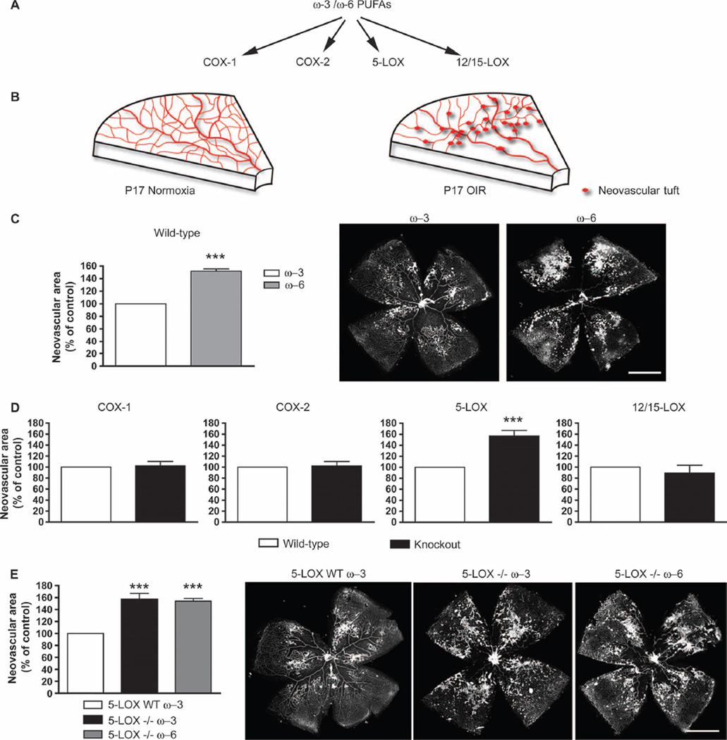
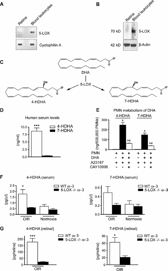
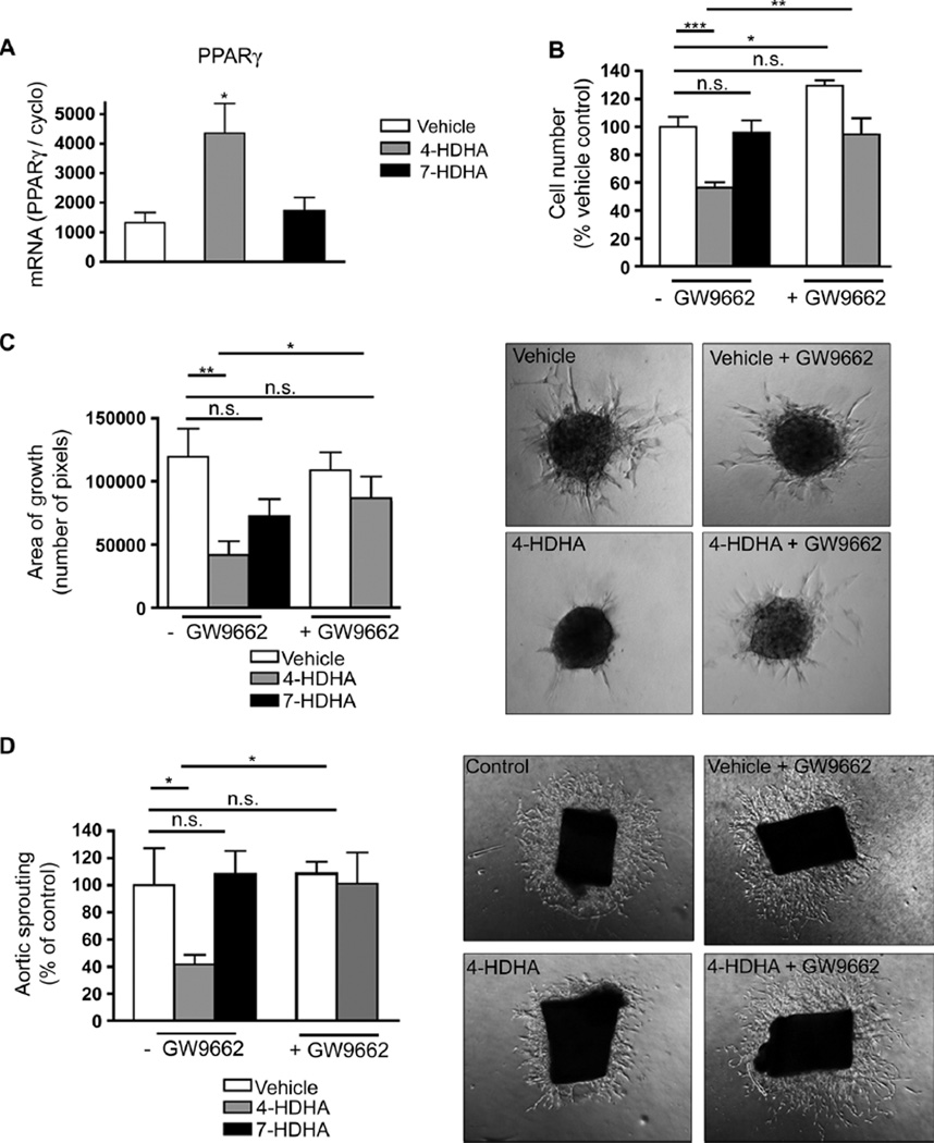
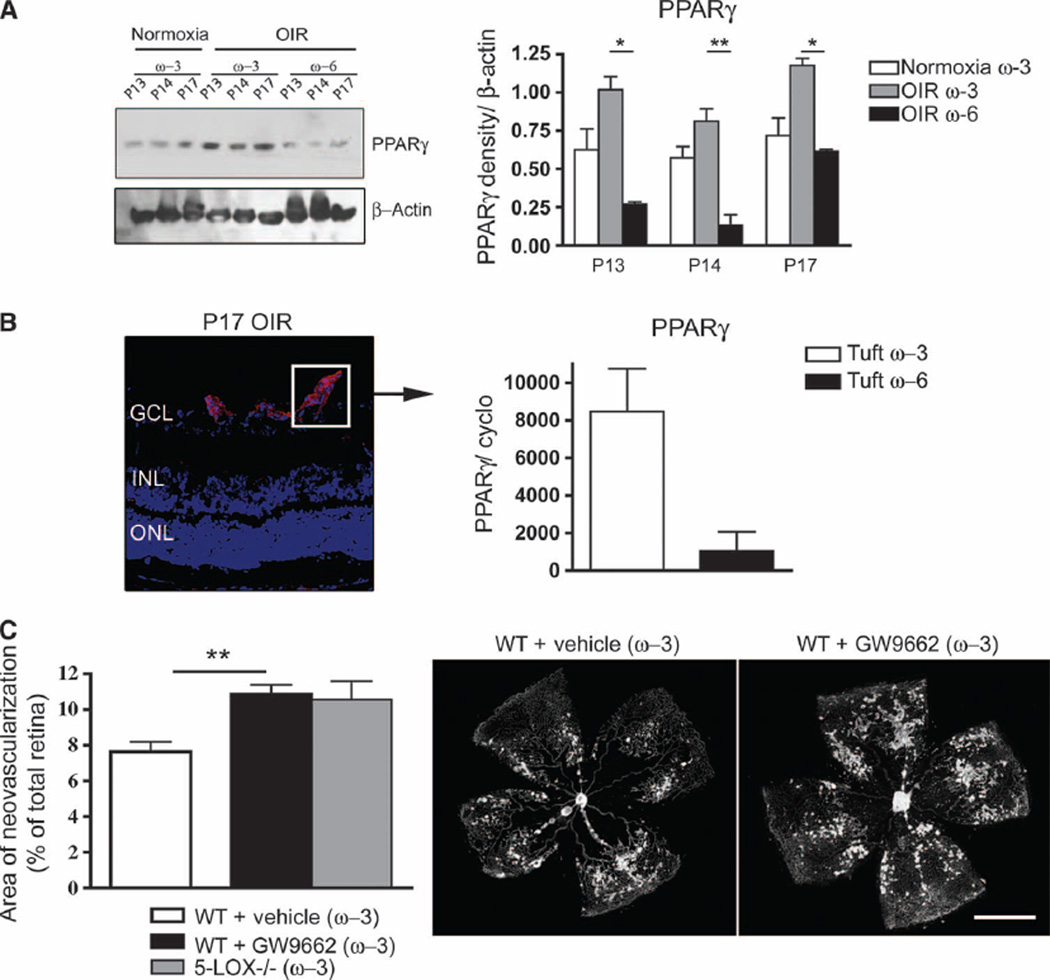
Lipid signaling is dysregulated in many diseases with vascular pathology, including cancer, diabetic retinopathy, retinopathy of prematurity, and age-related macular degeneration. We have previously demonstrated that diets enriched in ω-3 polyunsaturated fatty acids (PUFAs) effectively reduce pathological retinal neovascularization in a mouse model of oxygen-induced retinopathy, in part through metabolic products that suppress microglial-derived tumor necrosis factor-α. To better understand the protective effects of ω-3 PUFAs, we examined the relative importance of major lipid metabolic pathways and their products in contributing to this effect. ω-3 PUFA diets were fed to four lines of mice deficient in each key lipid-processing enzyme (cyclooxygenase 1 or 2, or lipoxygenase 5 or 12/15), retinopathy was induced by oxygen exposure; only loss of 5-lipoxygenase (5-LOX) abrogated the protection against retinopathy of dietary ω-3 PUFAs. This protective effect was due to 5-LOX oxidation of the ω-3 PUFA lipid docosahexaenoic acid to 4-hydroxy-docosahexaenoic acid (4-HDHA). 4-HDHA directly inhibited endothelial cell proliferation and sprouting angiogenesis via peroxisome proliferator-activated receptor γ (PPARγ), independent of 4-HDHA's anti-inflammatory effects. Our study suggests that ω-3 PUFAs may be profitably used as an alternative or supplement to current anti-vascular endothelial growth factor (VEGF) treatment for proliferative retinopathy and points to the therapeutic potential of ω-3 PUFAs and metabolites in other diseases of vasoproliferation. It also suggests that cyclooxygenase inhibitors such as aspirin and ibuprofen (but not lipoxygenase inhibitors such as zileuton) might be used without losing the beneficial effect of dietary ω-3 PUFA.


Figures

Fig. 1
Fig. 1
5-Lipoxygenase (5-LOX) mediates ω-3 PUFA suppression of neo-vascularization in oxygen-induced retinopathy (OIR). (A) Schematic of major ω-3 and ω-6 PUFA metabolism pathways. (B) Graphic depiction of vascular phenotypes from normoxic and OIR P17 mouse retinas. (C) Neovascularization at P17 in retinas of wild-type (WT) mice with OIR fed either ω-3 (n = 24) or ω −6 (n = 14) PUFAs from birth. Representative images of retinal flat mounts are displayed. (D) Quantified neovascularization in ω −3 PUFA–fed COX-1−/− (n = 16), COX-2−/− (n = 24), 5-LOX−/− (n = 24), and 12/15-LOX−/− (n = 12) mice compared to their respective ω-3 PUFA–fed WT controls at P17. (E) Quantification of neovascularization at P17 in 5-LOX WT mice fed ω-3 PUFAs compared to 5-LOX−/− mice fed either ω-3 or ω-6 PUFAs (n = 24 for all groups). n signifies number of eyes quantified. Representative images of retinal flat-mounts from each data set are shown. All values are percentage of neovascularization area over total retinal area and are expressed as a percentage of mean neovascularization in each strain-specific WT group; for absolute values, see fig. S1. Scale bars, 500 mm. ***P ≤ 0.0001.
Fig. 2
Fig. 2
The cysteinyl-leukotriene pathway does not contribute to the protective effects of 5-LOX on retinopathy. (A) Schematic of production of peptido-leukotrienes by 5-LOX. (B) Quantification of retinal neovascularization at P17 in LTC4S−/− and WT control mice fed ω-3–rich diets and subjected to OIR (P = 0.0929; n = 26). Representative images are shown. Scale bar, 500 µm.
Fig. 3
Fig. 3
VEGF levels in 5-LOX−/− mice remain unaffected. Real-time PCR analysis and quantification of VEGF mRNA reveal no change in expression at P17H expression in ω-3 PUFA–fed WT and 5-LOX−/− whole retinas, normalized to millions of copies of CyclophilinA mRNA (P = 0.1236; n = 3).
Fig. 4
Fig. 4
The leukocyte-derived 5-LOX metabolites 4-HDHA and 7-HDHA are enriched in serum and retina during OIR. (A) PCR analysis of 5-LOX mRNA from whole retina and blood leukocytes normalized to millions of copies of CyclophilinA mRNA (n = 3). (B) Western blot analysis of 5-LOX protein from whole retina and blood leukocytes (n = 3) reveals an immunoreactive band at ~75 kD. (C) Schematic depicting metabolism of DHA through 5-LOX into 4-HDHA and 7-HDHA. (D) 4-HDHA and 7-HDHAin human serum from healthy male subjects (n = 28) in a model of physiologically activated leukocytes (blood clotting). (E) 5-LOX–specific formation of 4-HDHA and 7-HDHA by mouse polymorphonuclear leukocytes (PMNs) after activation with a calcium ionophore (A23187; 2 µM) in presence or absence of DHA (10 µM), with or without a specific 5-LOX–reversible inhibitor (CAY10606; 50 µM) (n = 4). (F) Systemic serum concentrations of 4-HDHA (n = 5) and 7-HDHA (n = 6) in WT and 5-LOX−/− mice with OIR and in normoxic mice at P17. (G) Retinal concentrations of 4-HDHA (n = 6) and 7-HDHA (n = 4) in OIR WT and 5-LOX−/− mice. *P ≤ 0.05; **P ≤ 0.001; ***P ≤ 0.0005; #P < 0.05 versus PMN + DHA + A23187.
Fig. 5
Fig. 5
4-HDHA directly modulates endothelial cell proliferation and vascular sprouting through activation of PPARγ (A) qPCR quantification of PPARγ mRNA of cultured HRECs treated with 4-HDHA or 7-HDHA; all groups were normalized to millions of copies of CyclophilinA mRNA (n = 3). (B) Proliferation of HRECs treated with vehicle (n = 5), 4-HDHA (3 µM) (n = 10), or 7-HDHA (3 µM) (n = 9) with or without GW9662 (3 µM) present (n = 5). (C) Spheroid assay of microvascular sprouting: retinal endothelial cells treated with vehicle (n = 9), 4-HDHA (n = 10), or 7-HDHA (n = 17) in the presence and absence of GW9662 (3 µM) (n = 11); sprouting was quantified 24 hours after treatment. (D) Aortic ring assay of microvascular sprouting: Aortic rings were treated with vehicle, 4-HDHA, or 7-HDHA in the presence and absence of GW9662 (3 µM). Area of sprouting was quantified 48 hours after treatment (n = 5). For (C) and (D), images representative of the quantification are shown at the right. *P ≤ 0.05; **P ≤ 0.005; ***P ≤ 0.0001.
Fig. 6
Fig. 6
ω-3 PUFAs induce neovessel-specific expression of PPARγ, which mediates the antiangiogenic effects of w-3 PUFAs in retinopathy. (A) Western blot and densitometry analysis for PPARg in P13, P14, and P17 retinas from ω-3 PUFA–fed normoxic mice and mice with OIR fed either w-3 or w-6 PUFA diets. (B) qPCR quantification of PPARγ mRNA from laser-captured tufts (pathological neovessels) (at P17), normalized to a million copies of CyclophilinA mRNA. Left-hand panel indicates a tuft (neovessel sprout) representative of the captured cell population. (C) Quantification of neovascularization at P17 in ω-3 PUFA–fed WT mice injected with vehicle or GW9662 (3 µM) (n = 6) and ω-3–fed 5-LOX−/− mice. Scale bar, 500 mm. *P ≤ 0.05; **P ≤ 0.005.

References

    1. Chen J, Smith L. Retinopathy of prematurity. Angiogenesis. 2007;10:133–140. - PubMed
    1. Sapieha P, Hamel D, Shao Z, Rivera JC, Zaniolo K, Joyal JS, Chemtob S. Proliferative retinopathies: Angiogenesis that blinds. Int. J. Biochem. Cell Biol. 2010;42:5–12. - PubMed
    1. Sapieha P, Joyal JS, Rivera JC, Kermorvant-Duchemin E, Sennlaub F, Hardy P, Lachapelle P, Chemtob S. Retinopathy of prematurity: Understanding ischemic retinal vasculopathies at an extreme of life. J. Clin. Invest. 2010;120:3022–3032. - PMC - PubMed
    1. Smith LE. Pathogenesis of retinopathy of prematurity. Semin. Neonatol. 2003;8:469–473. - PubMed
    1. Connor KM, SanGiovanni JP, Lofqvist C, Aderman CM, Chen J, Higuchi A, Hong S, Pravda EA, Majchrzak S, Carper D, Hellstrom A, Kang JX, Chew EY, Salem CN, Jr., Serhan CN, Smith LE. Increased dietary intake of ω-3-polyunsaturated fatty acids reduces pathological retinal angiogenesis. Nat. Med. 2007;13:868–873. - PMC - PubMed

Publication types

MeSH terms