. 2011 May;25(5):1680-96.
doi: 10.1096/fj.10-174656. Epub 2011 Feb 9.

Salivary glands act as mucosal inductive sites via the formation of ectopic germinal centers after site-restricted MCMV infection

Affiliations

Salivary glands act as mucosal inductive sites via the formation of ectopic germinal centers after site-restricted MCMV infection

Jasvir S Grewal et al. FASEB J. 2011 May.

Abstract

We investigated the hypothesis that salivary gland inoculation stimulates formation of ectopic germinal centers (GCs), transforming the gland into a mucosal inductive site. Intraglandular infection of mice with murine cytomegalovirus (MCMV; control: UV-inactivated MCMV) induces salivary gland ectopic follicles comprising cognate interactions between CD4(+) and B220(+) lymphocytes, IgM(+) and isotype-switched IgG(+) and IgA(+) B cells, antigen presenting cells, and follicular dendritic cells. B cells coexpressed the GC markers GCT (57%) and GL7 (52%), and bound the lectin peanut agglutinin. Lymphoid follicles were characterized by a 2- to 3-fold increase in mRNA for CXCL13 (lymphoid neogenesis), syndecan-1 (plasma cells), Blimp-1 (plasma cell development/differentiation), and a 2- to 6-fold increase for activation-induced cytidine deaminase, PAX5, and the nonexcised rearranged DNA of an IgA class-switch event, supporting somatic hypermutation and class-switch recombination within the salivary follicles. Intraglandular inoculation also provided protection against a systemic MCMV challenge, as evidenced by decreased viral titers (10(5) plaque-forming units to undetectable), and restoration of normal salivary flow rates from a 6-fold decrease. Therefore, these features suggest that the salivary gland participates in oral mucosal immunity via generation of ectopic GCs, which function as ectopic mucosal inductive sites.


Figures

Figure 1.
Figure 1.
Infiltration of B and T cells in the salivary gland and PGLNs inoculated with tcMCMV. Single-cell suspensions were prepared from the salivary glands (A, B) and PGLNs (C, D) on d 14 after inoculation. A, C) Cell yields. B, D) Phenotype. Results are pooled data from 3 independent experiments (4 mice/experiment) except for the UV-tcMCMV group, which are pooled data from 2 independent experiments (4 mice/experiment). P < 0.05 vs. saline; ★★P < 0.05 vs. TC. Values are represented as means ± sd.
Figure 2.
Figure 2.
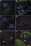
Development of ectopic lymphoid follicles, which exhibit characteristics of GCs. Salivary gland frozen sections (A–E) were stained with various fluorescently labeled antibodies. A, B) Intensely stained IgM+ (blue), IgG+ (green), and IgA+ (red) B cells (B, arrowheads). C, D) CD11b (MAC-1; green), B220 (red), and CD11c (blue). E) B220 (green), CD8 (red), and GCT (blue). F) PGLN frozen sections were stained with B220 (green), CD8 (red), and GCT (blue). GCT+ cells were often doubly stained with the CD8 marker (E, F; magenta, arrowheads). Panels B and D show magnified views of boxed areas in panels A and C, respectively. Views: ×20 (A, C, E); ×63 (B, D, F). Images are representative of 3 independent experiments demonstrating similar results.
Figure 3.
Figure 3.
Ectopic lymphoid follicles express markers of GC cells, including CXCL13, Ki67, and PNA. Salivary gland frozen sections on d 14 after inoculation were stained with fluorescently labeled antibodies specific for CXCL13 (red; A) and Ki-67, marker of proliferation (green; B), and binding to the lectin, PNA (C); ×20 view. Images are representative of 2 (CXCL13) or 3 (Ki67, PNA) independent experiments demonstrating similar results.
Figure 4.
Figure 4.
FDCs are present in the salivary gland within ectopic lymphoid follicles. Salivary glands were collected for frozen sections and stained with antibodies to FDC-M1 (top panel) or FDC-M2 (bottom panel). A) Mice inoculated with the equivalent of 105 pfu UV-tcMCMV. B) Mice inoculated with 105 pfu tcMCMV. Representative of 3 independent experiments demonstrating similar results.
Figure 5.
Figure 5.
Key cell surface markers of GC cells are present on B cells obtained from salivary glands. Lymphocytes were collected from the salivary glands 14 d after inoculation and were analyzed for coexpression of the B-cell surface marker B220, and GL7 or GCT, both markers of GC B cells. Dot plots (left panels) demonstrate lymphocytes stained with B220 (x axis) and GL7 or GCT (y axis). Percentage of cells in each quadrant is shown. Histograms (right panels) show percentage of the B220+-gated population expressing GL7 or GCT. A) Phenotype of cells after inoculation with UV-tcMCMV. B) Phenotype of cells after inoculation with tcMCMV. Representative of 3 independent experiments demonstrating similar results.
Figure 6.
Figure 6.
Key mRNA markers of GCs are present in the salivary glands. RNA was prepared from whole salivary glands after removal of the associated lymph nodes on d 14 after inoculation. A) RNA was analyzed for the presence of CXCL13, syndecan-1, Blimp-1, AID, PAX5, IμCα, and MCMV glycoprotein B. B) Histograms represent densitometric data from mean ± sd autoradiogram signals from 3 mice from the UV-tcMCMV group (□) and 5 mice from the tcMCMV group (■). P < 0.05 vs. UV-tcMCMV.
Figure 7.
Figure 7.
MCMV-specific antibodies and ASCs are generated after tcMCMV inoculation. A) Balb/cByJ mice were inoculated i.g. with 105 pfu tcMCMV (solid symbols), and saliva (top panel) and serum (bottom panel) samples were collected on d 7, 14, 21, and 28 after inoculation. Saliva and serum were also collected after i.g. inoculation with TC (control) at 7 d after inoculation. MCMV-specific antibodies of the IgM (■), IgG (♦), and IgA (▴) isotypes were determined via ELISA. Average peak antibody titers from mice inoculated i.p. with MCMV (open symbols) are shown for comparison. Results are pooled data from 4 mice analyzed in duplicate from 4 independent experiments. P < 0.05, #P < 0.01 vs. i.g. inoculation with TC on d 7. B) Cells were isolated from the spleen (□), salivary gland (■), and PGLNs (hatched bar) from mice inoculated i.g. with tcMCMV or TC on d 7, 14, 21, and 28. Cells were then stimulated in vitro in the presence of TC or tcMCMV. MCMV-specific ASC were detected using a standard ELISpot assay, and nonspecific, control antigen spots were subtracted. Results are pooled data from 3 independent experiments (4 mice/experiment). P < 0.05 vs. i.g. inoculated controls on d 7; #P < 0.05 vs. ASCs from spleen. Values are represented as means ± sd.
Figure 8.
Figure 8.
MCMV-specific proliferation in the salivary gland, PGLNs, and spleen is observed after inoculation with tcMCMV. Lymphocytes were isolated from the salivary gland (SG), PGLNs, and spleen following inoculation with TC or 105 pfu tcMCMV. A) BrdU incorporation was evaluated on d 4 following in vitro restimulation with TC (□) or tcMCMV (■). Percentage of total cells expressing BrdU is shown on left axis. Stimulation indices (●) are shown on right axis. B) BrdU+ cells were analyzed for the T-cell markers CD4 and CD8, and the B-cell marker CD19. Results are pooled data from 3 independent experiments, represented as means ± sd (3–8 mice/time point).
Figure 9.
Figure 9.
An i.g. inoculation with tcMCMV results in lower viral titers and prevents diminished salivary output following an MCMV challenge. A) Viral titers 14 d after challenge in salivary glands (SG), spleen, lungs (pfu/mg tissue), saliva, and serum (pfu/ml fluid). Results are pooled from 2–3 mice/group from 3 independent experiments. ●, Limit of detection. P < 0.05 vs. i.g. TC/i.p. MCMV. B) Saliva was collected and measured at 7 and 14 d after i.p. MCMV challenge from mice that had been inoculated i.g. with TC or tcMCMV. Results are pooled from 4–6 mice/group from 2 or 3 independent experiments. P < 0.05 vs. i.g. tcMCMV/i.p. TC.
Figure 10.
Figure 10.
Salivary gland immunization with tcMCMV protects mice from pathology associated with systemic MCMV infection. Balb/cByJ mice were uninoculated, inoculated i.g. with TC, or inoculated i.g. with 105 pfu tcMCMV. At 28 d after inoculation, mice were challenged i.p. with 105 pfu MCMV. Hematoxylin-and-eosin-stained, paraffin-embedded sections of the spleen (A, C, E) and salivary glands (B, D, F) were assessed 7 d after i.p. MCMV challenge for necrosis and cellular infiltrates, respectively. A, B) No inoculation, challenged i.p. with MCMV. C, D) Intraglandular TC, challenged i.p. with MCMV. E, F) Intraglandular tcMCMV, challenged i.p. with MCMV. Images are representative of 2 independent experiments with 3 mice/time point; ×4 view.

References

    1. Brandtzaeg P. (2007) Induction of secretory immunity and memory at mucosal surfaces. Vaccine 25, 5467–5484 - PubMed
    1. Takahashi I., Nochi T., Yuki Y., Kiyono H. (2009) New horizon of mucosal immunity and vaccines. Curr. Opin. Immunol. 21, 352–358 - PubMed
    1. Macpherson A. J., McCoy K. D., Johansen F. E., Brandtzaeg P. (2008) The immune geography of IgA induction and function. Mucosal Immunol. 1, 11–22 - PubMed
    1. Brandtzaeg P., Pabst R. (2004) Let's go mucosal: communication on slippery ground. Trends Immunol. 25, 570–577 - PubMed
    1. Mason K. L., Huffnagle G. B., Noverr M. C., Kao J. Y. (2008) Overview of gut immunology. Adv. Exp. Med. Biol. 635, 1–14 - PubMed

Publication types

Substances