. 2011 May;300(5):C979-88.
doi: 10.1152/ajpcell.00368.2010. Epub 2011 Feb 9.

Collagen IV contributes to nitric oxide-induced angiogenesis of lung endothelial cells

Affiliations

Collagen IV contributes to nitric oxide-induced angiogenesis of lung endothelial cells

Huafang Wang et al. Am J Physiol Cell Physiol. 2011 May.

Abstract

Nitric oxide (NO) mediates endothelial angiogenesis via inducing the expression of integrin α(v)β(3). During angiogenesis, endothelial cells adhere to and migrate into the extracellular matrix through integrins. Collagen IV binds to integrin α(v)β(3), leading to integrin activation, which affects a number of signaling processes in endothelial cells. In the present study, we evaluated the role of collagen IV in NO-induced angiogenesis. We found that NO donor 2,2'-(hydroxynitrosohydrazino)bis-ethanamine (NOC-18) causes increases in collagen IV mRNA and protein in lung endothelial cells and collagen IV release into the medium. Addition of collagen IV into the coating of endothelial culture increases endothelial monolayer wound repair, proliferation, and tube formation. Inhibition of collagen IV synthesis using gene silencing attenuates NOC-18-induced increases in monolayer wound repair, cell proliferation, and tube formation as well as in the phosphorylation of focal adhesion kinase (FAK). Integrin blocking antibody LM609 prevents NOC-18-induced increase in endothelial monolayer wound repair. Inhibition of protein kinase G (PKG) using the specific PKG inhibitor KT5823 or PKG small interfering RNA prevents NOC-18-induced increases in collagen IV protein and mRNA and endothelial angiogenesis. Together, these results indicate that NO promotes collagen IV synthesis via a PKG signaling pathway and that the increase in collagen IV synthesis contributes to NO-induced angiogenesis of lung endothelial cells through integrin-FAK signaling. Manipulation of collagen IV could be a novel approach for the prevention and treatment of diseases such as alveolar capillary dysplasia, severe pulmonary arterial hypertension, and tumor invasion.


Figures

Fig. 1.
Fig. 1.
2,2′-(Hydroxynitrosohydrazino)bis-ethanamine (NOC-18) increases collagen IV synthesis in pulmonary artery endothelial cells (PAEC). Lung endothelial cells were incubated with NOC-18 (1–100 μM) for 16 h, after which collagen IV mRNA (A) and collagen IV protein in the cells and culture medium (B and C) were determined as described in materials and methods. The blots shown are representative of 5 separate experiments. The bar graph in C depicts changes in collagen IV protein content in the cells and culture medium, quantified by scanning densitometry. Results are expressed as means ± SE; n = 5 experiments. *P < 0.05, vs. 0 (control); #P < 0.05, vs. 0 (control).
Fig. 2.
Fig. 2.
Collagen IV increases endothelial monolayer wound repair, cell proliferation, and tube formation. A: 24-well plates were coated with collagen IV (10–30 μg/ml) or BSA (10–30 μg/ml) and then PAEC were seeded. The monolayer wound repair was assayed after cells became confluent. B: 96-well culture plates were coated with collagen IV (30 μg/ml) or BSA (30 μg/ml) and then PAEC in suspension were seeded on 5 × 10³ cells/well. After 24 h, proliferation assay was evaluated after incubation of cells with 2′-deoxyuridine (BrdU; 10 μM). C and D: collagen IV (30 μg/ml) or BSA (30 μg/ml) was added to solidified matrigel in 96-well culture plates and then PAEC suspension was seeded for tube formation assay. After 8 h, tube length was measured. Data shown are representative images from 5 experiments (amplification, ×100). D: bar graph depicting the changes in tube length. Results are expressed as means ± SE; n = 5 experiments. *P < 0.05 vs. BSA group.
Fig. 3.
Fig. 3.
Knockdown of collagen IV attenuates NOC-18-induced increase in angiogenesis. PAEC were transfected with control small interfering RNA (siRNA) or siRNA against collagen IV mRNA. After 48 h, collagen IV protein (A and B), monolayer wound repair (C), cell proliferation (D), and endothelial tube formation (E and F) were assayed as described in materials and methods. A: representative immunoblot of collagen IV protein from 5 experiments. B: bar graph depicting the changes in intracellular collagen IV protein content. E: representative images from 5 tube formation assays (amplification, ×100). F: bar graph showing the changes in tube length in control PAEC and PAEC-transfected control siRNA or siRNA against collagen IV mRNA in the absence and presence of NOC-18 (10 μM). Results are expressed as means ± SE; n = 5 experiments. *P < 0.05 vs. vehicle group (without NOC-18); #P < 0.05 vs. vehicle group with control siRNA.
Fig. 4.
Fig. 4.
Specific PKG inhibitor KT5823 prevents NOC-18-induced increases in collagen IV synthesis, endothelial monolayer wound repair, cell proliferation, and tube formation. Intracellular collagen IV protein content (A and B), collagen IV mRNA (C), monolayer wound repair (D), cell proliferation (E), and tube formation (F and G) were evaluated in control PAEC and PAEC exposed to NOC-18 (10 μM) in the absence and presence of KT5823 (10 μM). A: representative immunoblot of collagen IV protein from 6 experiments. B: bar graph depicting the changes in intracellular collagen IV protein content. F: representative images from 5 tube formation assays (amplification, ×100). G: bar graph showing the changes in tube length. Results are expressed as means ± SE; n = 5 or 6 experiments. *P < 0.05 vs. vehicle group in control.
Fig. 5.
Fig. 5.
Knockdown of PKG prevents NOC-18-induced increase in angiogenesis. PAEC were transfected with control siRNA or siRNA against the mRNA of PKG. After 48 h, the cells were incubated with or without NOC-18 (10 μM) for 16 h and then intracellular protein content of PKG and collagen IV (A and B) and collagen IV mRNA (C) was assayed. The monolayer wound repair (D), cell proliferation (E), and tube formation (F and G) with or without NOC-18 (10 μM) were measured as described in materials and methods. A: representative immunoblot of collagen IV and PKG protein from 5 experiments. B: bar graph depicting the changes in intracellular protein content of PKG and collagen IV. F: representative images from 5 tube formation assays (amplification, ×100). G: bar graph showing the changes in tube length. Results are expressed as means ± SE; n = 5 experiments. *P < 0.05 vs. vehicle group (without NOC-18); #P < 0.05 vs. vehicle group with control siRNA.
Fig. 5.
Fig. 5.
Knockdown of PKG prevents NOC-18-induced increase in angiogenesis. PAEC were transfected with control siRNA or siRNA against the mRNA of PKG. After 48 h, the cells were incubated with or without NOC-18 (10 μM) for 16 h and then intracellular protein content of PKG and collagen IV (A and B) and collagen IV mRNA (C) was assayed. The monolayer wound repair (D), cell proliferation (E), and tube formation (F and G) with or without NOC-18 (10 μM) were measured as described in materials and methods. A: representative immunoblot of collagen IV and PKG protein from 5 experiments. B: bar graph depicting the changes in intracellular protein content of PKG and collagen IV. F: representative images from 5 tube formation assays (amplification, ×100). G: bar graph showing the changes in tube length. Results are expressed as means ± SE; n = 5 experiments. *P < 0.05 vs. vehicle group (without NOC-18); #P < 0.05 vs. vehicle group with control siRNA.
Fig. 6.
Fig. 6.
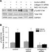
Knockdown of collagen IV attenuates NOC-18-induced increase in focal adhesion kinase (FAK) phosphorylation. PAEC were transfected with control siRNA or siRNA against collagen IV mRNA. After 48 h, protein content of phosphorylated FAK (pTyr397) and total FAK were measured as described in materials and methods. A: representative immunoblot of phosphorylated FAK and total FAK from 4 experiments. B: bar graph depicting the changes in protein content of phosphorylated FAK and total FAK. Results are expressed as means ± SE; n = 4 experiments. *P < 0.05 vs. vehicle group (without NOC-18); #P < 0.05 vs. vehicle group with control siRNA.
Fig. 7.
Fig. 7.
Integrin αvβ3 blocking antibody LM609 prevents NOC-18-induced increase in endothelial monolayer wound repair. PAEC were incubated with integrin αvβ3 blocking antibody LM609 (10 μg/ml) or nonimmune mouse IgG (10 μg/ml) for 16 h, after which endothelial monolayer wound repair was evaluated. Results are expressed as means ± SE; n = 4 experiments. *P < 0.05 vs. vehicle group.
Fig. 8.
Fig. 8.
A schematic pathway illustrating the role of collagen IV and PKG in nitric oxide (NO)-induced angiogenesis. NO activates PKG and induces the synthesis of collagen IV and integrin αvβ3. Collagen IV in the extracellular matrix (ECM) binds to integrin αvβ3, leading to the increase in FAK phosphorylation in the focal adhesion and angiogenesis.

Comment in

References

    1. Alvarez Y, Astudillo O, Jensen L, Reynolds AL, Waghorne N, Brazil DP, Cao Y, O'Connor JJ, Kennedy BN. Selective inhibition of retinal angiogenesis by targeting PI3 kinase. PLoS One 4: e7867, 2009 - PMC - PubMed
    1. Babaei S, Stewart DJ. Overexpression of endothelial NO synthase induces angiogenesis in a co-culture model. Cardiovasc Res 55: 190–200, 2002 - PubMed
    1. Fellner SK, Arendshorst WJ. Complex interactions of NO/cGMP/PKG systems on Ca2+ signaling in afferent arteriolar vascular smooth muscle. Am J Physiol Heart Circ Physiol 298: H144–H151, 2010 - PMC - PubMed
    1. Ferrara N, Gerber HP. The role of vascular endothelial growth factor in angiogenesis. Acta Haematol 106: 148–156, 2001 - PubMed
    1. Frelin C, Ladoux A, D'Angelo G. Vascular endothelial growth factors and angiogenesis. Ann Endocrinol (Paris) 61: 70–74, 2000 - PubMed

Publication types

MeSH terms