

Bone marrow stromal cells promote skilled motor recovery and enhance contralesional axonal connections after ischemic stroke in adult mice
- PMID: 21307396
- PMCID: PMC3060040
- DOI: 10.1161/STROKEAHA.110.607226
Bone marrow stromal cells promote skilled motor recovery and enhance contralesional axonal connections after ischemic stroke in adult mice
Abstract
Background and purpose: We tested the effect of bone marrow stromal cells (BMSCs) on neuronal remodeling of the corticospinal tract originating from the contralesional cortex in mice subjected to unilateral pyramidotomy (PT) followed by middle cerebral artery occlusion (MCAO).
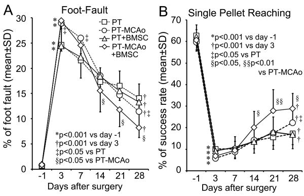
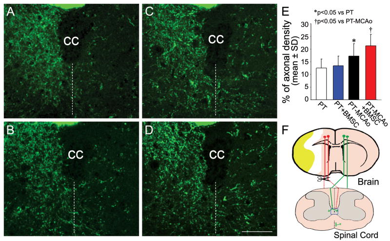
Methods: Adult mice with transgenic yellow fluorescent protein labeling in the corticospinal tract were subjected to right hemispheric PT and right permanent or sham MCAO. One day later, the mice were treated intravenously with BMSCs or phosphate-buffered saline. A Foot-Fault test and a single pellet-reaching test were performed before surgery, 3 days after MCAO, and weekly thereafter. Pseudorabies virus-614-monomeric red fluorescent protein was injected into the left forelimb flexor muscles 28 days after surgery (4 days before euthanasia). The brain and cervical cord were processed for fluorescent microscopy to detect red fluorescent protein and yellow fluorescent protein labeling, respectively.
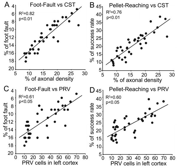
Results: Significant functional improvements were evident in PT-MCAO mice treated with BMSCs (n=9) compared with phosphate-buffered saline-treated mice (n=9, P<0.05), but not in mice with PT-sham MCAO treated with either phosphate-buffered saline (n=9) or BMSCs (n=10). Furthermore, in PT-MCAO mice, both corticospinal tract axonal density in the denervated side of the cervical gray matter and red fluorescent protein-labeled pyramidal neurons in the left intact cortex were significantly increased compared with PT-sham MCAO mice (P<0.05). BMSCs significantly enhanced both corticospinal tract density and red fluorescent protein labeling in PT-MCAO mice (P<0.05) only. The behavioral outcome was highly correlated with corticospinal tract density and red fluorescent protein labeling.
Conclusions: BMSCs amplify stroke-induced contralesional neuronal remodeling, which contributes to motor recovery after stroke.
Conflict of interest statement
Figures
References
-
- Twitchell TE. The restoration of motor function following hemiplegia in man. Brain. 1951;74:443–480. - PubMed
-
- Bareyre FM, Kerschensteiner M, Misgeld T, Sanes JR. Transgenic labeling of the corticospinal tract for monitoring axonal responses to spinal cord injury. Nat Med. 2005;11:1355–1360. - PubMed
Publication types
MeSH terms
Grants and funding
LinkOut - more resources
Full Text Sources
Other Literature Sources
Medical
