Review
. 2010 Nov-Dec;3(6):392-403.
doi: 10.4161/oxim.3.6.14786. Epub 2010 Nov 1.

Retinal redox stress and remodeling in cardiometabolic syndrome and diabetes

Affiliations
Review

Retinal redox stress and remodeling in cardiometabolic syndrome and diabetes

Ying Yang et al. Oxid Med Cell Longev. 2010 Nov-Dec.

Abstract

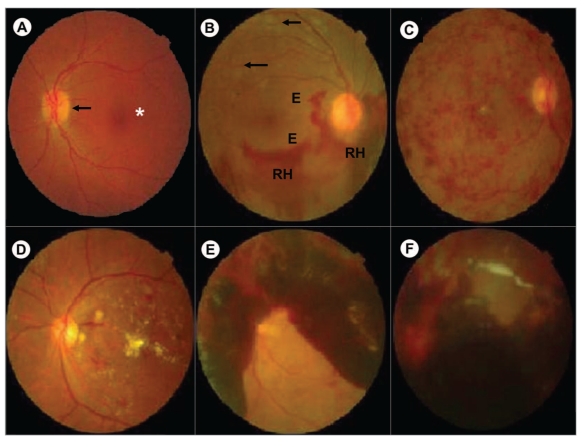
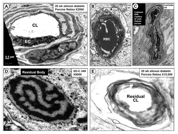
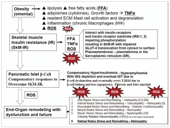
Diabetic retinopathy (DR) is a significant cause of global blindness; a major cause of blindness in the United States in people aged between 20-74. There is emerging evidence that retinopathy is initiated and propagated by multiple metabolic toxicities associated with excess production of reactive oxygen species (ROS). The four traditional metabolic pathways involved in the development of DR include: increased polyol pathway flux, advanced glycation end-product formation, activation of protein kinase C isoforms, and hexosamine pathway flux. These pathways individually and synergistically contribute to redox stress with excess ROS resulting in retinal tissue injury resulting in significant microvascular blood retinal barrier remodeling. The toxicity of hyperinsulinemia, hyperglycemia, hypertension, dyslipidemia, increased cytokines and growth factors, in conjunction with redox stress, contribute to the development and progression of DR. Redox stress contributes to the development and progression of abnormalities of endothelial cells and pericytes in DR. This review focuses on the ultrastructural observations of the blood retinal barrier including the relationship between the endothelial cell and pericyte remodeling in young nine week old Zucker obese (fa/fa) rat model of obesity; cardiometabolic syndrome, and the 20 week old alloxan induced diabetic porcine model. Preventing or delaying the blindness associated with these intersecting abnormal metabolic pathways may be approached through strategies targeted to reduction of tissue inflammation and oxidative - redox stress. Understanding these abnormal metabolic pathways and the accompanying redox stress and remodeling may provide both the clinician and researcher a new concept of approaching this complicated disease process.


Figures

Figure 1
Figure 1
Collage of the Porcine Retina. This collage of the retina allows one to follow the path of incident light as it passes through each layer of theretina and into the choroid coat. Each layer of the retina is prominently displayed and identified layer by layer.
Figure 2
Figure 2
Five Micron Choroid Capillary with Intact Pericyte. This image depicts the normal relationship of the pericyte (Pc) in the blood-retinal barrierin relation to the endothelial cell (EC). Note the red blood cell (RBC) within its capillary lumen. The inner and outer basement membranes (BM) areboxed in and identified. Inset depicts how endothelial uncoupling occurs.
Figure 3
Figure 3
Degenerative Pericyte “Ghost cell” in the 20 Week Alloxan Diabetic Porcine Model. (A) demonstrates the sharply demarcated pericyteprocess (PCP) sandwiched and encapsulated by an inner and outer basement membrane (BM) as it surrounds the endothelial cell (EC). (B) in contrastdepicts PCP degeneration and ghost cell changes typical of the 20 week alloxan diabetic porcine model. Note how the PcP is diminished in size andthere is a considerable decrease in electron density staining, typical of pericyte ghost cells. As this progresses the PcP will disappear and its remainsare thought to be incorporated within the inner and outer BM to result in BM thickening.
Figure 4
Figure 4
Characterization of Retina in Human Diabetic Retinopathy. (A) depicts the normal appearance of the retina. The macula is the darkened red spot in the center of the picture (asterisk). The yellow circle is the optic nerve (arrow). (B) displays dilated capillaries (microaneurysms), which leak red blood cells and plasma into the substance of the retina. This results in the appearance of retinal hemorrhages (RH), edema (E) and deposits or exudates (arrows). (C) demonstrates extensive intraretinal microvascular abnormalities, cotton wool spots, venous dilation and multiple extensive hemorrhages. (D) depicts macular edema with multiple hard exudates. (E) demonstrates vitreous hemorrhage as a result of neovascularization bleeding and hemorrhage. (F) depicts retina detachment and vitreous hemorrhage.
Figure 5
Figure 5
Schematic of the Hyperglycemia Mediated Retinal Redox Stress and Remodeling in Metabolic Syndrome and Type 2 Diabetes Mellitus. Note how hyperglycemia is important in activating the four major pathways involved in the production of excessive reactive oxygen species (ROS) to result in diabetic retinopathy (DR), eventual proliferative diabetic retinopathy (PDR) and diabetic macular edema (DME). AGE, advanced glycosylation endproducts; Ang II, angiotensin II; DAG/PKC, diacylglycerol protein kinase C; AR, aldose reductase; DME, diabetic macular edema; DR, diabetic retinopathy; eNOS, endothelial nitric oxide synthase; GSH, glutathione; Mt, mitochondria; NADPH, reduced nicotinamide adenine dinucleotidephosphate; NFκB, nuclear factor kappa B; Nox 2, NADPH Oxidase 2; RAAS, renin-angiotensin-aldosterone system; RAGE, receptor for advanced glycation endproducts; ROS, reactive oxygen species; TGFβ, transforming growth factor beta.
Figure 6
Figure 6
Morphological Differences between Peripheral Continuous Capillaries and Blood-Retinal Barrier Capillaries. Peripheral continuous capillaries do not share a BM; rather, they communicate with peg sockets (PS) and adherens junctions (AJ) as in (A and B). In the blood-retinal barrier this communication is shared via a common inner and outer basement membrane (BM) with the pericyte being uniquely sandwiched or encapsulated by a prominent inner and outer BM.
Figure 7
Figure 7
Fate of 20 Week Old Alloxan Diabetic Retinal-Blood Barrier Capillaries. (A) depicts the pericyte process remodeling and the thickened basement membranes (BM) found in the remaining and surviving retinal-blood barrier capillaries. Note the numerous enveloping—encircling pericyte processes (PcP) as it protects the surviving endothelial cell (EC) and it's thickened continuous BM. (B and C) illustrate capillary collapse of retinal-blood barrier capillaries in the 20 week alloxan diabetic porcine capillaries as compared to representative 9 week old Zucker lean and obese models. (D) illustrates illustrate a typical residual body and this was found in a podocyte of the 9 week old Zucker Obese kidney. (E) depicts a similar appearing residual body of what is thought to be a residual body of a demised-degenerated retinal-blood barrier capillary found in the 20 week alloxan diabetic porcine model.
Figure 8
Figure 8
Sequence of Events in the Development of Metabolic Syndrome and overt Type 2 Diabetes Mellitus. This figure illustrates the sequence of events leading to end-organ remodeling with dysfunction and eventual failure and begins with obesity and subsequent skeletal muscle insulin resistance (IR ) involving free fatty acids (FFA), tumor necrosis factor alpha (TNFα) and reactive oxygen species (ROS). This is followed by a compensatory islet beta-cell (β-cell) response in order to overcome skeletal muscle IR and is associated with eventual β-cell dysfunction and failure resulting in type 2 diabetes mellitus (T2DM). Additionally, there is glucotoxicity once impaired fasting glucose—prediabetes and overt T2DM develops contributing to additional ROS in addition to the ROS generated by the A-FLIGHT-U multiple metabolic toxicities and the ensuing end-organ damage resulting in the multiple end-organ failure and multiple diabetic pathologies including diabetic retinopathy.

Similar articles

Cited by

References

    1. Zhang X, Saaddine JB, Chou CF, et al. Prevalence of diabetic retinopathy in the United States, 2005–2008. JAMA–Journal of the American Medical Association. 2010;304(6):649–656. - PMC - PubMed
    1. Wong TY, Duncan BB, Golden SH, et al. Associations between the metabolic syndrome and retinal microvascular signs: the atherosclerosis risk in communities study. Investigative Ophthalmology and Visual Science. 2004;45(9):2949–2954. - PubMed
    1. Tikellis G, Anuradha S, Klein R, Wong TY. Association between physical activity and retinal microvascular signs: the atherosclerosis risk in communities (ARIC) study. Microcirculation. 2010;17(5):381–393. - PMC - PubMed
    1. Serne EH, Ijzerman RG, de Jongh RT, Stehouwer CD. Blood pressure and insulin resistance: role for microvascular function? Cardiovascular Research. 2002;53:271–276. - PubMed
    1. Hayden MR, Tyagi SC. Intimal redox stress: accelerated atherosclerosis in metabolic syndrome and type 2 diabetes mellitus. Atheroscleropathy. Cardiovascular Diabetology. 2002;1, article 3 - PMC - PubMed

Publication types