

Myofibroblast differentiation during fibrosis: role of NAD(P)H oxidases
- PMID: 21307839
- PMCID: PMC3675765
- DOI: 10.1038/ki.2010.516
Myofibroblast differentiation during fibrosis: role of NAD(P)H oxidases
Abstract
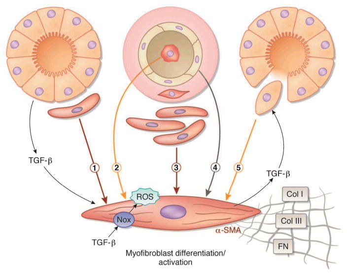
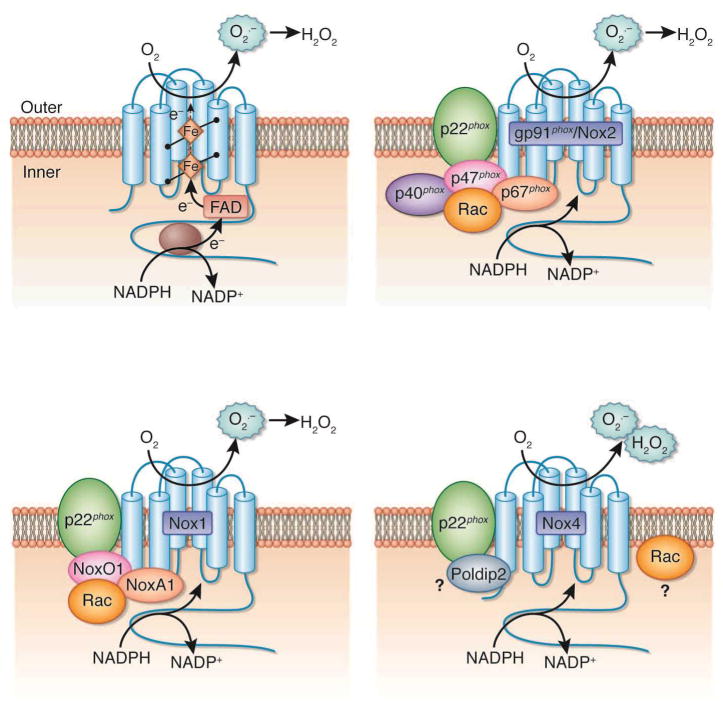
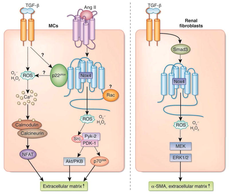
Progression of fibrosis involves interstitial hypercellularity, matrix accumulation, and atrophy of epithelial structures, resulting in loss of normal function and ultimately organ failure. There is common agreement that the fibroblast/myofibroblast is the cell type most responsible for interstitial matrix accumulation and consequent structural deformations associated with fibrosis. During wound healing and progressive fibrotic events, fibroblasts transform into myofibroblasts acquiring smooth muscle features, most notably the expression of alpha-smooth muscle actin and synthesis of mesenchymal cell-related matrix proteins. In renal disease, glomerular mesangial cells also acquire a myofibroblast phenotype and synthesize the same matrix proteins. The origin of interstitial myofibroblasts during fibrosis is a matter of debate, where the cells are proposed to derive from resident fibroblasts, pericytes, perivascular adventitial, epithelial, and/or endothelial sources. Regardless of the origin of the cells, transforming growth factor-beta1 (TGF-β1) is the principal growth factor responsible for myofibroblast differentiation to a profibrotic phenotype and exerts its effects via Smad signaling pathways involving mitogen-activated protein kinase and Akt/protein kinase B. Additionally, reactive oxygen species (ROS) have important roles in progression of fibrosis. ROS are derived from a variety of enzyme sources, of which the nicotinamide adenine dinucleotide phosphate (NAD(P)H) oxidase family has been identified as a major source of superoxide and hydrogen peroxide generation in the cardiovasculature and kidney during health and disease. Recent evidence indicates that the NAD(P)H oxidase homolog Nox4 is most accountable for ROS-induced fibroblast and mesangial cell activation, where it has an essential role in TGF-β1 signaling of fibroblast activation and differentiation into a profibrotic myofibroblast phenotype and matrix production. Information on the role of ROS in mesangial cell and fibroblast signaling is incomplete, and further research on myofibroblast differentiation during fibrosis is warranted.
Conflict of interest statement
The authors declared no competing interests.
Figures
References
-
- Strutz F, Muller GA. Mechanisms of renal fibrogenesis. In: Neilson EG, Couser WG, editors. Immunologic renal diseases. 2. Philadelphia: Lippincott Williams & Wilkins; 2001. pp. 73–101.
-
- Hewitson TD. Renal tubulointerstitial fibrosis: common but never simple. Am J Physiol - Renal Physiol. 2009;296:F1239–F1244. - PubMed
-
- Hughson MD. End-stage renal disease. In: Jennette JC, Olson JL, Schwartz MM, Silva FG, editors. Heptinstall’s Pathology of the Kidney. 6. Philadelphia: Lippincott-Raven Publishers; 2007. pp. 1307–1346.
Publication types
MeSH terms
Substances
Grants and funding
LinkOut - more resources
Full Text Sources
Other Literature Sources
