Review
. 2011 May;4(3):279-87.
doi: 10.1038/mi.2011.3. Epub 2011 Feb 9.

Apoptosis is an innate defense function of macrophages against Mycobacterium tuberculosis

Affiliations
Review

Apoptosis is an innate defense function of macrophages against Mycobacterium tuberculosis

S M Behar et al. Mucosal Immunol. 2011 May.

Abstract

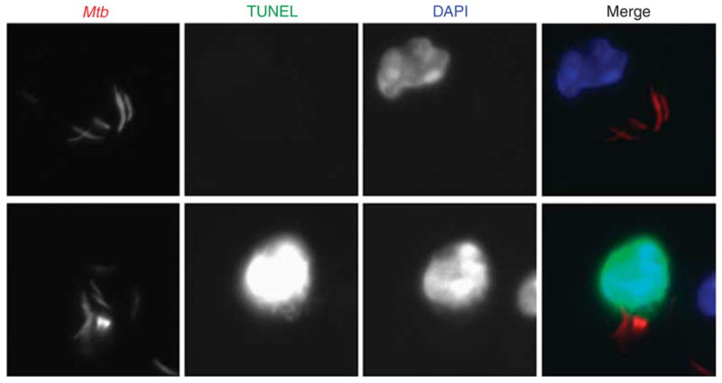
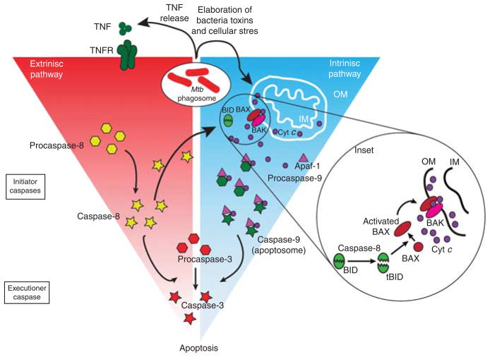
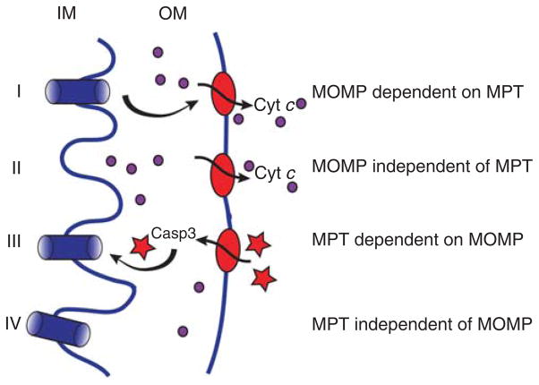
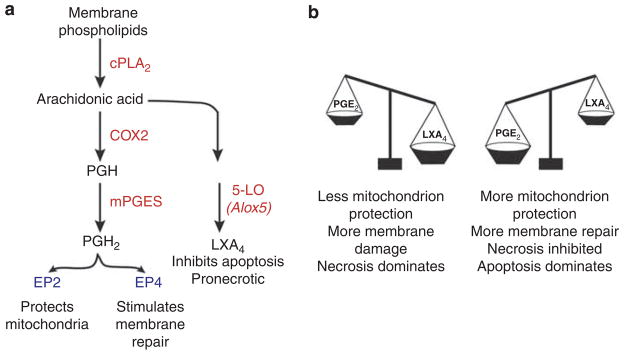
Two different forms of death are commonly observed when Mycobacterium tuberculosis (Mtb)-infected macrophages die: (i) necrosis, a death modality defined by cell lysis and (ii) apoptosis, a form of death that maintains an intact plasma membrane. Necrosis is a mechanism used by bacteria to exit the macrophage, evade host defenses, and spread. In contrast, apoptosis of infected macrophages is associated with diminished pathogen viability. Apoptosis occurs when tumor necrosis factor activates the extrinsic death domain pathway, leading to caspase-8 activation. In addition, mitochondrial outer membrane permeabilization leading to activation of the intrinsic apoptotic pathway is required. Both pathways lead to caspase-3 activation, which results in apoptosis. We have recently demonstrated that during mycobacterial infection, cell death is regulated by the eicosanoids, prostaglandin E(2) (proapoptotic) and lipoxin (LX)A(4) (pronecrotic). Although PGE(2) protects against necrosis, virulent Mtb induces LXA(4) and inhibits PGE(2) production. Under such conditions, mitochondrial inner membrane damage leads to macrophage necrosis. Thus, virulent Mtb subverts eicosanoid regulation of cell death to foil innate defense mechanisms of the macrophage.


Conflict of interest statement

DISCLOSURE

The authors declared no conflict of interest.

Figures

Figure 1
Figure 1
Apoptotic cell death following Mtb infection. CD11b+ thioglycolate-elicited peritoneal mouse macrophages were infected at an MOI of 10:1 with mCherry-expressing H37Rv. Cells were fixed, permeabilized, and TUNEL stained according to the In situ Cell Death Detection Kit (Roche, Mannheim, Germany) 2 days later. Coverslips were mounted on slides using ProLong Gold with DAPI (Invitrogen, Carlsbad, CA) to label nuclei. Macrophages containing intracellular bacteria were identified by wide-field microscopy using a Nikon Microscope (Nikon, Tokyo, Japan) at ×40 magnification and then the nuclear staining assessed. Both TUNEL-negative (top row) and TUNEL-positive (bottom row) infected cells are found. DAPI, 4′,6-diamidino-2-phenylindole; MOI, multiplicity of infection; Mtb, Mycobacterium tuberculosis; TUNEL, terminal deoxynucleotidyl transferase dUTP nick-end labeling.
Figure 2
Figure 2
Extrinsic and intrinsic pathways of apoptosis. During Mtb infection, several signals generated by the infected macrophage can trigger apoptosis. For example, TNF production can activate the extrinsic pathway resulting in caspase-8 activation. What triggers the intrinsic pathway is unknown. Cellular stress or caspase-8 activation can result in BID cleavage, leading to BAX activation, and the ensuing events lead to cytochrome c translocation from the mitochondrial intermembrane space into the cytosol where it activates caspase-9. Alternately, caspase-8 activation can directly lead to caspase-3 activation in certain cells (type I). cyt c, cytochrome c; IM, inner mitochondrial membrane; Mtb, Mycobacterium tuberculosis; OM, outer mitochondrial membrane; TNF, tumor necrosis factor; TNFR, TNF receptor.
Figure 3
Figure 3
Mitochondrial membrane damage leads to cell death. Four different scenarios of inner or outer mitochondrial membrane damage lead to mitochondrion dysfunction (see Table 1). Opening of the permeability transition pore in the inner mitochondrial membrane (IM), leads to MPT, and can occur in isolation (IV) or can increase outer membrane (OM) permeability (MOMP), leading to leaking of cytochrome c (I). In addition, MOMP leading to cytochrome c release into the cytosol can occur independently of MPT (II). Finally, under certain conditions, OM permeability might allow entry of activated caspase-3 into the mitochondrial intermembrane space, which can lead to damage of the IM and result in MPT. Casp3; caspase-3; cyt c, cytochrome c; IM, inner mitochondrial membrane; MOMP, mitochondrial outer membrane permeability; OM, outer mitochondrial membrane.
Figure 4
Figure 4
The role of eicosanoids in regulating death of infected macrophages. (a) Simplified version of eicosanoid biosynthesis. Substrates and products are indicated in black text. Enzymes are in red, and receptors in blue. (b) The balance of prostanoid and lipoxin production determines the death modality of Mtb-infected macrophages. Excess production of LXA4 leads to necrosis. In contrast, when PGE2 production predominates, necrosis is inhibited and more apoptosis is the consequence. COX2, cyclooxygenase-2; cPLA2, cytosolic phospholipase A2; 5-LO, 5-lipoxygenase; LXA4, lipoxin A4; Mtb, Mycobacterium tuberculosis; mPGES, membrane associated prostaglandin E synthase-1; PGE2, prostaglandin E2; PGH prostaglandin H.

Comment in

Similar articles

Cited by

References

    1. Kang BK, Schlesinger LS. Characterization of mannose receptor-dependent phagocytosis mediated by Mycobacterium tuberculosis lipoarabinomannan. Infect Immun. 1998;66:2769–2777. - PMC - PubMed
    1. Schlesinger LS, Kaufman TM, Iyer S, Hull SR, Marchiando LK. Differences in mannose receptor-mediated uptake of lipoarabinomannan from virulent and attenuated strains of Mycobacterium tuberculosis by human macrophages. J Immunol. 1996;157:4568–4575. - PubMed
    1. Sturgill-Koszycki S, et al. Lack of acidification in Mycobacterium phagosomes produced by exclusion of the vesicular proton-ATPase [see comments] [published erratum appears in Science 1994 11 March;263(5152):1359] Science. 1994;263:678–681. - PubMed
    1. Chen M, Gan H, Remold HG. A mechanism of virulence: virulent Mtb tuberculosis strain H37Rv but not attenuated H37Ra causes significant mitochondrial inner membrane disruption in macrophages leading to necrosis. J Immunol. 2006;176:3707–3716. - PubMed
    1. Molloy A, Laochumroonvorapong P, Kaplan G. Apoptosis, but not necrosis, of infected monocytes is coupled with killing of intracellular bacillus Calmette-Guerin. J Exp Med. 1994;180:1499–1509. - PMC - PubMed

Publication types

MeSH terms