

doi: 10.1038/jid.2011.21.
Epub 2011 Feb 10.
Superficial spreading-like melanoma in Arf(-/-)::Tyr-Nras(Q61K)::K14-Kitl mice: keratinocyte Kit ligand expression sufficient to "translocate" melanomas from dermis to epidermis
- PMID: 21307875
- PMCID: PMC3138531
- DOI: 10.1038/jid.2011.21
Item in Clipboard
Superficial spreading-like melanoma in Arf(-/-)::Tyr-Nras(Q61K)::K14-Kitl mice: keratinocyte Kit ligand expression sufficient to "translocate" melanomas from dermis to epidermis
J Invest Dermatol.
2011 Jun.
No abstract available
Conflict of interest statement
HPS is a shareholder and consultant for Molemap, Australia. The other authors state no conflict of interest.
Figures
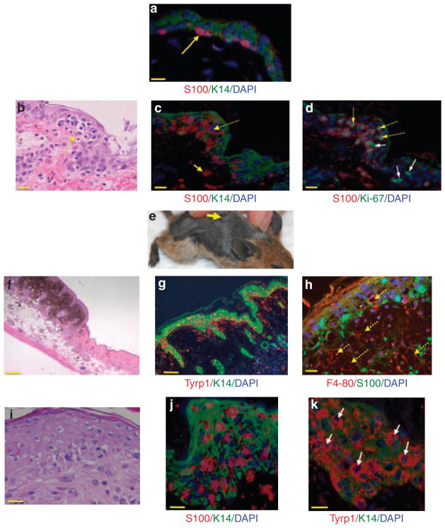
(a) Double-label immunofluorescence (IF) staining for S100 and K14 on “normal” Arf−/−::Tyr-Nras::K14-Kitl mouse skin (methods fully described in Supplementary Material online). Briefly, sections of formalin-fixed and paraffin-embedded skin were stained with anti-S100 (Dako, Eli, UK) primary antibody, followed by an AlexaFluor 555-labeled donkey anti-rabbit secondary antibody (Jackson ImmunoResearch Laboratories, West Grove, PA). After several washes, we stained keratinocytes using anti-K14 (Dako) followed with AlexaFluor 488-labeled donkey anti-rabbit (Jackson ImmunoResearch Laboratories). The yellow arrow points to red S100 staining MCs sitting at the dermoepidermal junction. Scale bar = 20 μM. (b) Hematoxylin and eosin (H&E) staining of bleached formalin-fixed, paraffin-embedded sections of Arf−/−::Tyr-Nras::K14-Kitl mouse skin. Bleaching is described in Supplementary Methods online. Note cluster (nest) of atypical MC cells in the epidermis, indicated by a dotted yellow arrow. Scale bar = 20 μM. (c) IF staining of a similar portion of epidermis with a dense cluster of S100-positive epidermal MCs (indicated by dotted arrows). Solid yellow arrow points to a dermal MC. Scale bar = 20 μM. (d) Epidermal MC cluster stained for S100 and Ki-67. Dotted yellow arrow shows MCs in the suprabasal epidermis positive for Ki-67. Solid white arrows denote keratinocytes (S100-negative cells) staining for Ki-67. Scale bar = 20 μM. (e) Arf−/−::Tyr-Nras::K14-Kitl mouse with a slightly elevated pigmented plaque on its back. Note that because of the heavy pigmentation of the whole skin, the melanoma is not easily discernible in this photograph. Mouse background: FVB, two generations down C57BL6. (f) H&E-stained section showing raised plaque. Scale bar = 200 μM. (g) IF staining of a Arf−/−::Tyr-Nras::K14-Kitl plaque for Tyrp1 (red) and K14 (green). Scale bar = 100 μM. (h) Double-layer IF image of a pigmented plaque staining for F4/80 (red) and S100 (green). Dotted yellow arrows denote macrophage lineage cells (melanophages) in a “cobblestone” pattern. The pigmented part of the tumor consists of many melanophages interspersed with some melanocytes/melanoma cells. Scale bar = 20 μM. (i) Higher-power H&E image of MC nesting in the thickened epidermis within a plaque. Scale bar = 20 μM. (j) Higher-power image of IF staining of S100-positive epidermal MCs within an epidermal melanoma. K14-positive keratinocytes are green. Scale bar = 20 μM. (k) Double-label IF staining for Tyrp1 using the PEP1 antibody, a gift from Dr Vince Hearing. K14-positive keratinocytes are green. Whereas S100 IF staining somewhat obscures the 4,6-diamidino-2-phenylindole (DAPI)-stained nuclei, Tyrp1 staining allows better visualization of MC nuclei of varying sizes and shapes (indicated by white arrows). Scale bar = 20 μM.
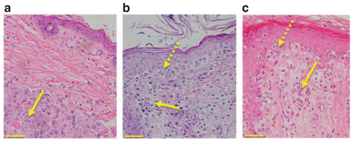
(a) Hematoxylin and eosin (H&E)-stained section showing a deep dermal malignant melanoma (MM) in an Arf−/−::Tyr-Nras mouse. The yellow arrow denotes a nodular melanoma composed of sheets of pleomorphic melanocytes (MCs). This nodular lesion is clearly separated from the epidermis by layers of collagen. (b) MM from an Arf−/−::Tyr-Nras::K14-Kitl mouse. Dotted yellow arrow denotes atypical MCs in the epidermis, and filled yellow arrow atypical MCs in the dermis. There is an increased number of MCs, varying in size and shape, not only at the dermoepidermal junction but also at all levels of the epidermis. (c) Human superficial spreading MM. Dotted yellow arrow denotes atypical MCs in the epidermis, and filled yellow arrow atypical MCs in the dermis. The morphological similarities between Figure 2b and c are striking. Scale bars = 100 μM.
Similar articles
-
(V600E)Braf::Tyr-CreERT2::K14-Kitl mice do not develop superficial spreading-like melanoma: keratinocyte Kit ligand is insufficient to "translocate" (V600E)Braf-driven melanoma to the epidermis.J Invest Dermatol. 2012 Feb;132(2):488-91. doi: 10.1038/jid.2011.341. Epub 2011 Nov 24. J Invest Dermatol. 2012. PMID: 22113477 No abstract available.
-
ARF functions as a melanoma tumor suppressor by inducing p53-independent senescence.Proc Natl Acad Sci U S A. 2007 Jun 26;104(26):10968-73. doi: 10.1073/pnas.0611638104. Epub 2007 Jun 19. Proc Natl Acad Sci U S A. 2007. PMID: 17576930 Free PMC article.
-
Oncogene-induced senescence does not require the p16(INK4a) or p14ARF melanoma tumor suppressors.J Invest Dermatol. 2009 Aug;129(8):1983-91. doi: 10.1038/jid.2009.5. Epub 2009 Feb 12. J Invest Dermatol. 2009. PMID: 19212341
-
The role of Kit-ligand in melanocyte development and epidermal homeostasis.Pigment Cell Res. 2003 Jun;16(3):287-96. doi: 10.1034/j.1600-0749.2003.00055.x. Pigment Cell Res. 2003. PMID: 12753403 Review.
-
Normal human melanocyte homeostasis as a paradigm for understanding melanoma.J Investig Dermatol Symp Proc. 2005 Nov;10(2):153-63. doi: 10.1111/j.1087-0024.2005.200407.x. J Investig Dermatol Symp Proc. 2005. PMID: 16358819 Review.
Cited by
-
Blinded by the light: why the treatment of metastatic melanoma has created a new paradigm for the management of cancer.Ther Adv Med Oncol. 2015 Mar;7(2):107-21. doi: 10.1177/1758834014566619. Ther Adv Med Oncol. 2015. PMID: 25755683 Free PMC article. Review.
-
A melanin-independent interaction between Mc1r and Met signaling pathways is required for HGF-dependent melanoma.Int J Cancer. 2015 Feb 15;136(4):752-60. doi: 10.1002/ijc.29050. Epub 2014 Jul 7. Int J Cancer. 2015. PMID: 24975581 Free PMC article.
-
Modeling melanoblast development.Cell Mol Life Sci. 2013 Mar;70(6):1067-79. doi: 10.1007/s00018-012-1112-4. Epub 2012 Aug 23. Cell Mol Life Sci. 2013. PMID: 22915137 Free PMC article. Review.
References
-
- Abdel-Daim M, Funasaka Y, Komoto M, et al. Pharmacogenomics of metabotropic glutamate receptor subtype 1 and in vivo malignant melanoma formation. J Dermatol. 2010;37:635–46. - PubMed
-
- Dhomen N, Reis-Filho JS, da Rocha Dias S, et al. Oncogenic Braf induces melanocyte senescence and melanoma in mice. Cancer Cell. 2009;15:294–303. - PubMed
-
- Ferguson B, Muller HK, Handoko HY, et al. Differential roles of the pRb and Arf/p53 pathways in murine naevus and melanoma genesis. Pigment Cell Melanoma Res. 2010;23:771–80. - PubMed
Publication types
MeSH terms
Substances
Grants and funding
LinkOut - more resources
Full Text Sources
Medical
Molecular Biology Databases
Miscellaneous
