. 2011 Jul;5(7):1178-90.
doi: 10.1038/ismej.2011.2. Epub 2011 Feb 10.

Comparative metagenomics of microbial traits within oceanic viral communities

Affiliations

Comparative metagenomics of microbial traits within oceanic viral communities

Itai Sharon et al. ISME J. 2011 Jul.

Abstract

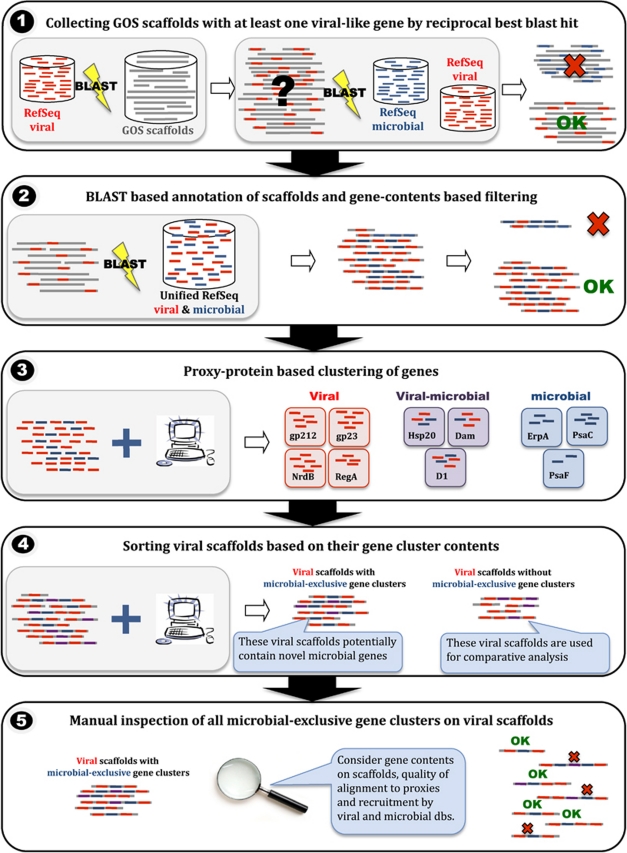
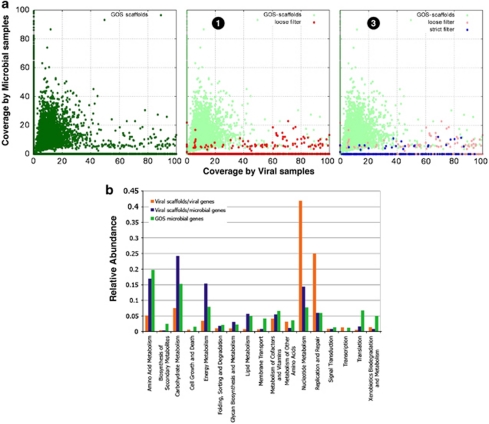
Viral genomes often contain genes recently acquired from microbes. In some cases (for example, psbA) the proteins encoded by these genes have been shown to be important for viral replication. In this study, using a unique search strategy on the Global Ocean Survey (GOS) metagenomes in combination with marine virome and microbiome pyrosequencing-based datasets, we characterize previously undetected microbial metabolic capabilities concealed within the genomes of uncultured marine viral communities. A total of 34 microbial gene families were detected on 452 viral GOS scaffolds. The majority of auxiliary metabolic genes found on these scaffolds have never been reported in phages. Host genes detected in viruses were mainly divided between genes encoding for different energy metabolism pathways, such as electron transport and newly identified photosystem genes, or translation and post-translation mechanism related. Our findings suggest previously undetected ways, in which marine phages adapt to their hosts and improve their fitness, including translation and post-translation level control over the host rather than the already known transcription level control.


Figures

Figure 1
Figure 1
Pipeline for the identification of viral scaffolds with microbial genes. (1) Candidate scaffolds containing at least one potential viral gene were identified using a bi-directional BLAST (Basic Local Alignment Search Tool) search of all proteins in the RefSeq-viral database of the National Center for Biotechnology Information against all GOS scaffolds, and then a BLAST search of all significant best hits against a combined RefSeq-viral and RefSeq-microbial proteins database. Scaffolds with best hits from RefSeq-viral were further considered. (2) Gene contents of all candidate scaffolds were determined using an iterative sequence similarity-based procedure against the combined dataset of RefSeq-viral and RefSeq-microbial proteins. (3) Genes were clustered based on sequence similarity of their RefSeq hits, with each cluster being tagged based on the origin of its RefSeq proteins (viral exclusive, microbial exclusive or viral microbial). For the purpose of clustering we consider all genes' proxy proteins rather the genes and their scaffolds. Scaffold tagging was determined based on gene contents and on recruitment against the Northern Line Islands datasets (Figure 2). (4) Viral scaffolds were sorted into scaffolds containing members from viral or viral-microbial clusters only, and scaffolds also containing members of microbial exclusive clusters. Purple genes refer to genes that belong to viral-microbial clusters, red and blue refer to members of viral-exclusive and microbial-exclusive clusters, respectively. (5) The latter set was inspected manually in order to validate their origin and annotation.
Figure 2
Figure 2
Enrichment scheme for microbial traits in viral landscapes. (a) Recruitment coverage of GOS scaffolds enriched through the different selection filters with Northern Line Islands marine viromes (x-axis) and microbiomes (y-axis; Dinsdale et al., 2008a, 2008b). Left panel, previous selection; middle panel, loose filter settings corresponding to box 1 in Figure 1; right panel, strict filter settings corresponding to box 2 in Figure 1. Coverage is defined as percentage of GOS scaffold length covered by at least one recruited read. (b) Distribution of viral (red) and microbial (blue) genes on viral scaffolds, as well as genes selected at random from other GOS scaffolds (green). Kyoto Encyclopedia of Genes and Genomes categories ‘energy metabolism' and ‘carbohydrate metabolism' are enriched in the microbial genes/viral scaffolds set with respect to the GOS gene set (P-values of 5e−10 and 4e−9, respectively). Refer to the Supplementary Information for a full description.
Figure 3
Figure 3
Marine viral PDFs do not display the conserved C-terminal helix of subtype IB PDF and constitute a new class of active PDFs. (a) A phylogenetic tree was constructed from an alignment of ∼200 PDFs recapitulating sequence and phylogenetic diversity (Supplementary Figure S2). Among marine viral PDFs, only representative members are displayed (colored in gray). These proteins are related to PDFs from cyanobacteria (shown in green) and photosynthetic planktonic picoeukaryotes (shown in blue). Proteins showing the closest similarities around the three conserved motifs 1, 2 and 3 (colored in yellow) and C-helix were selected and realigned, and the motifs are shown. Unlike type III inactive PDFs (colored in orange below), all required residues of the motifs are conserved, which is strongly suggestive of peptide deformylase activity. The closest structural models (that is, E. coli and Arabidopsis thaliana PDF1B) are indicated on top. In both cases, the C-terminus folds as a α helix. (b) A refined three-dimensional model for viral PDF (top) compared to thethree-dimensional crystal structure of the most relevant PDF (see panel a) from chloroplastic PDF1B (PDB code 3cpmA; bottom). The two structures are shown in the same orientation, that is, toward the entry of the active site crevice. Both N- and C-ends are indicated in white with N and C, respectively.
Figure 4
Figure 4
Viral NAD(P)H dehydrogenase subunits. (a) NdhI protein alignment. Synechococcus sequences are colored in cyan, Prochlorococcus in green and viral proteins in red. The Nqo9 sequence from T. thermophilus is shown for reference. The conserved cysteine residues coordinating Fe–S clusters in Nqo9 are marked with arrows (black for N6a and gray for N6b). For clarity, only part of the protein length is shown. (b) NdhD FastTree approximated maximum-likelihood phylogenetic tree. Synechococcus NdhD sequences are colored in cyan, Prochlorococcus in green and viral proteins in red. Synechocystis PCC6803 and Synechococcus PCC7002 NdhD1, 2, 3 and 4 sequences were used as references. Only bootstrap values above 80% are shown as black circles on the branches.
Figure 5
Figure 5
Viral PSI PsaJ protein. (a) PSI PsaJ protein and peptide alignment. Viral PsaJ proteins identified in this study and viral PsaJF (only the N-terminus, which contains the PsaJ portion, is shown) are labeled in red to the right of the alignment. (b) PsaJ FastTree approximated maximum-likelihood phylogenetic tree. Synechococcus PsaJ sequences are colored in cyan, Prochlorococcus in green, viral PsaJF and the newly identified viral PsaJ in red. Only bootstrap values above 80% are shown as black circles on the branches. (c) Schematic physical maps of selected viral clones or Prochlorococcus and Synechococcus genome fragments containing the PSI psaJ gene and a viral GOS clone containing the psaJF fusion gene. ndhI denotes NAD(P)H dehydrogenase I gene, speD a polyamine biosynthesis gene, talC a transaldolase gene and nrdB the ribonucleoside-diphoshate reductase-β 2 gene. Red-arrowed boxes mark viral genes and gray mark bacterial genes. PSI genes are colored in yellow, green and blue.

References

    1. Alperovitch A, Sharon I, Rohwer F, Aro E-M, Glaser F, Milo R, et al. Reconstructing a puzzle: existence of cyanophages containing both photosystem-I & photosystem-II gene suites inferred from oceanic matagenomic datasets. Environ Microbiol. 2010;13:24–32. - PubMed
    1. Angly F, Felts B, Breitbart M, Salamon P, Edwards R, Carlson C, et al. The marine viromes of four oceanic regions. PLoS Biol. 2006;4:e368. - PMC - PubMed
    1. Aono J, Kangawa K, Matsuo H, Horiuchi T. Coliphage 434 tof protein: NH2-terminal amino acid sequence and kinetic and equilibrium measurements of DNA binding. Mol Gen Genet. 1982;186:460–466. - PubMed
    1. Ayala-Castro C, Saini A, Outten FW.2008Fe-S cluster assembly pathways in bacteria Microbiol Mol Biol Rev 72110–125.table of contents. - PMC - PubMed
    1. Battchikova N, Aro E-M. Cyanobacterial NDH-1 complexes: multiplicity in function and subunit composition. Physiol Plant. 2007;131:22–32. - PubMed

Publication types