. 2011 Apr;112(4):1168-75.
doi: 10.1002/jcb.23032.

Role of C/EBP-β, p38 MAPK, and MKK6 in IL-1β-mediated C3 gene regulation in astrocytes

Affiliations

Role of C/EBP-β, p38 MAPK, and MKK6 in IL-1β-mediated C3 gene regulation in astrocytes

Jeffrey Maranto et al. J Cell Biochem. 2011 Apr.

Abstract

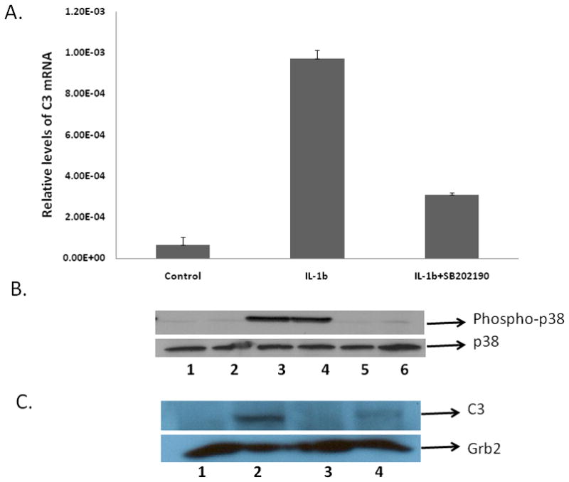
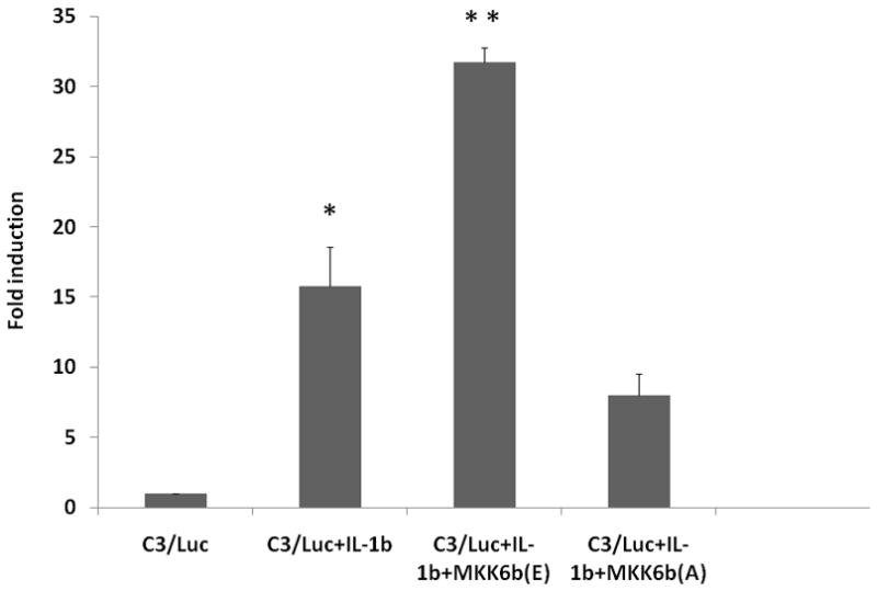
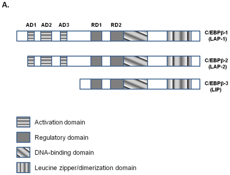
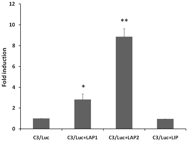
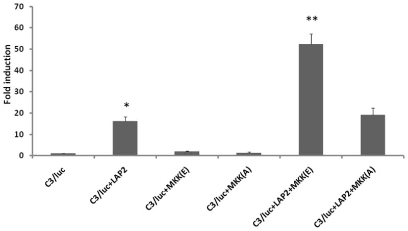
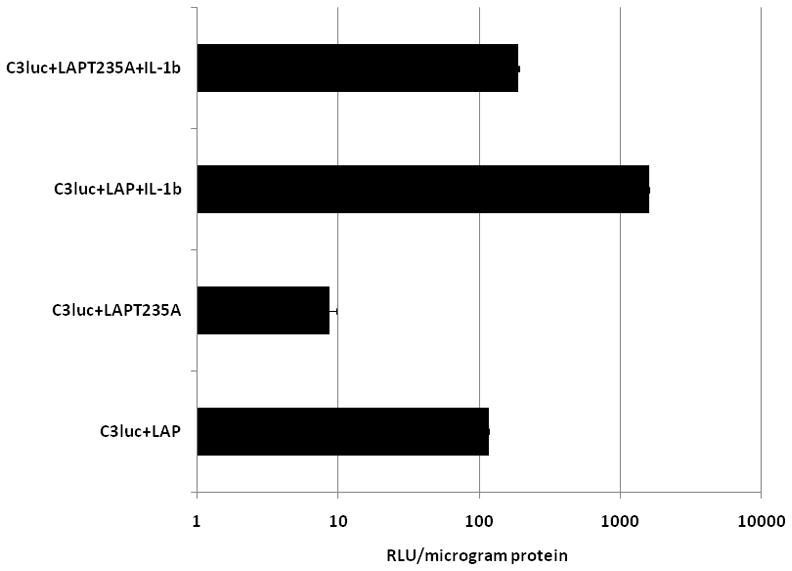
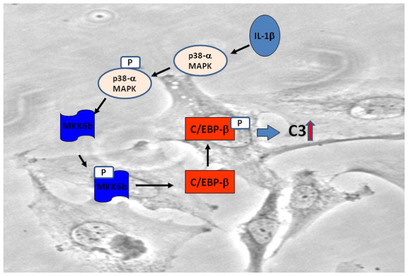
Complement component C3, the central player in the complement cascade and the pro-inflammatory cytokine IL-1β is expressed by activated glial cells and may contribute to neurodegeneration. This study examines the regulation of the expression of C3 by IL-1β in astroglial cells focusing on the role of the upstream kinase MKK6, p38-α MAPK, and C/EBP-β isoforms (LAP1, LAP2, or LIP) in astroglial cells. Activation of human astroglial cell line, U373 with IL-1β, led to the induction of C3 mRNA and protein expression as determined by real-time RT-PCR and Western blot analysis, respectively. This induction was suppressed by the pharmacological inhibitor of p38 MAPK (i.e., SB202190-HCl), suggesting the involvement of p38 MAPK in C3 gene expression. IL-1β also induced C3 promoter activity in U373 cells in a MAP kinase- and C/EBP-β-dependent manner. Cotransfection of C3 luciferase reporter construct with constitutively active form of the upstream kinase in the MAP kinase cascade, that is, MKK6 (the immediate upstream activator of p38 kinase) resulted in marked stimulation of the promoter activity, whereas overexpression of a dominant negative forms of MKK6 and p38α MAPK inhibited C3 promoter activity. Furthermore, a mutant form of C/EBP-β, LAP(T235A) showed reduction in IL-1β-mediated C3 promoter activation. These results suggest that the p38α, MAPK, and MKK6 play prominent roles in IL-1β and C/EBP-β-mediated C3 gene expression in astrocytes.


Figures

Figure 1
Figure 1. The effect of p38 MAPK inhibitor on IL-1β mediated C3 gene induction in U373 cells
A. Role of p38 MAPK in C3 mRNA regulation in IL-1β treated cells U373 cells were either cultured without addition (control) or were pretreated with 10 μM of SB202190-HCl for 30 min alone or were pretreated with 10 μM of SB202190-HCl for 30 min and then treated with IL-1β (10ng/ml) or treated with IL-1β (10ng/ml) alone for 4h. Total RNA was prepared, followed by RT-PCR and real-time PCR for C3 mRNA. Each sample was normalized for the corresponding values of β-actin mRNA as control. Relative quantification of the C3 mRNAs was calculated using the Roche analysis software (based on the 2nd derivative maximum; Roche, Indianapolis, IN, USA). B. Role of IL-1β on p38 MAPK phosphorylation. U373 cells were either cultured without addition (control, lane 1) or were pretreated with 10 μM of SB202190-HCl for 30 min alone (lane 2), or treated with IL-1β (10ng/ml) alone for 30 min (lanes 3–4), or were pretreated with 10μM of SB202190-HCl for 30 min and then treated with IL-1β (10ng/ml) (lanes 5–6). Protein lysates were prepared and levels of phosphorylated p38 and total p38 were determined by western blot analysis using phospho-p38 and total p38 antibodies (Cell Signaling Technology, MA). C. Role of p38 MAPK in C3 protein expression in IL-1β treated cells. U373 cells were either cultured without addition (control, lane 1); treated with IL-1β (10ng/ml) alone (lane 2); pretreated with 10 μM of SB202190-HCl for 30 min alone (lane 3); pretreated with 10 μM of SB202190-HCl for 30 min and then treated with IL-1β (10ng/ml) (lane 4). Protein lysates were prepared 24 h post treatment with IL-1β, followed by western blot analysis using C3 antibody. The membranes were probed with Grb2 antibody as loading control. The experiments were repeated twice.
Figure 2
Figure 2. Role of MKK6b in IL-1β mediated C3 promoter activation
Fold induction of C3 promoter activity in U373 cells transiently co-transfected with C3/Luc reporter vector and 1 μg of empty expression vector or expression vector for MKK6b(A) or MKK6b(E) and then treated 24h post transfection with IL-1β (10ng/ml). Cells were harvested 48h post transfection to assess luciferase activity. RLU/s was normalized to protein concentration and expressed as fold induction wherein the activity in C3/Luc alone transfected cells is set at 1. Statistical significance (*, p<0.05 between control and IL-1β treated cells; **, p<0.05 between IL-1β alone treated cells and IL-1β treated MKK6(E) transfected cells) was analyzed by Student’s t-test.
Figure 3
Figure 3. Role of C/EBP-β isoforms in C3 promoter activation
A. Schematic representation of the three isoforms of C/EBP-β generated as a result of alternate translation. B. Fold induction of C3 promoter activity in U373 cells transiently co-transfected with C3/Luc reporter vector and 1 μg of empty expression vector or expression vector for C/EBP-β1 (LAP1) or C/EBP-β2 (LAP2) or LIP. Cells were harvested 48h post transfection to assess luciferase activity. RLU/s was normalized to protein concentration and expressed as fold induction wherein the activity in C3/Luc alone transfected cells is set at 1. Statistical significance (*, p<0.05 between C3/Luc alone and C3/Luc plus LAP1 transfected cells; **, p<0.05 between between C3/Luc alone and C3/Luc plus LAP2 transfected cells) was analyzed by Student’s t-test.
Figure 4
Figure 4. Role of p38-α MAPK in C/EBP-β mediated C3 promoter activation
Fold induction of C3 promoter activity in U373 cells transiently co-transfected with C3/Luc reporter vector and 1 μg of empty expression vector or expression vector for (LAP2) and p38-α mutant (AF) or treated with p-38 MAPK inhibitor, SB202190-HCl (10 μM) 24hr after transfection. Cells were harvested 48h post transfection to assess luciferase activity. RLU/s was normalized to protein concentration and expressed as fold induction wherein the activity in C3/Luc alone transfected cells is set at 1. Statistical significance (*, p<0.05 between C3/Luc alone and C3/Luc plus LAP2 transfected cells) was analyzed by Student’s t-test.
Figure 5
Figure 5. Role of MKK6b in LAP2 mediated C3 promoter activation
Fold induction of C3 promoter activity in U373 cells transiently co-transfected with C3/Luc reporter vector and 1 μg of empty expression vector or expression vector for MKK6b(A) or MKK6b(E) and LAP2. Cells were harvested 48h post transfection to measure luciferase activity. RLU/s was normalized to protein concentration and expressed as fold induction wherein the activity in C3/Luc alone transfected cells is set at 1. Statistical significance (*, p<0.05 between C3/Luc alone and C3/Luc plus LAP2 transfected cells; **, p<0.05 between between C3/Luc plus MKK6b(E) alone and C3/Luc plus LAP2 and MKK6b(E) transfected cells) was analyzed by Student’s t-test.
Figure 6
Figure 6. Phosphorylation of C-EBP-β is essential for C3 promoter activation by IL-1β
Relative luciferase activity in U373 cells transiently co-transfected with C3/Luc reporter vector and 1μg of empty expression vector or expression vector for wild type LAP or mutant LAPT235A. 24h post transfection cells were treated with IL-1β (10ng/ml). Cells were harvested 48h post transfection to assess for luciferase activity.
Figure 7
Figure 7. Model illustrating the mechanism of IL-1β mediated C3 gene regulation in astrocytes
Binding of IL-1β to its cognate receptor induces phosphorylation of MKK6b which in turn activates p38-α MAPK. Phosphorylation of p38-α MAPK activates C/EBP-β to induce transcription of C3 gene.

Similar articles

Cited by

References

    1. Aldskogius H, Kozlova EN. Central neuron-glial and glial-glial interactions following axon injury. Prog Neurobiol. 1998;55:1–26. - PubMed
    1. Andoh A, Fujiyama Y, Shimada M, Bamba T. Modulation of complement component (C3 and factor B) biosynthesis by a histone deacetylase inhibitor in human intestinal epithelial cells. Int J Mol Med. 2000;6:51–54. - PubMed
    1. Aouadi M, Jager J, Laurent K, Gonzalez T, Cormont M, Binétruy B, Le Marchand-Brustel Y, Tanti JF, Bost F. p38MAP Kinase activity is required for human primary adipocyte differentiation. FEBS Lett. 2007;581:5591–5596. - PubMed
    1. Barnum SR, Jones JL, Benveniste EN. Interferon-gamma regulation of C3 gene expression in human astroglioma cells. J Neuroimmunol. 1992;38:275–282. - PubMed
    1. Barnum SR, Jones JL, Benveniste EN. Interleukin-1 and tumor necrosis factor-mediated regulation of C3 gene expression in human astroglioma cells. Glia. 1993;7:225–236. - PubMed

Publication types

MeSH terms