

Cyclosporine a mediates pathogenesis of aggressive cutaneous squamous cell carcinoma by augmenting epithelial-mesenchymal transition: role of TGFβ signaling pathway
- PMID: 21308804
- PMCID: PMC3110643
- DOI: 10.1002/mc.20744
Cyclosporine a mediates pathogenesis of aggressive cutaneous squamous cell carcinoma by augmenting epithelial-mesenchymal transition: role of TGFβ signaling pathway
Abstract
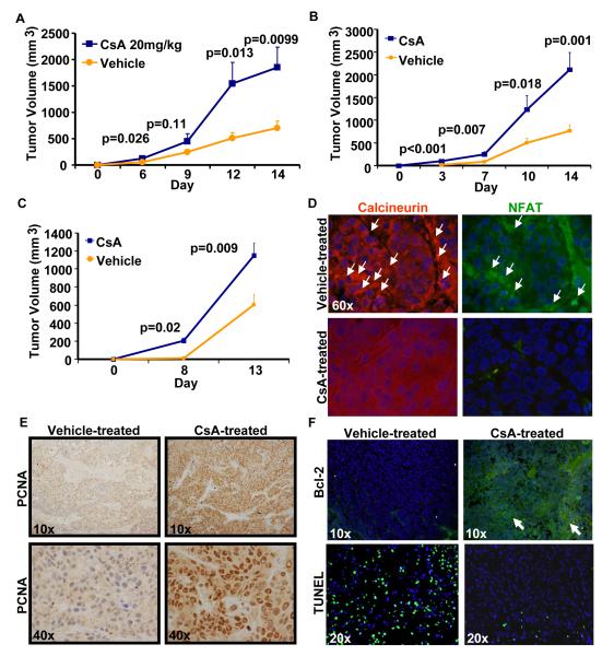
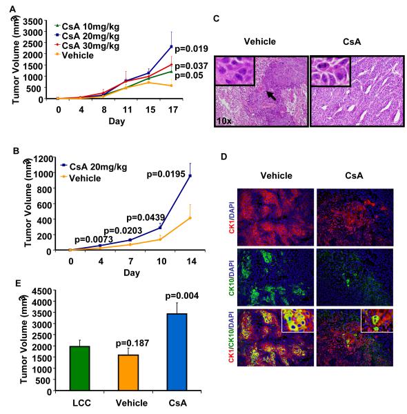
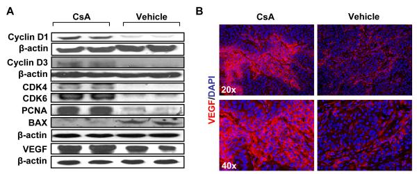
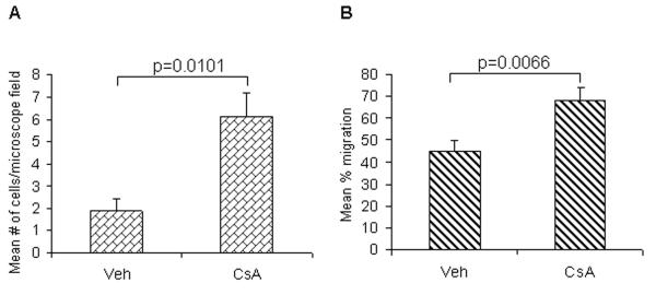
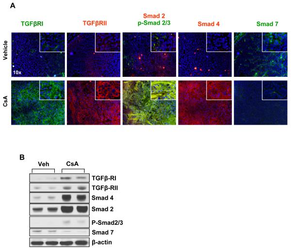
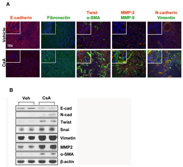
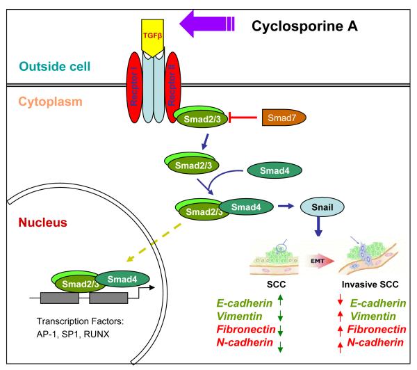
Organ transplant recipients (OTRs) develop multiple aggressive and metastatic non-melanoma skin cancers (NMSCs). Yet, the underlying mechanism remains elusive. Employing a variety of immune-compromised murine models, immunoblotting, immunohistochemical and immunofluorescence techniques, we show that human squamous xenograft tumors in nude mice grow faster and become significantly larger in size following treatment with the immunosuppressive drug, cyclosporine A (CsA). Re-injected tumor cells isolated from CsA-treated xenografts continued to form larger tumors in nude mice than those from vehicle-controls and retained the CsA-signatures of calcineurin signaling inhibition. Similar results were obtained when these tumors were grown in SCID-beige mice or in immuno-competent mice inoculated with syngeinic tumor cells. Consistently, tumors in the CsA group manifested enhanced cellular proliferation and decreased apoptosis. Tumors in CsA-treated animals also showed an augmented epithelial-mesenchymal transition (EMT) characterized by an increased expression of fibronectin, α-SMA, vimentin, N-cadherin, MMP-9/-2, snail and twist with a concomitant decrease in E-cadherin. CsA-treated xenograft tumors manifested increased TGFβ1 expression and TGFβ-dependent signaling characterized by increased nuclear p-Smad 2/3. Our data demonstrate that CsA alters the phenotype of skin SCCs to an invasive and aggressive tumor-type by enhancing expression of proteins regulating EMT acting through the TGFβ1 signaling pathway providing at least one unique mechanism by which multiple aggressive and metastatic NMSCs develop in OTRs.
Copyright © 2011 Wiley-Liss, Inc.
Figures
Similar articles
-
Combined inhibition of p38 and Akt signaling pathways abrogates cyclosporine A-mediated pathogenesis of aggressive skin SCCs.Biochem Biophys Res Commun. 2012 Aug 24;425(2):177-81. doi: 10.1016/j.bbrc.2012.07.062. Epub 2012 Jul 20. Biochem Biophys Res Commun. 2012. PMID: 22820192 Free PMC article.
-
Ruxolitinib inhibits cyclosporine-induced proliferation of cutaneous squamous cell carcinoma.JCI Insight. 2018 Sep 6;3(17):e120750. doi: 10.1172/jci.insight.120750. eCollection 2018 Sep 6. JCI Insight. 2018. PMID: 30185657 Free PMC article.
-
Esophageal Adenocarcinoma Cells and Xenograft Tumors Exposed to Erb-b2 Receptor Tyrosine Kinase 2 and 3 Inhibitors Activate Transforming Growth Factor Beta Signaling, Which Induces Epithelial to Mesenchymal Transition.Gastroenterology. 2017 Jul;153(1):63-76.e14. doi: 10.1053/j.gastro.2017.03.004. Epub 2017 Mar 9. Gastroenterology. 2017. PMID: 28286209
-
Metformin inhibits the radiation-induced invasive phenotype of esophageal squamous cell carcinoma.Int J Oncol. 2016 Nov;49(5):1890-1898. doi: 10.3892/ijo.2016.3676. Epub 2016 Aug 31. Int J Oncol. 2016. PMID: 27599468
-
Epithelial to mesenchymal transition in head and neck squamous cell carcinoma.Oral Oncol. 2013 Apr;49(4):287-92. doi: 10.1016/j.oraloncology.2012.10.009. Epub 2012 Nov 19. Oral Oncol. 2013. PMID: 23182398 Free PMC article. Review.
Cited by
-
Cutaneous Squamous Cell Carcinoma Arising in Immunosuppressed Patients: A Systematic Review of Tumor Profiling Studies.JID Innov. 2022 Mar 30;2(4):100126. doi: 10.1016/j.xjidi.2022.100126. eCollection 2022 Jul. JID Innov. 2022. PMID: 35620703 Free PMC article. Review.
-
Optical-Spectrometry-Based Method for Immunosuppressant Medicine Level Detection in Aqueous Solutions.Sensors (Basel). 2018 Jun 22;18(7):2001. doi: 10.3390/s18072001. Sensors (Basel). 2018. PMID: 29932121 Free PMC article.
-
Cutaneous Squamous Cell Carcinoma: From Biology to Therapy.Int J Mol Sci. 2020 Apr 22;21(8):2956. doi: 10.3390/ijms21082956. Int J Mol Sci. 2020. PMID: 32331425 Free PMC article. Review.
-
Neoplastic disease after liver transplantation: Focus on de novo neoplasms.World J Gastroenterol. 2015 Aug 7;21(29):8753-68. doi: 10.3748/wjg.v21.i29.8753. World J Gastroenterol. 2015. PMID: 26269665 Free PMC article. Review.
-
Unraveling cancer lineage drivers in squamous cell carcinomas.Pharmacol Ther. 2020 Feb;206:107448. doi: 10.1016/j.pharmthera.2019.107448. Epub 2019 Dec 11. Pharmacol Ther. 2020. PMID: 31836455 Free PMC article. Review.
References
-
- Neville JA, Welch E, Leffell DJ. Management of nonmelanoma skin cancer in 2007. Nat Clin Pract Oncol. 2007;4:462–9. - PubMed
-
- Jemal A, Siegel R, Ward E, Hao Y, Xu J, Thun MJ. Cancer statistics, 2009. CA Cancer J Clin. 2009;59:225–49. - PubMed
-
- Ulrich C, Kanitakis J, Stockfleth E, Euvrard S. Skin cancer in organ transplant recipients--where do we stand today? Am J Transplant. 2008;8:2192–8. - PubMed
-
- Euvrard S, Kanitakis J, Claudy A. Skin cancers after organ transplantation. N Engl J Med. 2003;348:1681–91. - PubMed
-
- Otley CC, Cherikh WS, Salasche SJ, McBride MA, Christenson LJ, Kauffman HM. Skin cancer in organ transplant recipients: effect of pretransplant end-organ disease. J Am Acad Dermatol. 2005;53:783–90. - PubMed
Publication types
MeSH terms
Substances
Grants and funding
LinkOut - more resources
Full Text Sources
Other Literature Sources
Medical
Research Materials
